|
Clear Cell Renal Cell Carcinoma With Smooth Muscle Stroma
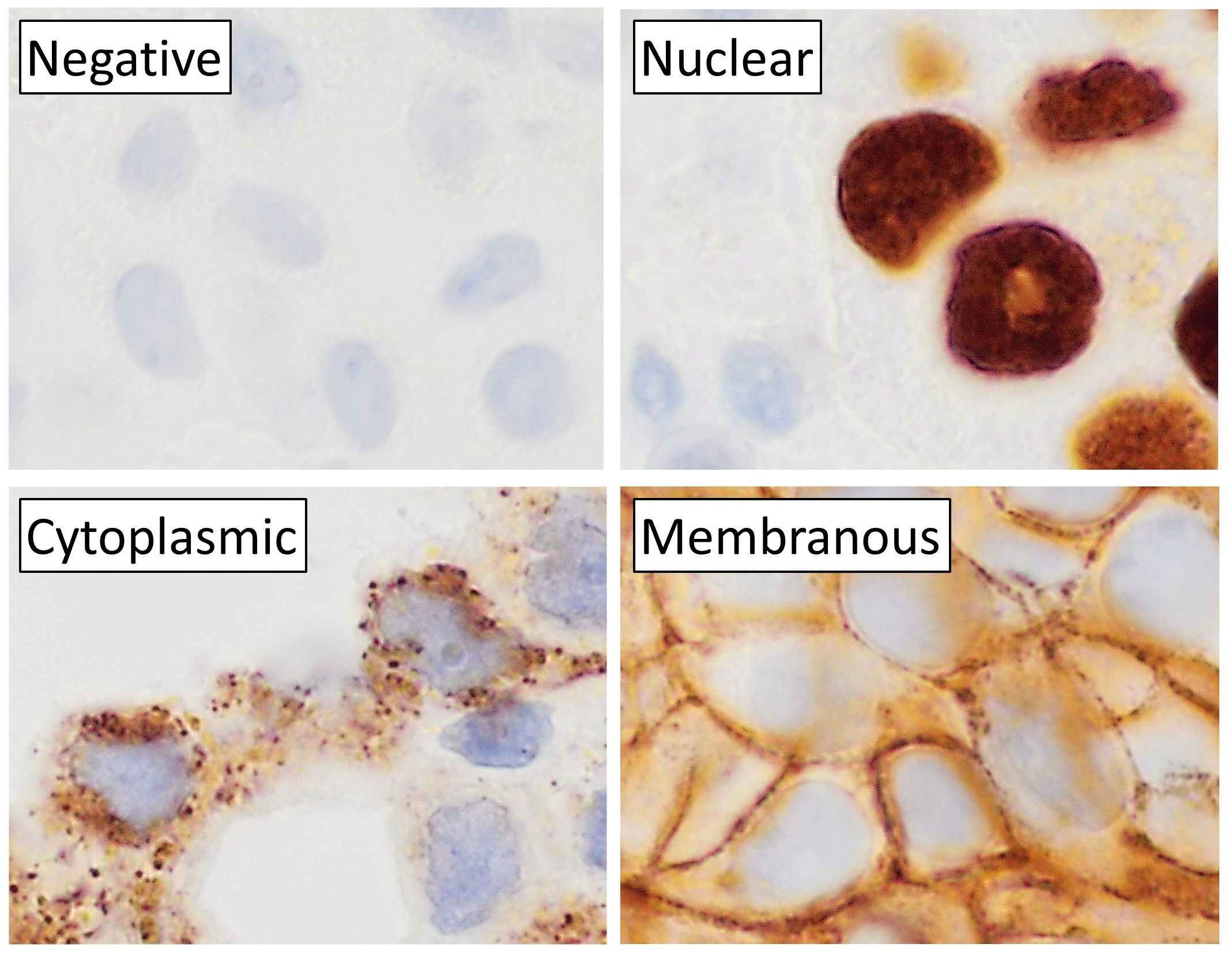
Clear cell papillary renal cell carcinoma (CCPRCC) is a rare subtype of renal cell carcinoma (RCC) that has histomorphology, microscopic morphologic features of papillary renal cell carcinoma and clear cell renal cell carcinoma, yet is pathologically distinct based on molecular pathology, molecular changes and immunohistochemistry. Pathology CCPRCC classically has apical nuclei, i.e. the nucleus is adjacent to the luminal aspect. In most glandular structures the nuclei are usually basally located, i.e. in the cytoplasm adjacent to the basement membrane. They typically immunostain, stain with CK7 and do not stain with TFE3 and AMACR. See also * Renal cancer * Renal cell carcinoma References {{Tumor morphology Kidney cancer ... [...More Info...] [...Related Items...] OR: [Wikipedia] [Google] [Baidu] |
Micrograph
A micrograph or photomicrograph is a photograph or digital image taken through a microscope or similar device to show a magnified image of an object. This is opposed to a macrograph or photomacrograph, an image which is also taken on a microscope but is only slightly magnified, usually less than 10 times. Micrography is the practice or art of using microscopes to make photographs. A micrograph contains extensive details of microstructure. A wealth of information can be obtained from a simple micrograph like behavior of the material under different conditions, the phases found in the system, failure analysis, grain size estimation, elemental analysis and so on. Micrographs are widely used in all fields of microscopy. Types Photomicrograph A light micrograph or photomicrograph is a micrograph prepared using an optical microscope, a process referred to as ''photomicroscopy''. At a basic level, photomicroscopy may be performed simply by connecting a camera to a microscope, th ... [...More Info...] [...Related Items...] OR: [Wikipedia] [Google] [Baidu] |
Immunohistochemistry
Immunohistochemistry (IHC) is the most common application of immunostaining. It involves the process of selectively identifying antigens (proteins) in cells of a tissue section by exploiting the principle of antibodies binding specifically to antigens in biological tissues. IHC takes its name from the roots "immuno", in reference to antibodies used in the procedure, and "histo", meaning tissue (compare to immunocytochemistry). Albert Coons conceptualized and first implemented the procedure in 1941. Visualising an antibody-antigen interaction can be accomplished in a number of ways, mainly either of the following: * ''Chromogenic immunohistochemistry'' (CIH), wherein an antibody is conjugated to an enzyme, such as peroxidase (the combination being termed immunoperoxidase), that can catalyse a colour-producing reaction. * '' Immunofluorescence'', where the antibody is tagged to a fluorophore, such as fluorescein or rhodamine. Immunohistochemical staining is widely used in the dia ... [...More Info...] [...Related Items...] OR: [Wikipedia] [Google] [Baidu] |
Renal Cancer
Kidney cancer, also known as renal cancer, is a group of cancers that starts in the kidney. Symptoms may include blood in the urine, lump in the abdomen, or back pain. Fever, weight loss, and tiredness may also occur. Complications can include spread to the lungs or brain. The main types of kidney cancer are renal cell cancer (RCC), transitional cell cancer (TCC), and Wilms tumor. RCC makes up approximately 80% of kidney cancers, and TCC accounts for most of the rest. Risk factors for RCC and TCC include smoking, certain pain medications, previous bladder cancer, being overweight, high blood pressure, certain chemicals, and a family history. Risk factors for Wilms tumor include a family history and certain genetic disorders such as WAGR syndrome. Diagnosis maybe suspected based on symptoms, urine testing, and medical imaging. It is confirmed by tissue biopsy. Treatment may include surgery, radiation therapy, chemotherapy, immunotherapy, and targeted therapy. Kidney cancer newly ... [...More Info...] [...Related Items...] OR: [Wikipedia] [Google] [Baidu] |
AMACR
α-Methylacyl-CoA racemase (AMACR, ) is an enzyme that in humans is encoded by the ''AMACR'' gene. AMACR catalysis, catalyzes the following chemical reaction: :(2''R'')-2-methylacyl-CoA \rightleftharpoons (2''S'')-2-methylacyl-CoA In mammalian cells, the enzyme is responsible for converting (2''R'')-methylacyl-CoA esters to their (2''S'')-methylacyl-CoA epimers and known substrates, including coenzyme A esters of pristanic acid (mostly derived from phytanic acid, a 3-methyl branched-chain fatty acid that is abundant in the diet) and bile acids derived from cholesterol. This transformation is required in order to degrade (2''R'')-methylacyl-CoA esters by β-oxidation, which process requires the (2''S'')-epimer. The enzyme is known to be localised in peroxisomes and mitochondria, both of which are known to β-oxidize 2-methylacyl-CoA esters. Nomenclature This enzyme belongs to the family of isomerases, specifically the racemases and epimerases which act on other compounds. The ... [...More Info...] [...Related Items...] OR: [Wikipedia] [Google] [Baidu] |
TFE3
Transcription factor E3 is a protein that in humans is encoded by the ''TFE3'' gene. Function TFE3, a member of the helix-loop-helix family of transcription factors, binds to the mu-E3 motif of the immunoglobulin heavy-chain enhancer and is expressed in many cell types (Henthorn et al., 1991). upplied by OMIMref name="entrez" /> Interactions TFE3 has been shown to interact with: * E2F3, * Microphthalmia-associated transcription factor, and * Mothers against decapentaplegic homolog 3 Mothers against decapentaplegic homolog 3 also known as SMAD family member 3 or SMAD3 is a protein that in humans is encoded by the SMAD3 gene. SMAD3 is a member of the SMAD family of proteins. It acts as a mediator of the signals initiated by t ... Translocations A proportion of renal carcinomas (RCC) that occur in young patients are associated with translocations involving the TFE3 gene at chromosome Xp11.2 PRCC References Further reading * * * * * * * * * * * * ... [...More Info...] [...Related Items...] OR: [Wikipedia] [Google] [Baidu] |
Immunostain
In biochemistry, immunostaining is any use of an antibody-based method to detect a specific protein in a sample. The term "immunostaining" was originally used to refer to the immunohistochemical staining of tissue sections, as first described by Albert Coons in 1941. However, immunostaining now encompasses a broad range of techniques used in histology, cell biology, and molecular biology that use antibody-based staining methods. Techniques Immunohistochemistry Immunohistochemistry or IHC staining of tissue sections (or immunocytochemistry, which is the staining of cells), is perhaps the most commonly applied immunostaining technique. While the first cases of IHC staining used fluorescent dyes (see ''immunofluorescence''), other non-fluorescent methods using enzymes such as peroxidase (see '' immunoperoxidase staining'') and alkaline phosphatase are now used. These enzymes are capable of catalysing reactions that give a coloured product that is easily detectable by light micros ... [...More Info...] [...Related Items...] OR: [Wikipedia] [Google] [Baidu] |
Basement Membrane
The basement membrane is a thin, pliable sheet-like type of extracellular matrix that provides cell and tissue support and acts as a platform for complex signalling. The basement membrane sits between Epithelium, epithelial tissues including mesothelium and endothelium, and the underlying connective tissue. Structure As seen with the electron microscope, the basement membrane is composed of two layers, the basal lamina and the reticular lamina. The underlying connective tissue attaches to the basal lamina with collagen VII anchoring fibrils and fibrillin microfibrils. The basal lamina layer can further be subdivided into two layers based on their visual appearance in electron microscopy. The lighter-colored layer closer to the epithelium is called the lamina lucida, while the denser-colored layer closer to the connective tissue is called the lamina densa. The Electron microscope, electron-dense lamina densa layer is about 30–70 nanometers thick and consists of an underlying ... [...More Info...] [...Related Items...] OR: [Wikipedia] [Google] [Baidu] |
Cytoplasm
In cell biology, the cytoplasm is all of the material within a eukaryotic cell, enclosed by the cell membrane, except for the cell nucleus. The material inside the nucleus and contained within the nuclear membrane is termed the nucleoplasm. The main components of the cytoplasm are cytosol (a gel-like substance), the organelles (the cell's internal sub-structures), and various cytoplasmic inclusions. The cytoplasm is about 80% water and is usually colorless. The submicroscopic ground cell substance or cytoplasmic matrix which remains after exclusion of the cell organelles and particles is groundplasm. It is the hyaloplasm of light microscopy, a highly complex, polyphasic system in which all resolvable cytoplasmic elements are suspended, including the larger organelles such as the ribosomes, mitochondria, the plant plastids, lipid droplets, and vacuoles. Most cellular activities take place within the cytoplasm, such as many metabolic pathways including glycolysis, and proces ... [...More Info...] [...Related Items...] OR: [Wikipedia] [Google] [Baidu] |
Molecular Pathology
Molecular pathology is an emerging discipline within pathology which is focused in the study and diagnosis of disease through the examination of molecules within organs, tissues or bodily fluids. Molecular pathology shares some aspects of practice with both anatomic pathology and clinical pathology, molecular biology, biochemistry, proteomics and genetics, and is sometimes considered a "crossover" discipline. It is multi-disciplinary in nature and focuses mainly on the sub-microscopic aspects of disease. A key consideration is that more accurate diagnosis is possible when the diagnosis is based on both the morphologic changes in tissues (traditional anatomic pathology) and on molecular testing. It is a scientific discipline that encompasses the development of molecular and genetic approaches to the diagnosis and classification of human diseases, the design and validation of predictive biomarkers for treatment response and disease progression, the susceptibility of individuals of ... [...More Info...] [...Related Items...] OR: [Wikipedia] [Google] [Baidu] |
Cell Nucleus
The cell nucleus (pl. nuclei; from Latin or , meaning ''kernel'' or ''seed'') is a membrane-bound organelle found in eukaryotic cells. Eukaryotic cells usually have a single nucleus, but a few cell types, such as mammalian red blood cells, have no nuclei, and a few others including osteoclasts have many. The main structures making up the nucleus are the nuclear envelope, a double membrane that encloses the entire organelle and isolates its contents from the cellular cytoplasm; and the nuclear matrix, a network within the nucleus that adds mechanical support. The cell nucleus contains nearly all of the cell's genome. Nuclear DNA is often organized into multiple chromosomes – long stands of DNA dotted with various proteins, such as histones, that protect and organize the DNA. The genes within these chromosomes are structured in such a way to promote cell function. The nucleus maintains the integrity of genes and controls the activities of the cell by regulating gene expres ... [...More Info...] [...Related Items...] OR: [Wikipedia] [Google] [Baidu] |
Clear Cell Renal Cell Carcinoma
Clear-cell renal-cell carcinoma (CCRCC) is a type of renal-cell carcinoma. Genetics Cytogenetics * Alterations of chromosome 3p segments occurs in 70–90% of CCRCCs * Inactivation of von Hippel–Lindau disease ( VHL) gene by gene mutation and promoter hypermethylation * Gain of chromosome 5q * Loss of chromosomes 8p, 9p, and 14q Molecular genetics Several frequently mutated genes were discovered in CCRCC: VHL, KDM6A/UTX, SETD2, KDM5C/JARID1C and MLL2. PBRM1 is also commonly mutated in CCRCC. Histogenesis CCRCC is derived from the proximal convoluted tubule. Microscopy Generally, the cells have a clear cytoplasm, are surrounded by a distinct cell membrane and contain round and uniform nuclei. Microscopically, CCRCCs are graded by the ISUP/WHO as follows: Updated: Jul 02, 2019 * Grade 1: Inconspicuous and basophilic nucleoli at magnification of 400 times * Grade 2: Clearly visible and eosinophilic nucleoli at magnification of 400 times * Grade 3: Clearly vis ... [...More Info...] [...Related Items...] OR: [Wikipedia] [Google] [Baidu] |
Papillary Renal Cell Carcinoma
Papillary renal cell carcinoma (PRCC) is a malignant, heterogeneous tumor originating from renal tubular epithelial cells of the kidney, which comprises approximately 10-15% of all kidney neoplasms. Based on its morphological features, PRCC can be classified into two main subtypes, which are type 1 (basophilic) and type 2 (eosinophilic). As with other types of renal cell cancer, most cases of PRCC are discovered incidentally without showing specific signs or symptoms of cancer. In advanced stages, hematuria, flank pain, and abdominal mass are the three classic manifestation. While a complete list of the causes of PRCC remains unclear, several risk factors were identified to affect PRCC development, such as genetic mutations, kidney-related disease, environmental and lifestyle risk factors. For pathogenesis, type 1 PRCC is mainly caused by MET gene mutation while type 2 PRCC is associated with several different genetic pathways. For diagnosis, PRCC is detectable through computed to ... [...More Info...] [...Related Items...] OR: [Wikipedia] [Google] [Baidu] |


