|
Cervical Sinus
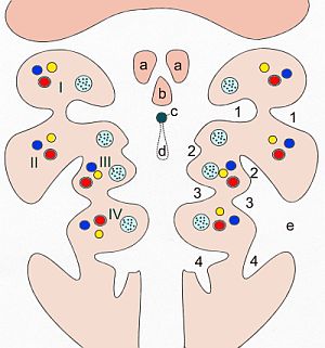
The cervical sinus is a structure formed during embryonic development. It is a deep depression found on each side of the neck. It is formed as the second pharyngeal arch (hyoid arch) grows faster than the other pharyngeal arches, so they become covered. The first pharyngeal arch (mandibular arch) also grows slightly faster. It may fail to obliterate, forming a branchial cleft cyst or fistula, which is prone to infection. Structure The cervical sinus is bounded in front by the second pharyngeal arch (hyoid arch), and behind by the thoracic wall. The second pharyngeal arch (hyoid arch) grows faster than the other pharyngeal arches, so they become covered. It is ultimately obliterated by the fusion of its walls by the 7th week of gestation. Clinical significance Sometimes, the cervical sinus can fail to obliterate and thus remains as a branchial cleft cyst. The second pharyngeal arch may also not grow over the lower pharyngeal arches. This may be found anterior to the sternocl ... [...More Info...] [...Related Items...] OR: [Wikipedia] [Google] [Baidu] |
Pharyngeal Pouch (embryology)
In the embryonic development of vertebrates, pharyngeal pouches form on the endodermal side between the pharyngeal arches. The pharyngeal grooves (or clefts) form the lateral ectodermal surface of the neck region to separate the arches. The pouches line up with the clefts, and these thin segments become gills in fish. Specific pouches First pouch The endoderm lines the future auditory tube (Pharyngotympanic Eustachian tube), middle ear, mastoid antrum, and inner layer of the tympanic membrane. Derivatives of this pouch are supplied by Mandibular nerve. Second pouch * Contributes the middle ear, palatine tonsils, supplied by the facial nerve. Third pouch * The third pouch possesses Dorsal and Ventral wings. Derivatives of the dorsal wings include the inferior parathyroid glands, while the ventral wings fuse to form the cytoreticular cells of the thymus. The main nerve supply to the derivatives of this pouch is Cranial Nerve IX, glossopharyngeal nerve. Fourth pouch Derivatives ... [...More Info...] [...Related Items...] OR: [Wikipedia] [Google] [Baidu] |
Pharyngeal Grooves
A pharyngeal groove (or branchial groove, or pharyngeal cleft) is made up of ectoderm unlike its counterpart the pharyngeal pouch on the endodermal side. The first pharyngeal groove produces the external auditory meatus (ear canal). The rest (2, 3, and 4) are overlapped by the growing 2nd pharyngeal arch, and form the floor of the depression termed the cervical sinus The cervical sinus is a structure formed during embryonic development. It is a deep depression found on each side of the neck. It is formed as the second pharyngeal arch (hyoid arch) grows faster than the other pharyngeal arches, so they become ..., which opens ventrally, and is finally obliterated. See also * Branchial cleft cyst References Animal developmental biology Pharyngeal arches {{developmental-biology-stub ... [...More Info...] [...Related Items...] OR: [Wikipedia] [Google] [Baidu] |
Tuberculum Laterale
The tongue is a muscular organ in the mouth of a typical tetrapod. It manipulates food for mastication and swallowing as part of the digestive process, and is the primary organ of taste. The tongue's upper surface (dorsum) is covered by taste buds housed in numerous lingual papillae. It is sensitive and kept moist by saliva and is richly supplied with nerves and blood vessels. The tongue also serves as a natural means of cleaning the teeth. A major function of the tongue is the enabling of speech in humans and vocalization in other animals. The human tongue is divided into two parts, an oral part at the front and a pharyngeal part at the back. The left and right sides are also separated along most of its length by a vertical section of fibrous tissue (the lingual septum) that results in a groove, the median sulcus, on the tongue's surface. There are two groups of muscles of the tongue. The four intrinsic muscles alter the shape of the tongue and are not attached to bone. The ... [...More Info...] [...Related Items...] OR: [Wikipedia] [Google] [Baidu] |
Tuberculum Impar
The median tongue bud (also tuberculum impar) marks the beginning of the development of the tongue. It appears as a midline swelling from the first pharyngeal arch late in the fourth week of embryogenesis. In the fifth week, a pair of lateral lingual swelling The tongue is a muscular organ (anatomy), organ in the mouth of a typical tetrapod. It manipulates food for mastication and swallowing as part of the digestive system, digestive process, and is the primary organ of taste. The tongue's upper surfa ...s (or ''distal tongue buds'') develop above and in line with the median tongue bud. These swellings grow downwards towards each other, quickly overgrowing the median tongue bud. The line of the fusion of the distal tongue buds is marked by the median sulcus. References External links * Embryology {{Portal bar, Anatomy ... [...More Info...] [...Related Items...] OR: [Wikipedia] [Google] [Baidu] |
Foramen Cecum (tongue)
The tongue is a muscular organ in the mouth of a typical tetrapod. It manipulates food for mastication and swallowing as part of the digestive process, and is the primary organ of taste. The tongue's upper surface (dorsum) is covered by taste buds housed in numerous lingual papillae. It is sensitive and kept moist by saliva and is richly supplied with nerves and blood vessels. The tongue also serves as a natural means of cleaning the teeth. A major function of the tongue is the enabling of speech in humans and vocalization in other animals. The human tongue is divided into two parts, an oral part at the front and a pharyngeal part at the back. The left and right sides are also separated along most of its length by a vertical section of fibrous tissue (the lingual septum) that results in a groove, the median sulcus, on the tongue's surface. There are two groups of muscles of the tongue. The four intrinsic muscles alter the shape of the tongue and are not attached to bone ... [...More Info...] [...Related Items...] OR: [Wikipedia] [Google] [Baidu] |
Ductus Thyreoglossus
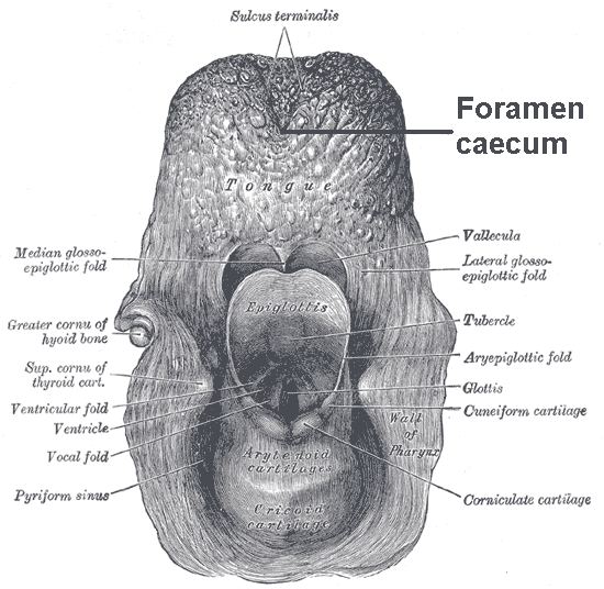
The thyroglossal duct is an embryological anatomical structure forming an open connection between the initial area of development of the thyroid gland and its final position. It is located exactly mid-line, between the anterior 2/3 and posterior 1/3 of the tongue. The thyroid gland starts developing in the oropharynx in the fetus and descends to its final position taking a path through the tongue, hyoid bone and neck muscles. The connection between its original position and its final position is the thyroglossal duct. This duct normally atrophies and closes off as the foramen cecum before birth but can remain open in some people. Clinical significance A thyroglossal duct that fails to atrophy is called a persistent thyroglossal duct, a condition that may lead to the formation of a thyroglossal duct cyst A thyroglossal cyst is a fibrous cyst that forms from a persistent thyroglossal duct. Thyroglossal cysts can be defined as an irregular neck mass or a lump which develops fro ... [...More Info...] [...Related Items...] OR: [Wikipedia] [Google] [Baidu] |
Sinus Cervicalis
The cervical sinus is a structure formed during embryonic development. It is a deep depression found on each side of the neck. It is formed as the second pharyngeal arch (hyoid arch) grows faster than the other pharyngeal arches, so they become covered. The first pharyngeal arch (mandibular arch) also grows slightly faster. It may fail to obliterate, forming a branchial cleft cyst or fistula, which is prone to infection. Structure The cervical sinus is bounded in front by the second pharyngeal arch (hyoid arch), and behind by the thoracic wall. The second pharyngeal arch (hyoid arch) grows faster than the other pharyngeal arches, so they become covered. It is ultimately obliterated by the fusion of its walls by the 7th week of gestation. Clinical significance Sometimes, the cervical sinus can fail to obliterate and thus remains as a branchial cleft cyst. The second pharyngeal arch may also not grow over the lower pharyngeal arches. This may be found anterior to the sternocl ... [...More Info...] [...Related Items...] OR: [Wikipedia] [Google] [Baidu] |
Human Embryonic Development
Human embryonic development, or human embryogenesis, is the development and formation of the human embryo. It is characterised by the processes of cell division and cellular differentiation of the embryo that occurs during the early stages of development. In biological terms, the development of the human body entails growth from a one-celled zygote to an adult human being. Fertilization occurs when the sperm cell successfully enters and fuses with an egg cell (ovum). The genetic material of the sperm and egg then combine to form the single cell zygote and the germinal stage of development commences. Embryonic development in the human, covers the first eight weeks of development; at the beginning of the ninth week the embryo is termed a fetus. Human embryology is the study of this development during the first eight weeks after fertilization. The normal period of gestation (pregnancy) is about nine months or 40 weeks. The germinal stage refers to the time from fertilization through ... [...More Info...] [...Related Items...] OR: [Wikipedia] [Google] [Baidu] |
Neck
The neck is the part of the body on many vertebrates that connects the head with the torso. The neck supports the weight of the head and protects the nerves that carry sensory and motor information from the brain down to the rest of the body. In addition, the neck is highly flexible and allows the head to turn and flex in all directions. The structures of the human neck are anatomically grouped into four compartments; vertebral, visceral and two vascular compartments. Within these compartments, the neck houses the cervical vertebrae and cervical part of the spinal cord, upper parts of the respiratory and digestive tracts, endocrine glands, nerves, arteries and veins. Muscles of the neck are described separately from the compartments. They bound the neck triangles. In anatomy, the neck is also called by its Latin names, or , although when used alone, in context, the word ''cervix'' more often refers to the uterine cervix, the neck of the uterus. Thus the adjective ''cervical'' ma ... [...More Info...] [...Related Items...] OR: [Wikipedia] [Google] [Baidu] |
Second Pharyngeal Arch
The pharyngeal arches, also known as visceral arches'','' are structures seen in the embryonic development of vertebrates that are recognisable precursors for many structures. In fish, the arches are known as the branchial arches, or gill arches. In the human embryo, the arches are first seen during the fourth week of development. They appear as a series of outpouchings of mesoderm on both sides of the developing pharynx. The vasculature of the pharyngeal arches is known as the aortic arches. In fish, the branchial arches support the gills. Structure In vertebrates, the pharyngeal arches are derived from all three germ layers (the primary layers of cells that form during embryogenesis). Neural crest cells enter these arches where they contribute to features of the skull and facial skeleton such as bone and cartilage. However, the existence of pharyngeal structures before neural crest cells evolved is indicated by the existence of neural crest-independent mechanisms of phar ... [...More Info...] [...Related Items...] OR: [Wikipedia] [Google] [Baidu] |
Pharyngeal Arch
The pharyngeal arches, also known as visceral arches'','' are structures seen in the embryonic development of vertebrates that are recognisable precursors for many structures. In fish, the arches are known as the branchial arches, or gill arches. In the human embryo, the arches are first seen during the fourth week of development. They appear as a series of outpouchings of mesoderm on both sides of the developing pharynx. The vasculature of the pharyngeal arches is known as the aortic arches. In fish, the branchial arches support the gills. Structure In vertebrates, the pharyngeal arches are derived from all three germ layers (the primary layers of cells that form during embryogenesis). Neural crest cells enter these arches where they contribute to features of the skull and facial skeleton such as bone and cartilage. However, the existence of pharyngeal structures before neural crest cells evolved is indicated by the existence of neural crest-independent mechanisms of pharyng ... [...More Info...] [...Related Items...] OR: [Wikipedia] [Google] [Baidu] |
Branchial Cleft Cyst
A branchial cleft cyst is a cyst as a swelling in the upper part of neck anterior to sternocleidomastoid. It can, but does not necessarily, have an opening to the skin surface, called a fistula. The cause is usually a developmental abnormality arising in the early prenatal period, typically failure of obliteration of the second, third, and fourth branchial cleft, i.e. failure of fusion of the second branchial arches and epicardial ridge in lower part of the neck. Branchial cleft cysts account for almost 20% of neck masses in children. Less commonly, the cysts can develop from the first, third, or fourth clefts, and their location and the location of associated fistulas differs accordingly. Symptoms and signs Most branchial cleft cysts present in late childhood or early adulthood as a solitary, painless mass, which went previously unnoticed, that has now become infected (typically after an upper respiratory tract infection). Fistulas, if present, are asymptomatic until infection ari ... [...More Info...] [...Related Items...] OR: [Wikipedia] [Google] [Baidu] |


