|
Ductus Thyreoglossus
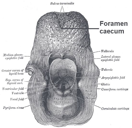
The thyroglossal duct is an embryological anatomical structure forming an open connection between the initial area of development of the thyroid gland and its final position. It is located exactly mid-line, between the anterior 2/3 and posterior 1/3 of the tongue. The thyroid gland starts developing in the oropharynx in the fetus and descends to its final position taking a path through the tongue, hyoid bone and neck muscles. The connection between its original position and its final position is the thyroglossal duct. This duct normally atrophies and closes off as the foramen cecum before birth but can remain open in some people. Clinical significance A thyroglossal duct that fails to atrophy is called a persistent thyroglossal duct, a condition that may lead to the formation of a thyroglossal duct cyst A thyroglossal cyst is a fibrous cyst that forms from a persistent thyroglossal duct. Thyroglossal cysts can be defined as an irregular neck mass or a lump which develops fro ... [...More Info...] [...Related Items...] OR: [Wikipedia] [Google] [Baidu] |
Branchial Pouches
In the embryonic development of vertebrates, pharyngeal pouches form on the endodermal side between the pharyngeal arches. The pharyngeal grooves (or clefts) form the lateral ectodermal surface of the neck region to separate the arches. The pouches line up with the clefts, and these thin segments become gills in fish. Specific pouches First pouch The endoderm lines the future auditory tube (Pharyngotympanic Eustachian tube), middle ear, mastoid antrum, and inner layer of the tympanic membrane. Derivatives of this pouch are supplied by Mandibular nerve. Second pouch * Contributes the middle ear, palatine tonsils, supplied by the facial nerve. Third pouch * The third pouch possesses Dorsal and Ventral wings. Derivatives of the dorsal wings include the inferior parathyroid glands, while the ventral wings fuse to form the cytoreticular cells of the thymus. The main nerve supply to the derivatives of this pouch is Cranial Nerve IX, glossopharyngeal nerve. Fourth pouch Derivatives i ... [...More Info...] [...Related Items...] OR: [Wikipedia] [Google] [Baidu] |
Fetus
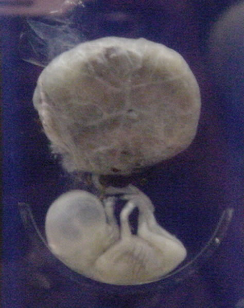
A fetus or foetus (; plural fetuses, feti, foetuses, or foeti) is the unborn offspring that develops from an animal embryo. Following embryonic development the fetal stage of development takes place. In human prenatal development, fetal development begins from the ninth week after fertilization (or eleventh week gestational age) and continues until birth. Prenatal development is a continuum, with no clear defining feature distinguishing an embryo from a fetus. However, a fetus is characterized by the presence of all the major body organs, though they will not yet be fully developed and functional and some not yet situated in their final anatomical location. Etymology The word ''fetus'' (plural ''fetuses'' or '' feti'') is related to the Latin '' fētus'' ("offspring", "bringing forth", "hatching of young") and the Greek "φυτώ" to plant. The word "fetus" was used by Ovid in Metamorphoses, book 1, line 104. The predominant British, Irish, and Commonwealth spelling is '' ... [...More Info...] [...Related Items...] OR: [Wikipedia] [Google] [Baidu] |
Thyroglossal Duct Cyst
A thyroglossal cyst is a fibrous cyst that forms from a persistent thyroglossal duct. Thyroglossal cysts can be defined as an irregular neck mass or a lump which develops from cells and tissues left over after the formation of the thyroid gland during developmental stages.University of Rochester Medical Center. (2015). Thyroglossal duct cyst. Retrieved from http://www.urmc.rochester.edu Thyroglossal cysts are the most common cause of midline neck masses and are generally located caudal to (below) the hyoid bone. These neck masses can occur anywhere along the path of the thyroglossal duct, from the base of the tongue to the suprasternal notch. Other common causes of midline neck masses include lymphadenopathy, dermoid cysts, and various odontogenic anomalies. Thyroglossal cysts develop at birth. Many diagnostic procedures may be used to establish the degree of the cyst. Signs and symptoms Thyroglossal duct cysts most often present with a palpable asymptomatic midline neck mass us ... [...More Info...] [...Related Items...] OR: [Wikipedia] [Google] [Baidu] |
Persistent Thyroglossal Duct
A persistent thyroglossal duct is a usually benign medical condition in which the thyroglossal duct, a structure usually only found during embryology, embryonic development, fails to atrophy. The duct persists as a midline structure forming an open connection between the back of the tongue and the thyroid gland. This opening can lead to fluid accumulation and infection, which necessitate the removal of the duct. Signs and symptoms Studies done on cadavers claim persistent thyroglossal ducts can be completely asymptomatic and found in 7% of the human adult population. However, the continued presence of the duct can often lead to complications due to infections and fluid buildup. The glands in the mucosa of the duct will continue their secretions until the fluid forms a cyst or exit the duct via the opening in the foramen cecum. Local infections, such as colds, tonsillitis, or inflammation of the lymph nodes in the area can also lead to the accumulation of fluid within the duct. Even ... [...More Info...] [...Related Items...] OR: [Wikipedia] [Google] [Baidu] |
Childbirth
Childbirth, also known as labour and delivery, is the ending of pregnancy where one or more babies exits the internal environment of the mother via vaginal delivery or caesarean section. In 2019, there were about 140.11 million births globally. In the developed countries, most deliveries occur in hospitals, while in the developing countries most are home births. The most common childbirth method worldwide is vaginal delivery. It involves four stages of labour: the shortening and opening of the cervix during the first stage, descent and birth of the baby during the second, the delivery of the placenta during the third, and the recovery of the mother and infant during the fourth stage, which is referred to as the postpartum. The first stage is characterized by abdominal cramping or back pain that typically lasts half a minute and occurs every 10 to 30 minutes. Contractions gradually becomes stronger and closer together. Since the pain of childbirth correlates with contractions ... [...More Info...] [...Related Items...] OR: [Wikipedia] [Google] [Baidu] |
Tongue
The tongue is a muscular organ (anatomy), organ in the mouth of a typical tetrapod. It manipulates food for mastication and swallowing as part of the digestive system, digestive process, and is the primary organ of taste. The tongue's upper surface (dorsum) is covered by taste buds housed in numerous lingual papillae. It is sensitive and kept moist by saliva and is richly supplied with nerves and blood vessels. The tongue also serves as a natural means of oral hygiene, cleaning the teeth. A major function of the tongue is the enabling of speech in humans and animal communication, vocalization in other animals. The human tongue is divided into two parts, an oral cavity, oral part at the front and a pharynx, pharyngeal part at the back. The left and right sides are also separated along most of its length by a vertical section of connective tissue, fibrous tissue (the lingual septum) that results in a groove, the median sulcus, on the tongue's surface. There are two groups of muscle ... [...More Info...] [...Related Items...] OR: [Wikipedia] [Google] [Baidu] |
Atrophy
Atrophy is the partial or complete wasting away of a part of the body. Causes of atrophy include mutations (which can destroy the gene to build up the organ), poor nourishment, poor circulation, loss of hormonal support, loss of nerve supply to the target organ, excessive amount of apoptosis of cells, and disuse or lack of exercise or disease intrinsic to the tissue itself. In medical practice, hormonal and nerve inputs that maintain an organ or body part are said to have ''trophic'' effects. A diminished muscular trophic condition is designated as ''atrophy''. Atrophy is reduction in size of cell, organ or tissue, after attaining its normal mature growth. In contrast, hypoplasia is the reduction in the cellular numbers of an organ, or tissue that has not attained normal maturity. Atrophy is the general physiological process of reabsorption and breakdown of tissues, involving apoptosis. When it occurs as a result of disease or loss of trophic support because of other diseases ... [...More Info...] [...Related Items...] OR: [Wikipedia] [Google] [Baidu] |
Muscles Of The Neck
Skeletal muscles (commonly referred to as muscles) are Organ (biology), organs of the vertebrate muscular system and typically are attached by tendons to bones of a skeleton. The muscle cells of skeletal muscles are much longer than in the other types of muscle tissue, and are often known as Skeletal muscle#Skeletal muscle cells, muscle fibers. The muscle tissue of a skeletal muscle is striated muscle tissue, striated – having a striped appearance due to the arrangement of the sarcomeres. Skeletal muscles are voluntary muscles under the control of the somatic nervous system. The other types of muscle are cardiac muscle which is also striated and smooth muscle which is non-striated; both of these types of muscle tissue are classified as involuntary, or, under the control of the autonomic nervous system. A skeletal muscle contains multiple muscle fascicle, fascicles – bundles of muscle fibers. Each individual fiber, and each muscle is surrounded by a type of connective tissue ... [...More Info...] [...Related Items...] OR: [Wikipedia] [Google] [Baidu] |
Hyoid Bone
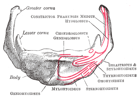
The hyoid bone (lingual bone or tongue-bone) () is a horseshoe-shaped bone situated in the anterior midline of the neck between the chin and the thyroid cartilage. At rest, it lies between the base of the mandible and the third cervical vertebra. Unlike other bones, the hyoid is only distantly articulated to other bones by muscles or ligaments. It is the only bone in the human body that is not connected to any other bones nearby. The hyoid is anchored by muscles from the anterior, posterior and inferior directions, and aids in tongue movement and swallowing. The hyoid bone provides attachment to the muscles of the floor of the mouth and the tongue above, the larynx below, and the epiglottis and pharynx behind. Its name is derived . Structure The hyoid bone is classed as an irregular bone and consists of a central part called the body, and two pairs of horns, the greater and lesser horns. Body The body of the hyoid bone is the central part of the hyoid bone. *At the fro ... [...More Info...] [...Related Items...] OR: [Wikipedia] [Google] [Baidu] |
Oropharynx
The pharynx (plural: pharynges) is the part of the throat behind the mouth and nasal cavity, and above the oesophagus and trachea (the tubes going down to the stomach and the lungs). It is found in vertebrates and invertebrates, though its structure varies across species. The pharynx carries food and air to the esophagus and larynx respectively. The flap of cartilage called the epiglottis stops food from entering the larynx. In humans, the pharynx is part of the digestive system and the conducting zone of the respiratory system. (The conducting zone—which also includes the nostrils of the nose, the larynx, trachea, bronchi, and bronchioles—filters, warms and moistens air and conducts it into the lungs). The human pharynx is conventionally divided into three sections: the nasopharynx, oropharynx, and laryngopharynx. It is also important in vocalization. In humans, two sets of pharyngeal muscles form the pharynx and determine the shape of its lumen. They are arranged as an in ... [...More Info...] [...Related Items...] OR: [Wikipedia] [Google] [Baidu] |
Pharyngeal Grooves
A pharyngeal groove (or branchial groove, or pharyngeal cleft) is made up of ectoderm unlike its counterpart the pharyngeal pouch on the endodermal side. The first pharyngeal groove produces the external auditory meatus (ear canal). The rest (2, 3, and 4) are overlapped by the growing 2nd pharyngeal arch, and form the floor of the depression termed the cervical sinus The cervical sinus is a structure formed during embryonic development. It is a deep depression found on each side of the neck. It is formed as the second pharyngeal arch (hyoid arch) grows faster than the other pharyngeal arches, so they become ..., which opens ventrally, and is finally obliterated. See also * Branchial cleft cyst References Animal developmental biology Pharyngeal arches {{developmental-biology-stub ... [...More Info...] [...Related Items...] OR: [Wikipedia] [Google] [Baidu] |
Tongue
The tongue is a muscular organ (anatomy), organ in the mouth of a typical tetrapod. It manipulates food for mastication and swallowing as part of the digestive system, digestive process, and is the primary organ of taste. The tongue's upper surface (dorsum) is covered by taste buds housed in numerous lingual papillae. It is sensitive and kept moist by saliva and is richly supplied with nerves and blood vessels. The tongue also serves as a natural means of oral hygiene, cleaning the teeth. A major function of the tongue is the enabling of speech in humans and animal communication, vocalization in other animals. The human tongue is divided into two parts, an oral cavity, oral part at the front and a pharynx, pharyngeal part at the back. The left and right sides are also separated along most of its length by a vertical section of connective tissue, fibrous tissue (the lingual septum) that results in a groove, the median sulcus, on the tongue's surface. There are two groups of muscle ... [...More Info...] [...Related Items...] OR: [Wikipedia] [Google] [Baidu] |

.jpg)


