|
Carotid Triangle
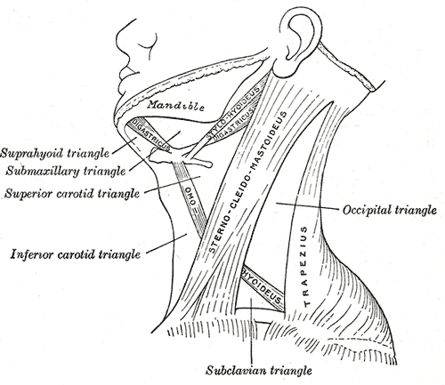
The carotid triangle (or superior carotid triangle) is a portion of the anterior triangle of the neck. Coverings and boundaries It is bounded: * Posteriorly by the anterior border of the Sternocleidomastoid; * Anteroinferiorly, by the superior belly of the Omohyoid muscle. * Superiorly by the posterior belly of the digastric muscle. It is (covered) by the integument, superficial fascia, Platysma and deep fascia; ramifying in which are branches of the facial and cutaneous cervical nerves. Its floor is formed by parts of the *Thyrohyoid membrane, *Hyoglossus, and the * Constrictores pharyngis medius and inferior. Arteries The external and internal carotids lie side by side, the external being the more anterior of the two. The following branches of the external carotid are also met with in this space: * the superior thyroid artery, running forward and downward; * the lingual artery, directly forward; * the facial artery, forward and upward; * the occipital artery, backward a ... [...More Info...] [...Related Items...] OR: [Wikipedia] [Google] [Baidu] |
Anterior Triangle Of The Neck
The anterior triangle is a region of the neck. Structure The triangle is inverted with its apex inferior to its base which is under the chin. Investing fascia covers the roof of the triangle while visceral fascia covers the floor. Anatomy Muscles: * Suprahyoid muscles - Digastric (Ant and Post Belly), mylohyoid, geniohyoid and Stylohyoid. * Infrahyoid muscles - Omohyoid, Sternohyoid, Sternothyroid, and Thyrohyoid. Nerve supply 2 Bellies of Digastric * Anterior: Mylohyoid nerve * Posterior: Facial nerve Stylohyoid: by the facial nerve, by a branch from that to the posterior belly of digastric. Mylohyoid: by its own nerve, a branch of the inferior alveolar ( from the mandibular division of trigeminal nerve), which arises just before the parent nerve enters the mandibular foramen, pierces the sphenomandibular ligament, and runs forward on the inferior surface of the mylohyoid, supplying it and the anterior belly of the digastric. Geniohyoid: by a branch from the hypoglossal ... [...More Info...] [...Related Items...] OR: [Wikipedia] [Google] [Baidu] |
Ascending Pharyngeal
The ascending pharyngeal artery is an artery in the neck that supplies the pharynx, developing from the proximal part of the embryonic second aortic arch. It is the smallest branch of the external carotid and is a long, slender vessel, deeply seated in the neck, beneath the other branches of the external carotid and under the stylopharyngeus muscle. It lies just superior to the bifurcation of the common carotid arteries. The artery most typically bifurcates into embryologically distinct pharyngeal and neuromeningeal trunks. The pharyngeal trunk usually consists of several branches which supply the middle and inferior pharyngeal constrictor muscles and the stylopharyngeus, ramifying in their substance and in the mucous membranes lining them. These branches are in hemodynamic equilibrium with contributors from the internal maxillary artery. The neuromeningeal trunk classically consists of jugular and hypoglossal divisions, which enter the jugular and hypoglossal foramina to supp ... [...More Info...] [...Related Items...] OR: [Wikipedia] [Google] [Baidu] |
Anterior Triangle Of The Neck
The anterior triangle is a region of the neck. Structure The triangle is inverted with its apex inferior to its base which is under the chin. Investing fascia covers the roof of the triangle while visceral fascia covers the floor. Anatomy Muscles: * Suprahyoid muscles - Digastric (Ant and Post Belly), mylohyoid, geniohyoid and Stylohyoid. * Infrahyoid muscles - Omohyoid, Sternohyoid, Sternothyroid, and Thyrohyoid. Nerve supply 2 Bellies of Digastric * Anterior: Mylohyoid nerve * Posterior: Facial nerve Stylohyoid: by the facial nerve, by a branch from that to the posterior belly of digastric. Mylohyoid: by its own nerve, a branch of the inferior alveolar ( from the mandibular division of trigeminal nerve), which arises just before the parent nerve enters the mandibular foramen, pierces the sphenomandibular ligament, and runs forward on the inferior surface of the mylohyoid, supplying it and the anterior belly of the digastric. Geniohyoid: by a branch from the hypoglossal ... [...More Info...] [...Related Items...] OR: [Wikipedia] [Google] [Baidu] |
Superior Laryngeal Nerve
The superior laryngeal nerve is a branch of the vagus nerve. It arises from the middle of the inferior ganglion of vagus nerve and in its course receives a branch from the superior cervical ganglion of the sympathetic nervous system. The superior laryngeal nerve consists of two branches: the internal laryngeal nerve (sensory), which supplies sensory fibers to the laryngeal mucosa, and the external laryngeal nerve (motor), which innervates the cricothyroid muscle. The superior laryngeal nerve innervates the two cricothyroid muscles. The recurrent laryngeal nerve gets its name from the fact that it loops below the aorta on its way to the intrinsic muscles of the larynx. The left recurrent laryngeal nerve passes under and around the aorta on its way to the larynx, whereas the right recurrent laryngeal nerve passes under and around the subclavian artery. Because the aorta is inferior to the subclavian artery, the left recurrent laryngeal nerve is a bit longer than the right recurre ... [...More Info...] [...Related Items...] OR: [Wikipedia] [Google] [Baidu] |
Hyoid Bone
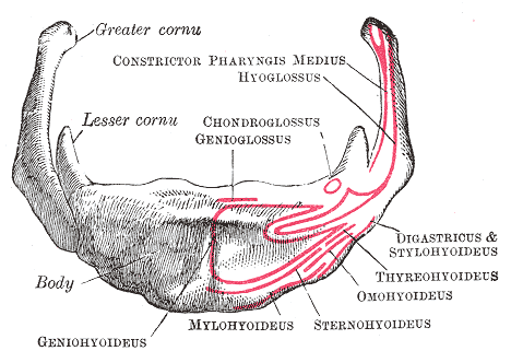
The hyoid bone (lingual bone or tongue-bone) () is a horseshoe-shaped bone situated in the anterior midline of the neck between the chin and the thyroid cartilage. At rest, it lies between the base of the mandible and the third cervical vertebra. Unlike other bones, the hyoid is only distantly articulated to other bones by muscles or ligaments. It is the only bone in the human body that is not connected to any other bones nearby. The hyoid is anchored by muscles from the anterior, posterior and inferior directions, and aids in tongue movement and swallowing. The hyoid bone provides attachment to the muscles of the floor of the mouth and the tongue above, the larynx below, and the epiglottis and pharynx behind. Its name is derived . Structure The hyoid bone is classed as an irregular bone and consists of a central part called the body, and two pairs of horns, the greater and lesser horns. Body The body of the hyoid bone is the central part of the hyoid bone. *At the fro ... [...More Info...] [...Related Items...] OR: [Wikipedia] [Google] [Baidu] |
External Carotid
The external carotid artery is a major artery of the head and neck. It arises from the common carotid artery when it splits into the external and internal carotid artery. External carotid artery supplies blood to the face and neck. Structure The external carotid artery begins at the upper border of thyroid cartilage, and curves, passing forward and upward, and then inclining backward to the space behind the neck of the mandible, where it divides into the superficial temporal and maxillary artery within the parotid gland. It rapidly diminishes in size as it travels up the neck, owing to the number and large size of its branches. At its origin, this artery is closer to the skin and more medial than the internal carotid, and is situated within the carotid triangle. Development In children, the external carotid artery is somewhat smaller than the internal carotid; but in the adult, the two vessels are of nearly equal size. Relations At the origin, external carotid artery is mor ... [...More Info...] [...Related Items...] OR: [Wikipedia] [Google] [Baidu] |
Sternocleidomastoideus
The sternocleidomastoid muscle is one of the largest and most superficial cervical muscles. The primary actions of the muscle are rotation of the head to the opposite side and flexion of the neck. The sternocleidomastoid is innervated by the accessory nerve. Etymology and location It is given the name ''sternocleidomastoid'' because it originates at the manubrium of the sternum (''sterno-'') and the clavicle (''cleido-'') and has an insertion at the mastoid process of the temporal bone of the skull. Structure The sternocleidomastoid muscle originates from two locations: the manubrium of the sternum and the clavicle. It travels obliquely across the side of the neck and inserts at the mastoid process of the temporal bone of the skull by a thin aponeurosis. The sternocleidomastoid is thick and narrow at its centre, and broader and thinner at either end. The sternal head is a round fasciculus, tendinous in front, fleshy behind, arising from the upper part of the front of the manubri ... [...More Info...] [...Related Items...] OR: [Wikipedia] [Google] [Baidu] |
Accessory Nerve
The accessory nerve, also known as the eleventh cranial nerve, cranial nerve XI, or simply CN XI, is a cranial nerve that supplies the sternocleidomastoid and trapezius muscles. It is classified as the eleventh of twelve pairs of cranial nerves because part of it was formerly believed to originate in the brain. The sternocleidomastoid muscle tilts and rotates the head, whereas the trapezius muscle, connecting to the scapula, acts to shrug the shoulder. Traditional descriptions of the accessory nerve divide it into a spinal part and a cranial part. The cranial component rapidly joins the vagus nerve, and there is ongoing debate about whether the cranial part should be considered part of the accessory nerve proper. Consequently, the term "accessory nerve" usually refers only to nerve supplying the sternocleidomastoid and trapezius muscles, also called the spinal accessory nerve. Strength testing of these muscles can be measured during a neurological examination to assess funct ... [...More Info...] [...Related Items...] OR: [Wikipedia] [Google] [Baidu] |
Sympathetic Trunk
The sympathetic trunks (sympathetic chain, gangliated cord) are a paired bundle of nerve fibers that run from the base of the skull to the coccyx. They are a major component of the sympathetic nervous system. Structure The sympathetic trunk lies just lateral to the vertebral bodies for the entire length of the vertebral column. It interacts with the anterior rami of spinal nerves by way of rami communicantes. The sympathetic trunk permits preganglionic fibers of the sympathetic nervous system to ascend to spinal levels superior to T1 and descend to spinal levels inferior to L2/3.Greenstein B., Greenstein A. (2002): Color atlas of neuroscience – Neuroanatomy and neurophysiology. Thieme, Stuttgart – New York, . The superior end of it is continued upward through the carotid canal into the skull, and forms a plexus on the internal carotid artery; the inferior part travels in front of the coccyx, where it converges with the other trunk at a structure known as the ganglion impar. ... [...More Info...] [...Related Items...] OR: [Wikipedia] [Google] [Baidu] |
Vagus Nerve
The vagus nerve, also known as the tenth cranial nerve, cranial nerve X, or simply CN X, is a cranial nerve that interfaces with the parasympathetic control of the heart, lungs, and digestive tract. It comprises two nerves—the left and right vagus nerves—but they are typically referred to collectively as a single subsystem. The vagus is the longest nerve of the autonomic nervous system in the human body and comprises both sensory and motor fibers. The sensory fibers originate from neurons of the nodose ganglion, whereas the motor fibers come from neurons of the dorsal motor nucleus of the vagus and the nucleus ambiguus. The vagus was also historically called the pneumogastric nerve. Structure Upon leaving the medulla oblongata between the olive and the inferior cerebellar peduncle, the vagus nerve extends through the jugular foramen, then passes into the carotid sheath between the internal carotid artery and the internal jugular vein down to the neck, chest, and abdom ... [...More Info...] [...Related Items...] OR: [Wikipedia] [Google] [Baidu] |
Cervical Plexus
The cervical plexus is a plexus of the anterior rami of the first four cervical spinal nerves which arise from C1 to C4 cervical segment in the neck. They are located laterally to the transverse processes between prevertebral muscles from the medial side and vertebral (m. scalenus, m. levator scapulae, m. splenius cervicis) from lateral side. There is anastomosis with accessory nerve, hypoglossal nerve and sympathetic trunk. It is located in the neck, deep to the sternocleidomastoid muscle. Nerves formed from the cervical plexus innervate the back of the head, as well as some neck muscles. The branches of the cervical plexus emerge from the posterior triangle at the nerve point, a point which lies midway on the posterior border of the sternocleidomastoid. Branches The cervical plexus has two types of branches: cutaneous and muscular. *Cutaneous (4 branches): **Lesser occipital nerve - innervates the skin and the scalp posterosuperior to the auricle (C2) **Great auricular nerve - ... [...More Info...] [...Related Items...] OR: [Wikipedia] [Google] [Baidu] |
Ansa Cervicalis
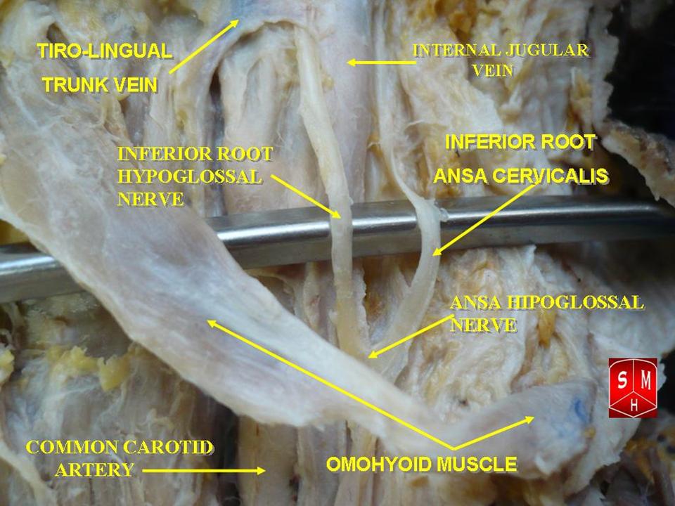
The ansa cervicalis (or ansa hypoglossi in older literature) is a loop of nerves that are part of the cervical plexus. It lies superficial to the internal jugular vein in the carotid triangle. Its name means "handle of the neck" in Latin. Branches from the ansa cervicalis innervate most of the infrahyoid muscles, including the sternothyroid muscle, sternohyoid muscle and the omohyoid muscle. Note that the thyrohyoid muscle, which is also an infrahyoid muscle and the geniohyoid muscle which is a suprahyoid muscle are innervated by cervical spinal nerve 1 via the hypoglossal nerve. Roots Two roots make up the ansa cervicalis, a superior root, and an inferior root. The superior root of the ansa cervicalis is formed from cervical spinal nerve 1 of the cervical plexus. These nerve fibers travel in the hypoglossal nerve before separating in the carotid triangle to form the superior root. The superior root goes around the occipital artery and then descends on the carotid sheath. I ... [...More Info...] [...Related Items...] OR: [Wikipedia] [Google] [Baidu] |


