|
External Carotid
The external carotid artery is a major artery of the head and neck. It arises from the common carotid artery when it splits into the external and internal carotid artery. External carotid artery supplies blood to the face and neck. Structure The external carotid artery begins at the upper border of thyroid cartilage, and curves, passing forward and upward, and then inclining backward to the space behind the neck of the mandible, where it divides into the superficial temporal and maxillary artery within the parotid gland. It rapidly diminishes in size as it travels up the neck, owing to the number and large size of its branches. At its origin, this artery is closer to the skin and more medial than the internal carotid, and is situated within the carotid triangle. Development In children, the external carotid artery is somewhat smaller than the internal carotid; but in the adult, the two vessels are of nearly equal size. Relations At the origin, external carotid artery is mor ... [...More Info...] [...Related Items...] OR: [Wikipedia] [Google] [Baidu] |
Common Carotid Artery
In anatomy, the left and right common carotid arteries (carotids) ( in Merriam-Webster Online Dictionary '.) are that supply the head and neck with ; they divide in the neck to form the and |
Carotid Triangle
The carotid triangle (or superior carotid triangle) is a portion of the anterior triangle of the neck. Coverings and boundaries It is bounded: * Posteriorly by the anterior border of the Sternocleidomastoid; * Anteroinferiorly, by the superior belly of the Omohyoid muscle. * Superiorly by the posterior belly of the digastric muscle. It is (covered) by the integument, superficial fascia, Platysma and deep fascia; ramifying in which are branches of the facial and cutaneous cervical nerves. Its floor is formed by parts of the *Thyrohyoid membrane, *Hyoglossus, and the * Constrictores pharyngis medius and inferior. Arteries The external and internal carotids lie side by side, the external being the more anterior of the two. The following branches of the external carotid are also met with in this space: * the superior thyroid artery, running forward and downward; * the lingual artery, directly forward; * the facial artery, forward and upward; * the occipital artery, backward a ... [...More Info...] [...Related Items...] OR: [Wikipedia] [Google] [Baidu] |
Human Pharynx
The pharynx (plural: pharynges) is the part of the throat behind the mouth and nasal cavity, and above the oesophagus and trachea (the tubes going down to the stomach and the lungs). It is found in vertebrates and invertebrates, though its structure varies across species. The pharynx carries food and air to the esophagus and larynx respectively. The flap of cartilage called the epiglottis stops food from entering the larynx. In humans, the pharynx is part of the digestive system and the conducting zone of the respiratory system. (The conducting zone—which also includes the nostrils of the nose, the larynx, trachea, bronchi, and bronchioles—filters, warms and moistens air and conducts it into the lungs). The human pharynx is conventionally divided into three sections: the nasopharynx, oropharynx, and laryngopharynx. It is also important in vocalization. In humans, two sets of pharyngeal muscles form the pharynx and determine the shape of its lumen. They are arranged as an i ... [...More Info...] [...Related Items...] OR: [Wikipedia] [Google] [Baidu] |
Hyoid Bone
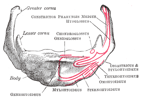
The hyoid bone (lingual bone or tongue-bone) () is a horseshoe-shaped bone situated in the anterior midline of the neck between the chin and the thyroid cartilage. At rest, it lies between the base of the mandible and the third cervical vertebra. Unlike other bones, the hyoid is only distantly articulated to other bones by muscles or ligaments. It is the only bone in the human body that is not connected to any other bones nearby. The hyoid is anchored by muscles from the anterior, posterior and inferior directions, and aids in tongue movement and swallowing. The hyoid bone provides attachment to the muscles of the floor of the mouth and the tongue above, the larynx below, and the epiglottis and pharynx behind. Its name is derived . Structure The hyoid bone is classed as an irregular bone and consists of a central part called the body, and two pairs of horns, the greater and lesser horns. Body The body of the hyoid bone is the central part of the hyoid bone. *At the fro ... [...More Info...] [...Related Items...] OR: [Wikipedia] [Google] [Baidu] |
Facial Nerve
The facial nerve, also known as the seventh cranial nerve, cranial nerve VII, or simply CN VII, is a cranial nerve that emerges from the pons of the brainstem, controls the muscles of facial expression, and functions in the conveyance of taste sensations from the anterior two-thirds of the tongue. The nerve typically travels from the pons through the facial canal in the temporal bone and exits the skull at the stylomastoid foramen. It arises from the brainstem from an area posterior to the cranial nerve VI (abducens nerve) and anterior to cranial nerve VIII (vestibulocochlear nerve). The facial nerve also supplies preganglionic parasympathetic fibers to several head and neck ganglia. The facial and intermediate nerves can be collectively referred to as the nervus intermediofacialis. The path of the facial nerve can be divided into six segments: # intracranial (cisternal) segment # meatal (canalicular) segment (within the internal auditory canal) # labyrinthine segment ... [...More Info...] [...Related Items...] OR: [Wikipedia] [Google] [Baidu] |
Stylohyoideus Muscles
The stylohyoid muscle is a slender muscle, lying anterior and superior of the posterior belly of the digastric muscle. It is one of the suprahyoid muscles. It shares this muscle's innervation by the facial nerve, and functions to draw the hyoid bone backwards and elevate the tongue. Its origin is the styloid process of the temporal bone. It inserts on the body of the hyoid. Structure The stylohyoid muscle originates from the posterior and lateral surface of the styloid process of the temporal bone, near the base. Passing inferior and anterior, it inserts into the body of the hyoid bone, at its junction with the greater cornu, and just superior to the omohyoid muscle. It belongs to the group of suprahyoid muscles. It is perforated, near its insertion, by the intermediate tendon of the digastric muscle. The stylohyoid muscle has vascular supply from the lingual artery, a branch of the external carotid artery. Nerve supply A branch of the facial nerve (CN VII) innervates the st ... [...More Info...] [...Related Items...] OR: [Wikipedia] [Google] [Baidu] |
Digastricus
The digastric muscle (also digastricus) (named ''digastric'' as it has two 'bellies') is a small muscle located under the jaw. The term "digastric muscle" refers to this specific muscle. However, other muscles that have two separate muscle bellies include the suspensory muscle of duodenum, omohyoid, occipitofrontalis. It lies below the body of the mandible, and extends, in a curved form, from the mastoid notch to the mandibular symphysis. It belongs to the suprahyoid muscles group. A broad aponeurotic layer is given off from the tendon of the digastric muscle on either side, to be attached to the body and greater cornu of the hyoid bone; this is termed the suprahyoid aponeurosis. Structure The digastricus (digastric muscle) consists of two muscular bellies united by an intermediate rounded tendon. The two bellies of the digastric muscle have different embryological origins, and are supplied by different cranial nerves. Each person has a right and left digastric muscle. In mo ... [...More Info...] [...Related Items...] OR: [Wikipedia] [Google] [Baidu] |
Superior Thyroid Vein
The superior thyroid vein begins in the substance and on the surface of the thyroid gland, by tributaries corresponding with the branches of the superior thyroid artery, and ends in the upper part of the internal jugular vein. It receives the superior laryngeal vein, superior laryngeal and cricothyroid veins. Additional images File:Gray577.png, The venae cavae and azygos veins with their tributaries. File:Gray1178.png, The thymus of a full-time fetus. References Veins of the head and neck Thyroid {{circulatory-stub ... [...More Info...] [...Related Items...] OR: [Wikipedia] [Google] [Baidu] |
Common Facial
The facial vein usually unites with the anterior branch of the retromandibular vein to form the common facial vein, which crosses the external carotid artery and enters the internal jugular vein at a variable point below the hyoid bone. From near its termination a communicating branch often runs down the anterior border of the sternocleidomastoideus to join the lower part of the anterior jugular vein The anterior jugular vein is a vein in the neck. Structure The anterior jugular vein lies lateral to the cricothyroid ligament. It begins near the hyoid bone by the confluence of several superficial veins from the submandibular region. Its trib .... The common facial vein is not present in all individuals. References External links * () Veins of the head and neck Common vein {{Portal bar, Anatomy ... [...More Info...] [...Related Items...] OR: [Wikipedia] [Google] [Baidu] |
Ranine
The lingual veins begin on the dorsum, sides, and under surface of the tongue, and, passing backward along the course of the lingual artery, end in the internal jugular vein. The vena comitans of the hypoglossal nerve (ranine vein), a branch of considerable size, begins below the tip of the tongue, and may join the lingual; generally, however, it passes backward on the hyoglossus, and joins the common facial. The lingual veins are important clinically as they are capable of rapid absorption of drugs; for this reason, nitroglycerin is given under the tongue to patients suspected of having angina pectoris. Tributaries # Sublingual vein # Deep lingual vein # Dorsal lingual veins # Suprahyoid vein The suprahyoid muscles are four muscles located above the hyoid bone in the neck. They are the digastric, stylohyoid, geniohyoid, and mylohyoid muscles. They are all pharyngeal muscles, with the exception of the geniohyoid muscle. The digastric is ... External links Photo of model (frog ... [...More Info...] [...Related Items...] OR: [Wikipedia] [Google] [Baidu] |
Lingual Veins
The lingual veins begin on the dorsum, sides, and under surface of the tongue, and, passing backward along the course of the lingual artery, end in the internal jugular vein. The vena comitans of the hypoglossal nerve (ranine vein), a branch of considerable size, begins below the tip of the tongue, and may join the lingual; generally, however, it passes backward on the hyoglossus, and joins the common facial. The lingual veins are important clinically as they are capable of rapid absorption of drugs; for this reason, nitroglycerin is given under the tongue to patients suspected of having angina pectoris. Tributaries # Sublingual vein # Deep lingual vein # Dorsal lingual veins # Suprahyoid vein The suprahyoid muscles are four muscles located above the hyoid bone in the neck. They are the digastric, stylohyoid, geniohyoid, and mylohyoid muscles. They are all pharyngeal muscles, with the exception of the geniohyoid muscle. The digastric is ... External links Photo of model (frog ... [...More Info...] [...Related Items...] OR: [Wikipedia] [Google] [Baidu] |
Hypoglossal Nerve
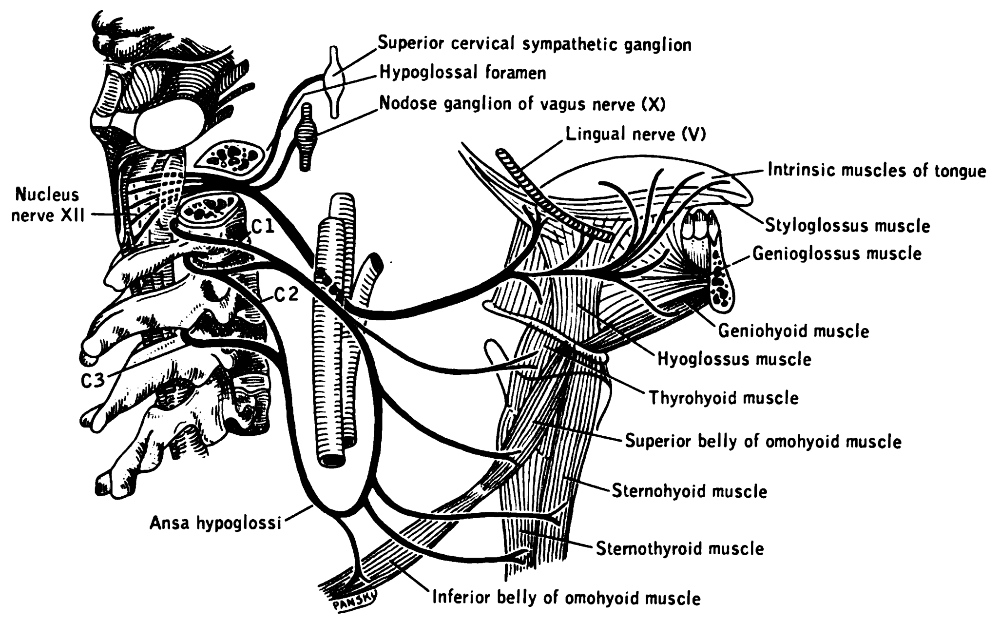
The hypoglossal nerve, also known as the twelfth cranial nerve, cranial nerve XII, or simply CN XII, is a cranial nerve that innervates all the extrinsic and intrinsic muscles of the tongue except for the palatoglossus, which is innervated by the vagus nerve. CN XII is a nerve with a solely motor function. The nerve arises from the hypoglossal nucleus in the medulla as a number of small rootlets, passes through the hypoglossal canal and down through the neck, and eventually passes up again over the tongue muscles it supplies into the tongue. The nerve is involved in controlling tongue movements required for speech and swallowing, including sticking out the tongue and moving it from side to side. Damage to the nerve or the neural pathways which control it can affect the ability of the tongue to move and its appearance, with the most common sources of damage being injury from trauma or surgery, and motor neuron disease. The first recorded description of the nerve is by Herophil ... [...More Info...] [...Related Items...] OR: [Wikipedia] [Google] [Baidu] |


