|
Cardinal Ligament
The cardinal ligament (or Mackenrodt's ligament, lateral cervical ligament, or transverse cervical ligament) is a major ligament of the uterus. It is located at the base of the broad ligament of the uterus. There are a pair of cardinal ligaments in the female human body. Structure The cardinal ligament is a paired structure on the lateral side of the uterus. It originates from the lateral part of the cervix. It attaches to the uterosacral ligament. It attaches the cervix to the lateral pelvic wall by its attachment to the Obturator fascia of the Obturator internus muscle, and is continuous externally with the fibrous tissue that surrounds the pelvic blood vessels. It thus provides support to the uterus. It carries the uterine arteries to provide the primary blood supply to the uterus. Clinical significance The cardinal ligament may be affected in hysterectomy. Due to its close proximity to the ureters, it can get damaged during ligation of the ligament. It is routinely ... [...More Info...] [...Related Items...] OR: [Wikipedia] [Google] [Baidu] |
Major Ligament Of The Uterus
The uterus (from Latin ''uterus'', plural ''uteri'') or womb () is the organ in the reproductive system of most female mammals, including humans that accommodates the embryonic and fetal development of one or more embryos until birth. The uterus is a hormone-responsive sex organ that contains glands in its lining that secrete uterine milk for embryonic nourishment. In the human, the lower end of the uterus, is a narrow part known as the isthmus that connects to the cervix, leading to the vagina. The upper end, the body of the uterus, is connected to the fallopian tubes, at the uterine horns, and the rounded part above the openings to the fallopian tubes is the fundus. The connection of the uterine cavity with a fallopian tube is called the uterotubal junction. The fertilized egg is carried to the uterus along the fallopian tube. It will have divided on its journey to form a blastocyst that will implant itself into the lining of the uterus – the endometrium, where it w ... [...More Info...] [...Related Items...] OR: [Wikipedia] [Google] [Baidu] |
Broad Ligament Of The Uterus
The broad ligament of the uterus is the wide fold of peritoneum that connects the sides of the uterus to the walls and floor of the pelvis. Structure Subdivisions Contents The contents of the broad ligament include the following: * Reproductive ** uterine tubes (or Fallopian tube) ** ovary (some sources consider the ovary to be on the broad ligament, but not in it.) * vessels ** ovarian artery (in the suspensory ligament) ** uterine artery (in reality, travels in the cardinal ligament) * ligaments ** ovarian ligament ** round ligament of uterus ** suspensory ligament of the ovary (Some sources consider it a part of the broad ligament, while other sources just consider it a "termination" of the ligament.) Relations The peritoneum surrounds the uterus like a flat sheet that folds over its fundus, covering it anteriorly and posteriorly; on the sides of the uterus, this sheet of peritoneum comes in direct contact with itself, forming the double layer of peritoneum known as ... [...More Info...] [...Related Items...] OR: [Wikipedia] [Google] [Baidu] |
Female
Female (Venus symbol, symbol: ♀) is the sex of an organism that produces the large non-motile ovum, ova (egg cells), the type of gamete (sex cell) that fuses with the Sperm, male gamete during sexual reproduction. A female has larger gametes than a male. Females and males are results of the anisogamous reproduction system, wherein gametes are of different sizes, unlike isogamy where they are the same size. The exact mechanism of female gamete evolution remains unknown. In species that have males and females, Sex-determination system, sex-determination may be based on either sex chromosomes, or environmental conditions. Most female mammals, including female humans, have two X chromosomes. Female characteristics vary between different species with some species having pronounced Secondary sex characteristic, secondary female sex characteristics, such as the presence of pronounced mammary glands in mammals. In humans, the word ''female'' can also be used to refer to gender i ... [...More Info...] [...Related Items...] OR: [Wikipedia] [Google] [Baidu] |
Uterus
The uterus (from Latin ''uterus'', plural ''uteri'') or womb () is the organ in the reproductive system of most female mammals, including humans that accommodates the embryonic and fetal development of one or more embryos until birth. The uterus is a hormone-responsive sex organ that contains glands in its lining that secrete uterine milk for embryonic nourishment. In the human, the lower end of the uterus, is a narrow part known as the isthmus that connects to the cervix, leading to the vagina. The upper end, the body of the uterus, is connected to the fallopian tubes, at the uterine horns, and the rounded part above the openings to the fallopian tubes is the fundus. The connection of the uterine cavity with a fallopian tube is called the uterotubal junction. The fertilized egg is carried to the uterus along the fallopian tube. It will have divided on its journey to form a blastocyst that will implant itself into the lining of the uterus – the endometrium, where it will ... [...More Info...] [...Related Items...] OR: [Wikipedia] [Google] [Baidu] |
Cervix
The cervix or cervix uteri (Latin, 'neck of the uterus') is the lower part of the uterus (womb) in the human female reproductive system. The cervix is usually 2 to 3 cm long (~1 inch) and roughly cylindrical in shape, which changes during pregnancy. The narrow, central cervical canal runs along its entire length, connecting the uterine cavity and the lumen of the vagina. The opening into the uterus is called the internal os, and the opening into the vagina is called the external os. The lower part of the cervix, known as the vaginal portion of the cervix (or ectocervix), bulges into the top of the vagina. The cervix has been documented anatomically since at least the time of Hippocrates, over 2,000 years ago. The cervical canal is a passage through which sperm must travel to fertilize an egg cell after sexual intercourse. Several methods of contraception, including cervical caps and cervical diaphragms, aim to block or prevent the passage of sperm through the cervical ... [...More Info...] [...Related Items...] OR: [Wikipedia] [Google] [Baidu] |
Uterosacral Ligament
The uterosacral ligaments (or rectouterine ligaments) belong to the major ligaments of uterus. Structure The rectouterine folds contain a considerable amount of fibrous tissue and non-striped muscular fibers which are attached to the front of the sacrum and constitute the uterosacral ligaments.* Netter, Atlas of Human Anatomy, 371 (4th Edition) These ligaments travel from the uterus to the anterior aspect of the sacrum. The pelvic splanchnic nerves Pelvic splanchnic nerves or nervi erigentes are splanchnic nerves that arise from sacral spinal nerves S2, S3, S4 to provide parasympathetic innervation to the organs of the pelvic cavity. Structure The pelvic splanchnic nerves arise from t ... run on top of the ligament. References {{Authority control Ligaments Pelvis Uterus ... [...More Info...] [...Related Items...] OR: [Wikipedia] [Google] [Baidu] |
Hysterectomy
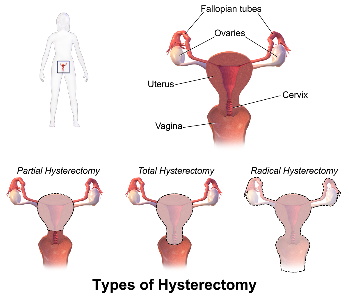
Hysterectomy is the surgical removal of the uterus. It may also involve removal of the cervix, ovaries (oophorectomy), Fallopian tubes (salpingectomy), and other surrounding structures. Usually performed by a gynecologist, a hysterectomy may be total (removing the body, fundus, and cervix of the uterus; often called "complete") or partial (removal of the uterine body while leaving the cervix intact; also called "supracervical"). Removal of the uterus renders the patient unable to bear children (as does removal of ovaries and fallopian tubes) and has surgical risks as well as long-term effects, so the surgery is normally recommended only when other treatment options are not available or have failed. It is the second most commonly performed gynecological surgical procedure, after cesarean section, in the United States. Nearly 68 percent were performed for conditions such as endometriosis, irregular bleeding, and uterine fibroids. It is expected that the frequency of hysterectom ... [...More Info...] [...Related Items...] OR: [Wikipedia] [Google] [Baidu] |
Pelvic Splanchnic Nerves
Pelvic splanchnic nerves or nervi erigentes are splanchnic nerves that arise from sacral spinal nerves S2, S3, S4 to provide parasympathetic innervation to the organs of the pelvic cavity. Structure The pelvic splanchnic nerves arise from the anterior rami of the sacral spinal nerves S2, S3, and S4, and enter the sacral plexus. They travel to their side's corresponding inferior hypogastric plexus, located bilaterally on the walls of the rectum. They contain both preganglionic parasympathetic fibers as well as visceral afferent fibers. Visceral afferent fibers go to spinal cord following pathway of pelvic splanchnic nerve fibers. The parasympathetic nervous system is referred to as the ''craniosacral outflow''; the pelvic splanchnic nerves are the ''sacral'' component. They are in the same region as the sacral splanchnic nerves, which arise from the sympathetic trunk and provide sympathetic efferent fibers. Function The pelvic splanchnic nerves contribute to the inner ... [...More Info...] [...Related Items...] OR: [Wikipedia] [Google] [Baidu] |
Mammal Female Reproductive System
Most mammals are viviparous, giving birth to live young. However, the five species of monotreme, the platypuses and the echidnas, lay eggs. The monotremes have a sex determination system different from that of most other mammals. In particular, the sex chromosomes of a platypus are more like those of a chicken than those of a therian mammal. The mammary glands of mammals are specialized to produce milk, a liquid used by newborns as their primary source of nutrition. The monotremes branched early from other mammals and do not have the teats seen in most mammals, but they do have mammary glands. The young lick the milk from a mammary patch on the mother's belly. Viviparous mammals are in the subclass Theria; those living today are in the Marsupialia and Placentalia infraclasses. A marsupial has a short gestation period, typically shorter than its estrous cycle, and gives birth to an underdeveloped (altricial) newborn that then undergoes further development; in many species, this ta ... [...More Info...] [...Related Items...] OR: [Wikipedia] [Google] [Baidu] |


