|
Carcinocythemia
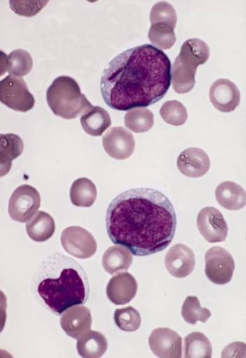
Carcinocythemia, also known as carcinoma cell leukemia, is a condition in which cells from malignant tumours of non-hematopoietic origin are visible on the peripheral blood smear. It is an extremely rare condition, with 33 cases identified in the literature from 1960 to 2018. Carcinocythemia typically occurs secondary to infiltration of the bone marrow by metastatic cancer and carries a very poor prognosis. Presentation Carcinocythemia occurs most commonly in breast cancer, followed by small cell lung cancer, and usually appears late in the course of the disease. Thrombosis and disseminated intravascular coagulation are frequently reported in association with carcinocythemia. The prognosis is poor: a review of 26 patients found that 85% died within 6 months of the diagnosis, with an average time of 6.1 weeks between diagnosis and death. The amount of tumour cells on the blood smear can range from 1 to 80 percent of the total white blood cell count, with lower percentages being ... [...More Info...] [...Related Items...] OR: [Wikipedia] [Google] [Baidu] |
Circulating Tumour Cells
A circulating tumor cell (CTC) is a cell that has shed into the vasculature or lymphatics from a primary tumor and is carried around the body in the blood circulation. CTCs can extravasate and become ''seeds'' for the subsequent growth of additional tumors (metastases) in distant organs, a mechanism that is responsible for the vast majority of cancer-related deaths. The detection and analysis of CTCs can assist early patient prognoses and determine appropriate tailored treatments. Currently, there is one FDA-approved method for CTC detection, CellSearch, which is used to diagnose breast, colorectal and prostate cancer. The detection of CTCs, or liquid biopsy, presents several advantages over traditional tissue biopsies. They are non-invasive, can be used repeatedly, and provide more useful information on metastatic risk, disease progression, and treatment effectiveness. For example, analysis of blood samples from cancer patients has found a propensity for increased CTC detection as t ... [...More Info...] [...Related Items...] OR: [Wikipedia] [Google] [Baidu] |
Manual Differential
A white blood cell differential is a medical laboratory test that provides information about the types and amounts of white blood cells in a person's blood. The test, which is usually ordered as part of a complete blood count (CBC), measures the amounts of the five normal white blood cell typesneutrophils, lymphocytes, monocytes, eosinophils and basophilsas well as abnormal cell types if they are present. These results are reported as percentages and absolute values, and compared against reference ranges to determine whether the values are normal, low, or high. Changes in the amounts of white blood cells can aid in the diagnosis of many health conditions, including viral, bacterial, and parasitic infections and blood disorders such as leukemia. White blood cell differentials may be performed by an automated analyzera machine designed to run laboratory tests – or manually, by examining blood smears under a microscope. The test was performed manually until white blood cell dif ... [...More Info...] [...Related Items...] OR: [Wikipedia] [Google] [Baidu] |
Breast Cancer
Breast cancer is cancer that develops from breast tissue. Signs of breast cancer may include a lump in the breast, a change in breast shape, dimpling of the skin, milk rejection, fluid coming from the nipple, a newly inverted nipple, or a red or scaly patch of skin. In those with distant spread of the disease, there may be bone pain, swollen lymph nodes, shortness of breath, or yellow skin. Risk factors for developing breast cancer include obesity, a lack of physical exercise, alcoholism, hormone replacement therapy during menopause, ionizing radiation, an early age at first menstruation, having children late in life or not at all, older age, having a prior history of breast cancer, and a family history of breast cancer. About 5–10% of cases are the result of a genetic predisposition inherited from a person's parents, including BRCA1 and BRCA2 among others. Breast cancer most commonly develops in cells from the lining of milk ducts and the lobules that supply these ... [...More Info...] [...Related Items...] OR: [Wikipedia] [Google] [Baidu] |
Disseminated Intravascular Coagulation
Disseminated intravascular coagulation (DIC) is a condition in which blood clots form throughout the body, blocking small blood vessels. Symptoms may include chest pain, shortness of breath, leg pain, problems speaking, or problems moving parts of the body. As clotting factors and platelets are used up, bleeding may occur. This may include blood in the urine, blood in the stool, or bleeding into the skin. Complications may include organ failure. Relatively common causes include sepsis, surgery, major trauma, cancer, and complications of pregnancy. Less common causes include snake bites, frostbite, and burns. There are two main types: acute (rapid onset) and chronic (slow onset). Diagnosis is typically based on blood tests. Findings may include low platelets, low fibrinogen, high INR, or high D-dimer. Treatment is mainly directed towards the underlying condition. Other measures may include giving platelets, cryoprecipitate, or fresh frozen plasma. Evidence to support these trea ... [...More Info...] [...Related Items...] OR: [Wikipedia] [Google] [Baidu] |
Monoblast
Monoblasts are the committed progenitor cells that differentiated from a committed macrophage or dendritic cell precursor (MDP) in the process of hematopoiesis. They are the first developmental stage in the monocyte series leading to a macrophage. Their myeloid cell fate is induced by the concentration of cytokines they are surrounded by during development. These cytokines induce the activation of transcription factors which push completion of the monoblast's myeloid cell fate. Monoblasts are normally found in bone marrow and do not appear in the normal peripheral blood. They mature into monocytes which, in turn, develop into macrophages. They then are seen as macrophages in the normal peripheral blood and many different tissues of the body. Macrophages can produce a variety of effector molecules that initiate local, systemic inflammatory responses. These monoblast differentiated cells are equipped to fight off foreign invaders using pattern recognition receptors to detect antigen as ... [...More Info...] [...Related Items...] OR: [Wikipedia] [Google] [Baidu] |
Megakaryoblast
A megakaryoblast is a precursor cell to a promegakaryocyte, which in turn becomes a megakaryocyte during haematopoiesis. It is the beginning of the thrombocytic series. Development The megakaryoblast derives from a CFU-Meg colony unit of pluripotential hemopoietic stem cells. (Some sources use the term "CFU-Meg" to identify the CFU.) The CFU-Meg derives from the CFU-GEMM CFU-GEMM is a colony forming unit that generates myeloid cells. CFU-GEMM cells are the oligopotential progenitor cells for myeloid cells; they are thus also called common myeloid progenitor cells or myeloid stem cells. "GEMM" stands for granulocyte ... (common myeloid progenitor). Structure These cells tend to range from 8μm to 30μm, owing to the variation in size between different megakaryoblasts. The nucleus is three to five times the size of the cytoplasm, and is generally round or oval in shape. Several nucleoli are visible, while the chromatin varies from cell to cell, ranging from fine to heavy and dense. ... [...More Info...] [...Related Items...] OR: [Wikipedia] [Google] [Baidu] |
Cytoplasm
In cell biology, the cytoplasm is all of the material within a eukaryotic cell, enclosed by the cell membrane, except for the cell nucleus. The material inside the nucleus and contained within the nuclear membrane is termed the nucleoplasm. The main components of the cytoplasm are cytosol (a gel-like substance), the organelles (the cell's internal sub-structures), and various cytoplasmic inclusions. The cytoplasm is about 80% water and is usually colorless. The submicroscopic ground cell substance or cytoplasmic matrix which remains after exclusion of the cell organelles and particles is groundplasm. It is the hyaloplasm of light microscopy, a highly complex, polyphasic system in which all resolvable cytoplasmic elements are suspended, including the larger organelles such as the ribosomes, mitochondria, the plant plastids, lipid droplets, and vacuoles. Most cellular activities take place within the cytoplasm, such as many metabolic pathways including glycolysis, and proces ... [...More Info...] [...Related Items...] OR: [Wikipedia] [Google] [Baidu] |
Vacuole
A vacuole () is a membrane-bound organelle which is present in plant and fungal cells and some protist, animal, and bacterial cells. Vacuoles are essentially enclosed compartments which are filled with water containing inorganic and organic molecules including enzymes in solution, though in certain cases they may contain solids which have been engulfed. Vacuoles are formed by the fusion of multiple membrane vesicles and are effectively just larger forms of these. The organelle has no basic shape or size; its structure varies according to the requirements of the cell. Discovery Contractile vacuoles ("stars") were first observed by Spallanzani (1776) in protozoa, although mistaken for respiratory organs. Dujardin (1841) named these "stars" as ''vacuoles''. In 1842, Schleiden applied the term for plant cells, to distinguish the structure with cell sap from the rest of the protoplasm. In 1885, de Vries named the vacuole membrane as tonoplast. Function The function and signifi ... [...More Info...] [...Related Items...] OR: [Wikipedia] [Google] [Baidu] |
Chromatin
Chromatin is a complex of DNA and protein found in eukaryotic cells. The primary function is to package long DNA molecules into more compact, denser structures. This prevents the strands from becoming tangled and also plays important roles in reinforcing the DNA during cell division, preventing DNA damage, and regulating gene expression and DNA replication. During mitosis and meiosis, chromatin facilitates proper segregation of the chromosomes in anaphase; the characteristic shapes of chromosomes visible during this stage are the result of DNA being coiled into highly condensed chromatin. The primary protein components of chromatin are histones. An octamer of two sets of four histone cores (Histone H2A, Histone H2B, Histone H3, and Histone H4) bind to DNA and function as "anchors" around which the strands are wound.Maeshima, K., Ide, S., & Babokhov, M. (2019). Dynamic chromatin organization without the 30-nm fiber. ''Current opinion in cell biology, 58,'' 95–104. https://doi.o ... [...More Info...] [...Related Items...] OR: [Wikipedia] [Google] [Baidu] |
Blast Cell
In cell biology, a precursor cell, also called a blast cell or simply blast, is a partially differentiated cell, usually referred to as a unipotent cell that has lost most of its stem cell properties. A precursor cell is also known as a progenitor cell but progenitor cells are multipotent. Precursor cells are known as the intermediate cell before they become differentiated after being a stem cell. Usually, a precursor cell is a stem cell with the capacity to differentiate into only one cell type. Sometimes, ''precursor cell'' is used as an alternative term for unipotent stem cells. In embryology, precursor cells are a group of cells that later differentiate into one organ. A blastoma is any cancer created by malignancies of precursor cells. Precursor cells, and progenitor cells, have many potential uses in medicine. , there is research being done to use these cells to build heart valves, blood vessels and other tissues, by using blood and muscle precursor, or progenitor cel ... [...More Info...] [...Related Items...] OR: [Wikipedia] [Google] [Baidu] |
Buffy Coat
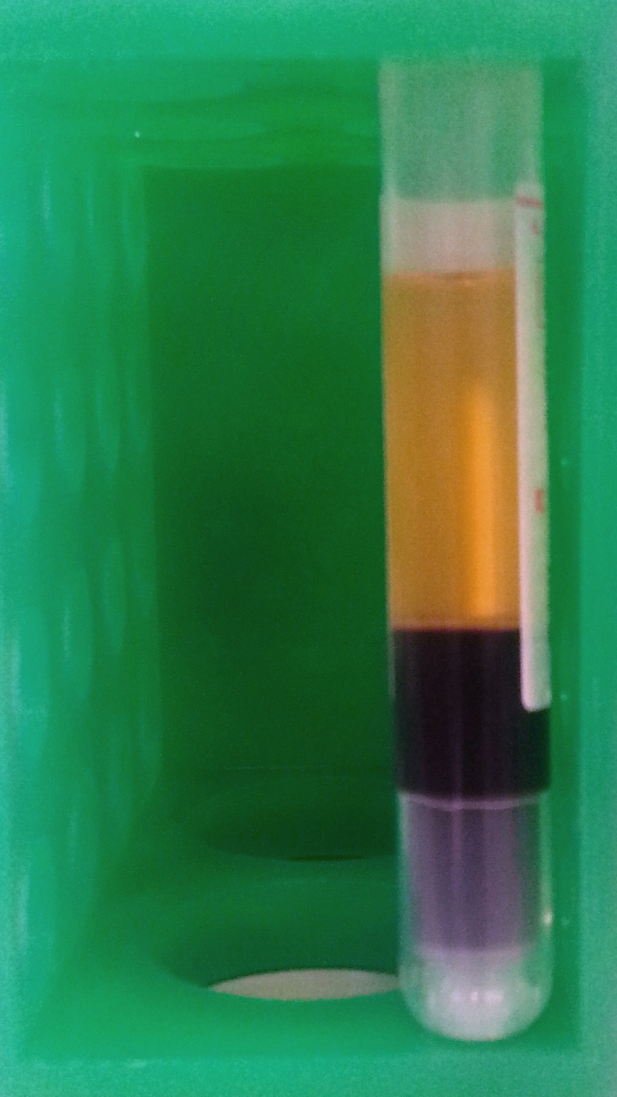
The buffy coat is the fraction of an anticoagulated blood sample that contains most of the white blood cells and platelets following centrifugation. It is rich in a number of immune cells including leukocytes, granulocytes. Description After centrifugation, one can distinguish a layer of clear fluid (the plasma), a layer of red fluid containing most of the red blood cells, and a thin layer in between. Composing less than 1% of the total volume of the blood sample, the buffy coat (so-called because it is usually buff in hue), contains most of the white blood cells and platelets. The buffy coat is usually whitish in color, but is sometimes green if the blood sample contains large amounts of neutrophils, which are high in green-colored myeloperoxidase. The layer beneath the buffy coat contains granulocytes and red blood cells. The buffy coat is commonly used for DNA extraction, with white blood cells providing approximately 10 times more concentrated sources of nucleated c ... [...More Info...] [...Related Items...] OR: [Wikipedia] [Google] [Baidu] |
Blood Smear
A blood smear, peripheral blood smear or blood film is a thin layer of blood smeared on a glass microscope slide and then stained in such a way as to allow the various blood cells to be examined microscopically. Blood smears are examined in the investigation of hematology, hematological (blood) disorders and are routinely employed to look for blood Apicomplexa, parasites, such as those of malaria and filariasis. Preparation A blood smear is made by placing a drop of blood on one end of a slide, and using a ''spreader slide'' to disperse the blood over the slide's length. The aim is to get a region, called a monolayer, where the cells are spaced far enough apart to be counted and differentiated. The monolayer is found in the "feathered edge" created by the spreader slide as it draws the blood forward. The slide is left to air dry, after which the blood is fixation (histology), fixed to the slide by immersing it briefly in methanol. The fixative is essential for good staining a ... [...More Info...] [...Related Items...] OR: [Wikipedia] [Google] [Baidu] |


