|
Cytochemistry
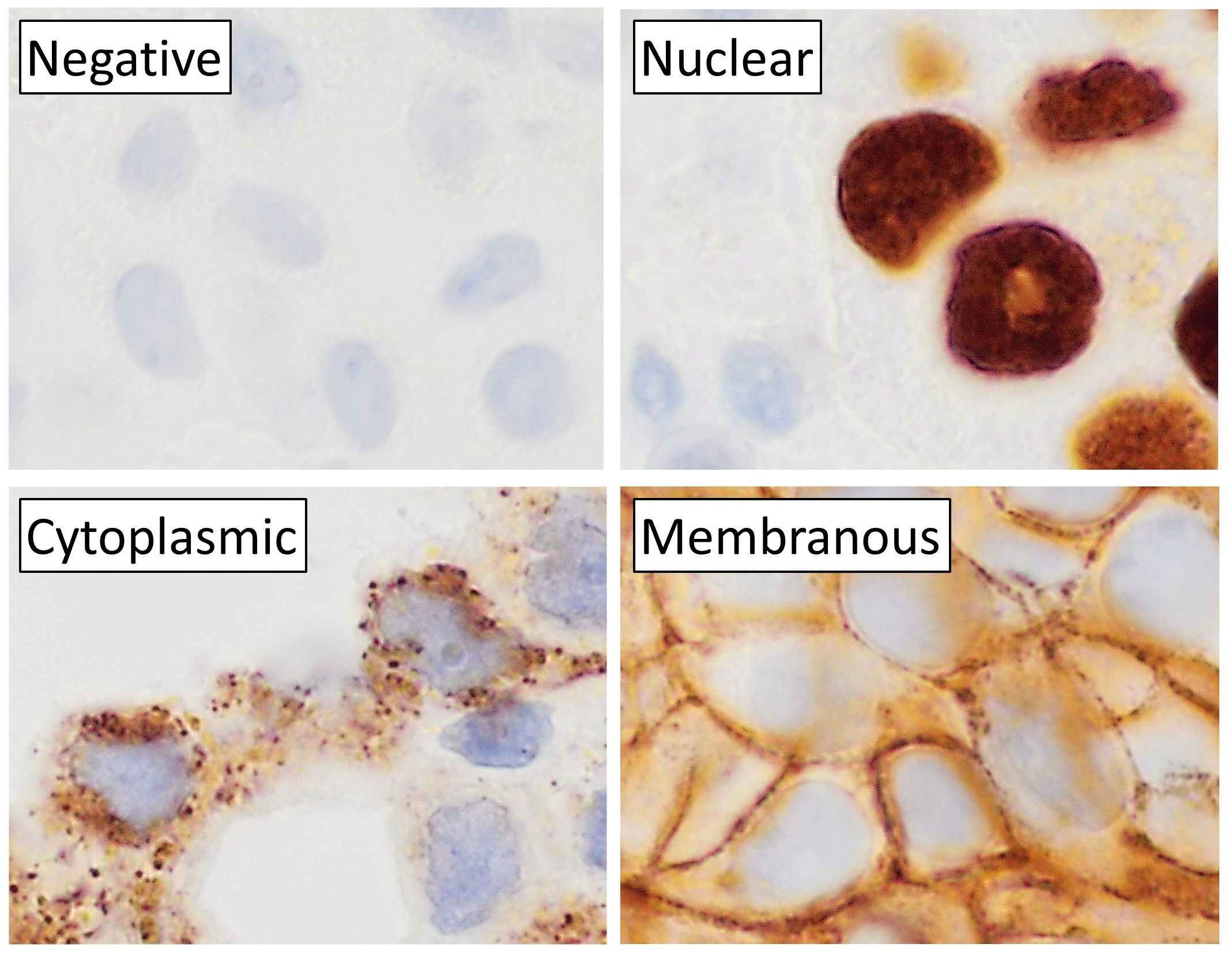
Cytochemistry is the branch of cell biology dealing with the detection of cell constituents by means of biochemical analysis and visualization techniques. This is the study of the localization of cellular components through the use of staining methods. The term is also used to describe a process of identification of the biochemical content of cells. ''Cytochemistry'' is a science of localizing chemical components of cells and cell organelles on thin histological sections by using several techniques like enzyme localization, micro-incineration, micro-spectrophotometry, radioautography, cryo-electron microscopy, X-ray microanalysis by energy-dispersive X-ray spectroscopy Energy-dispersive X-ray spectroscopy (EDS, EDX, EDXS or XEDS), sometimes called energy dispersive X-ray analysis (EDXA or EDAX) or energy dispersive X-ray microanalysis (EDXMA), is an analytical technique used for the elemental analysis or chemi ..., immunohistochemistry and cytochemistry, etc. Freeze Fracture ... [...More Info...] [...Related Items...] OR: [Wikipedia] [Google] [Baidu] |
Micro-incineration
Micro-incineration or microincineration is a technique to determine the manner and distribution of mineral elements in biological cells, biological tissues and organs. Slide preparation of tissues can be used. Examples include calcium (Ca), potassium (K), sodium (Na), magnesium (Mg), iron (Fe), and silicion (Si). The organic matter is vaporised by heating. The nature and position of the mineral ash is determined microscopically. Aqueous or cryo-EM fixed tissue materials can also be examined under transmission and scanning electron microscopy (TEM & SEM). The ashing procedure produces cellular oxidised-residues rich in Na2O, CaO, MgO, Fe2O3, SiO2, Ca(PO4)2, Mg(PO4)2, etc., which are detected by X-ray microanalysis Microanalysis is the chemical identification and quantitative analysis of very small amounts of chemical substances (generally less than 10 mg or 1 ml) or very small surfaces of material (generally less than 1 cm2). One of the pioneer ... with 2-4 t ... [...More Info...] [...Related Items...] OR: [Wikipedia] [Google] [Baidu] |
Micro-spectrophotometry
Microspectrophotometry is the measure of the spectra of microscopic samples using different wavelengths of electromagnetic radiation (e.g. ultraviolet, visible and near infrared, etc.) It is accomplished with ''microspectrophotometers'', ''cytospectrophotometers'', ''microfluorometers'', ''Raman microspectrophotometers'', etc. A microspectrophotometer can be configured to measure transmittance, absorbance, reflectance, light polarization, fluorescence (or other types of luminescence such as photoluminescence) of sample areas less than a micrometer in diameter through a modified optical microscope. Applications The main reason to use microspectrophotometry is the ability to measure the optical spectra of samples with a spatial resolution on the micron scale. Optical spectra may be acquired of either microscopic samples or larger samples with a micron-scale spatial resolution. Another reason microspectrophotometry is useful is that measurements are made without destroying the samp ... [...More Info...] [...Related Items...] OR: [Wikipedia] [Google] [Baidu] |
Radioautography
An autoradiograph is an image on an X-ray film or nuclear emulsion produced by the pattern of decay emissions (e.g., beta particles or gamma rays) from a distribution of a radioactive substance. Alternatively, the autoradiograph is also available as a digital image (digital autoradiography), due to the recent development of scintillation gas detectors or rare earth phosphorimaging systems. The film or emulsion is apposed to the labeled tissue section to obtain the autoradiograph (also called an autoradiogram). The ''auto-'' prefix indicates that the radioactive substance is within the sample, as distinguished from the case of historadiography or microradiography, in which the sample is marked using an external source. Some autoradiographs can be examined microscopically for localization of silver grains (such as on the interiors or exteriors of cells or organelles) in which the process is termed micro-autoradiography. For example, micro-autoradiography was used to examine whether ... [...More Info...] [...Related Items...] OR: [Wikipedia] [Google] [Baidu] |
Cryo-electron Microscopy
Cryogenic electron microscopy (cryo-EM) is a cryomicroscopy technique applied on samples cooled to cryogenic temperatures. For biological specimens, the structure is preserved by embedding in an environment of vitreous ice. An aqueous sample solution is applied to a grid-mesh and plunge-frozen in liquid ethane or a mixture of liquid ethane and propane. While development of the technique began in the 1970s, recent advances in detector technology and software algorithms have allowed for the determination of biomolecular structures at near-atomic resolution. This has attracted wide attention to the approach as an alternative to X-ray crystallography or NMR spectroscopy for macromolecular structure determination without the need for crystallization. In 2017, the Nobel Prize in Chemistry was awarded to Jacques Dubochet, Joachim Frank, and Richard Henderson "for developing cryo-electron microscopy for the high-resolution structure determination of biomolecules in solution." ''Nature M ... [...More Info...] [...Related Items...] OR: [Wikipedia] [Google] [Baidu] |
Energy-dispersive X-ray Spectroscopy
Energy-dispersive X-ray spectroscopy (EDS, EDX, EDXS or XEDS), sometimes called energy dispersive X-ray analysis (EDXA or EDAX) or energy dispersive X-ray microanalysis (EDXMA), is an analytical technique used for the elemental analysis or chemical characterization of a sample. It relies on an interaction of some source of X-ray excitation and a sample. Its characterization capabilities are due in large part to the fundamental principle that each element has a unique atomic structure allowing a unique set of peaks on its electromagnetic emission spectrum (which is the main principle of spectroscopy). The peak positions are predicted by the Moseley's law with accuracy much better than experimental resolution of a typical EDX instrument. To stimulate the emission of characteristic X-rays from a specimen a beam of electrons is focused into the sample being studied. At rest, an atom within the sample contains ground state (or unexcited) electrons in discrete energy levels or electron ... [...More Info...] [...Related Items...] OR: [Wikipedia] [Google] [Baidu] |
Immunohistochemistry
Immunohistochemistry (IHC) is the most common application of immunostaining. It involves the process of selectively identifying antigens (proteins) in cells of a tissue section by exploiting the principle of antibodies binding specifically to antigens in biological tissues. IHC takes its name from the roots "immuno", in reference to antibodies used in the procedure, and "histo", meaning tissue (compare to immunocytochemistry). Albert Coons conceptualized and first implemented the procedure in 1941. Visualising an antibody-antigen interaction can be accomplished in a number of ways, mainly either of the following: * ''Chromogenic immunohistochemistry'' (CIH), wherein an antibody is conjugated to an enzyme, such as peroxidase (the combination being termed immunoperoxidase), that can catalyse a colour-producing reaction. * '' Immunofluorescence'', where the antibody is tagged to a fluorophore, such as fluorescein or rhodamine. Immunohistochemical staining is widely used in the dia ... [...More Info...] [...Related Items...] OR: [Wikipedia] [Google] [Baidu] |
Biochemistry
Biochemistry or biological chemistry is the study of chemical processes within and relating to living organisms. A sub-discipline of both chemistry and biology, biochemistry may be divided into three fields: structural biology, enzymology and metabolism. Over the last decades of the 20th century, biochemistry has become successful at explaining living processes through these three disciplines. Almost all areas of the life sciences are being uncovered and developed through biochemical methodology and research. Voet (2005), p. 3. Biochemistry focuses on understanding the chemical basis which allows biological molecules to give rise to the processes that occur within living cells and between cells,Karp (2009), p. 2. in turn relating greatly to the understanding of tissues and organs, as well as organism structure and function.Miller (2012). p. 62. Biochemistry is closely related to molecular biology, which is the study of the molecular mechanisms of biological phenomena.As ... [...More Info...] [...Related Items...] OR: [Wikipedia] [Google] [Baidu] |


