|
Cruciate Ligaments
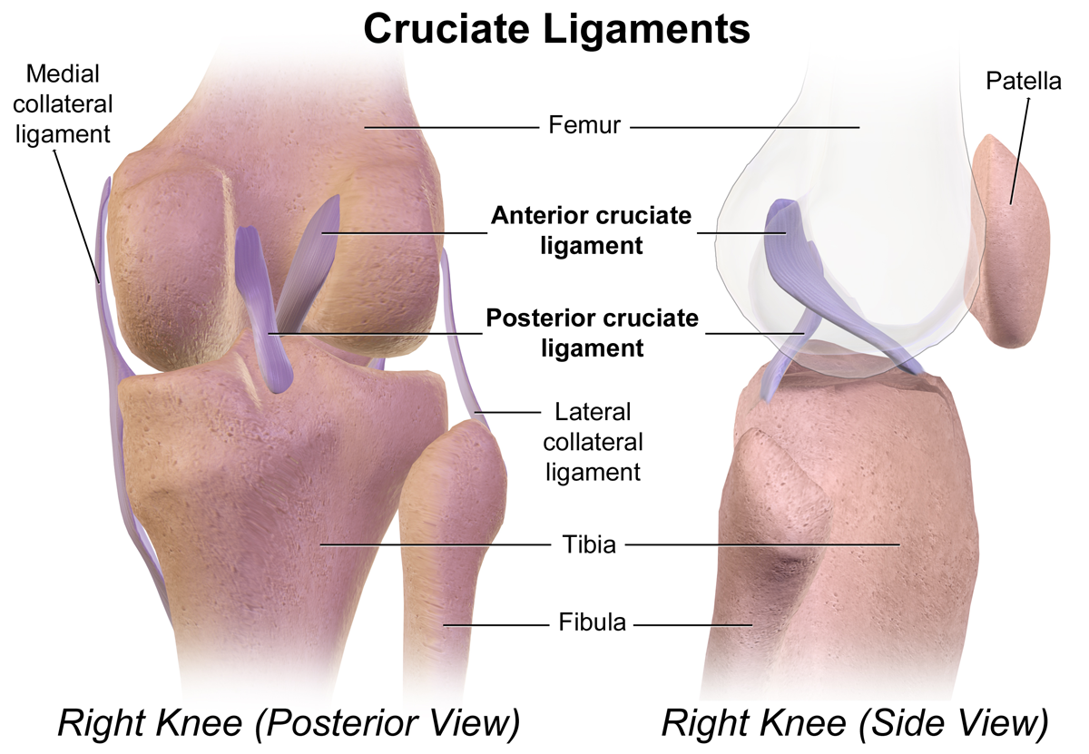
Cruciate ligaments (also cruciform ligaments) are pairs of ligaments arranged like a letter X. They occur in several joints of the body, such as the knee joint and the atlantoaxial joint, atlanto-axial joint. In a fashion similar to the cords in a toy Jacob's ladder (toy), Jacob's ladder, the crossed ligaments stabilize the joint while allowing a very large range of motion. Knee Structure Cruciate ligaments occur in the knee of humans and other bipedal animals and the corresponding Stifle joint, stifle of quadrupedal animals, and in the neck, fingers, and foot. * The cruciate ligaments of the knee are the anterior cruciate ligament (ACL) and the posterior cruciate ligament (PCL). These ligaments are two strong, rounded bands that extend from the head of the tibia to the intercondyloid notch of the femur. The ACL is lateral and the PCL is medial. They cross each other like the limbs of an X. They are named for their insertion into the tibia: the ACL attaches to the anterior ... [...More Info...] [...Related Items...] OR: [Wikipedia] [Google] [Baidu] |
Ligament
A ligament is the fibrous connective tissue that connects bones to other bones. It is also known as ''articular ligament'', ''articular larua'', ''fibrous ligament'', or ''true ligament''. Other ligaments in the body include the: * Peritoneal ligament: a fold of peritoneum or other membranes. * Fetal remnant ligament: the remnants of a fetal tubular structure. * Periodontal ligament: a group of fibers that attach the cementum of teeth to the surrounding alveolar bone. Ligaments are similar to tendons and fasciae as they are all made of connective tissue. The differences among them are in the connections that they make: ligaments connect one bone to another bone, tendons connect muscle to bone, and fasciae connect muscles to other muscles. These are all found in the skeletal system of the human body. Ligaments cannot usually be regenerated naturally; however, there are periodontal ligament stem cells located near the periodontal ligament which are involved in the adult regener ... [...More Info...] [...Related Items...] OR: [Wikipedia] [Google] [Baidu] |
Flexor Digitorum Superficialis Muscle
Flexor digitorum superficialis (''flexor digitorum sublimis'') is an extrinsic flexor muscle of the fingers at the proximal interphalangeal joints. It is in the anterior compartment of the forearm. It is sometimes considered to be the deepest part of the superficial layer of this compartment, and sometimes considered to be a distinct, "intermediate layer" of this compartment. It is relatively common for the Flexor digitorum superficialis to be missing from the little finger, bilaterally and unilaterally, which can cause problems when diagnosing a little finger injury. Structure The muscle has two classically described heads – the humeroulnar and radial – and it is between these heads that the median nerve and ulnar artery pass. The ulnar collateral ligament of elbow joint gives its origin to part of this muscle. Four long tendons come off this muscle near the wrist and travel through the carpal tunnel formed by the flexor retinaculum. These tendons, along with those of flex ... [...More Info...] [...Related Items...] OR: [Wikipedia] [Google] [Baidu] |
Terminologia Anatomica
''Terminologia Anatomica'' is the international standard for human anatomical terminology. It is developed by the Federative International Programme on Anatomical Terminology, a program of the International Federation of Associations of Anatomists (IFAA). The second edition was released in 2019 and approved and adopted by the IFAA General Assembly in 2020. ''Terminologia Anatomica'' supersedes the previous standard, ''Nomina Anatomica''. It contains terminology for about 7500 human anatomical structures. Categories of anatomical structures ''Terminologia Anatomica'' is divided into 16 chapters grouped into five parts. The official terms are in Latin. Although equivalent English-language terms are provided, as shown below, only the official Latin terms are used as the basis for creating lists of equivalent terms in other languages. Part I Chapter 1: General anatomy # General terms # Reference planes # Reference lines # Human body positions # Movements # Parts of human body ... [...More Info...] [...Related Items...] OR: [Wikipedia] [Google] [Baidu] |
Nomina Anatomica
''Nomina Anatomica'' (''NA'') was the international standard on human anatomic terminology from 1895 until it was replaced by ''Terminologia Anatomica'' in 1998. In the late nineteenth century some 30,000 terms for various body parts were in use. The same structures were described by different names, depending (among other things) on the anatomist's school and national tradition. Vernacular translations of Latin and Greek, as well as various eponymous terms, were barriers to effective international communication. There was disagreement and confusion among anatomists regarding anatomical terminology. Editions The first and last entries in the following table are not NA editions, but they are included for the sake of continuity. Although these early editions were authorized by different bodies, they are sometimes considered part of the same series. Before these codes of terminology, approved at anatomists congresses, the usage of anatomical terms was based on authoritative work ... [...More Info...] [...Related Items...] OR: [Wikipedia] [Google] [Baidu] |
Cruciate Ligament Of Atlas
The cruciate ligament of the atlas (cruciate may substitute for cruciform) is a ligament in the neck. It forms part of the atlanto-axial joint. The ligament is named after its cross shape. It consists of transverse and longitudinal components. The posterior longitudinal band may be absent in some people. The cruciate ligament of the atlas prevents abnormal movement of the atlanto-axial joint. It may be torn, such as by fractures of the atlas bone. Structure The cruciate ligament of the atlas consists of the transverse ligament of the atlas, a superior longitudinal band, and an inferior longitudinal band. The superior longitudinal band connects the transverse ligament to the anterior side of the foramen magnum (near the basilar part) in the occipital bone of the skull. The inferior longitudinal band connects the transverse ligament to the body of the axis bone (C2). Gerber's ligament runs deep to the superior band of the cruciate ligament of the atlas. Variation The inferior ... [...More Info...] [...Related Items...] OR: [Wikipedia] [Google] [Baidu] |
Annular And Cruciform Parts Of Fibrous Sheath Over Flexor Tendon Sheaths
Annulus (or anulus) or annular indicates a ring- or donut-shaped area or structure. It may refer to: Human anatomy * ''Anulus fibrosus disci intervertebralis'', spinal structure * Annulus of Zinn, a.k.a. annular tendon or ''anulus tendineus communis'', around the optic nerve * Annular ligament (other) * ''Digitus anularis'', a.k.a. ring finger * ''Anulus ciliaris'', a.k.a. ciliary body * ''Anulus femoralis'', a.k.a. femoral ring * ''Anulus inguinalis superficialis'', a.k.a. superficial inguinal ring * ''Anulus inguinalis profundus'', a.k.a. deep inguinal ring * ''Anuli fibrosi cordis'', a.k.a. fibrous rings of heart * ''Anulus umbilicalis '', a.k.a. umbilical ring Other * Annulus (construction), outer gear ring in an epicyclic gearing * Annulus (botany), structure on fern and moss sporangia * Annular lake, a ring-shaped lake caused by meteor impact * Annulus (mathematics), the shape between two concentric circles * Annulus (mycology), structure on mushroom * Annulus ... [...More Info...] [...Related Items...] OR: [Wikipedia] [Google] [Baidu] |
Stifle Joint
The stifle joint (often simply stifle) is a complex joint in the hind limbs of quadruped mammals such as the sheep, horse or dog. It is the equivalent of the human knee and is often the largest synovial joint in the animal's body. The stifle joint joins three bones: the femur, patella, and tibia. The joint consists of three smaller ones: the femoropatellar joint, medial joint, and lateral femorotibial joint. The stifle joint consists of the femorotibial articulation ( femoral and tibial condyles), femoropatellar articulation (femoral trochlea and the patella), and the proximal articulation. The joint is stabilized by paired collateral ligaments which act to prevent abduction/adduction at the joint, as well as paired cruciate ligaments. The cranial cruciate ligament and the caudal cruciate ligament restrict cranial and caudal translation (respectively) of the tibia on the femur. The cranial cruciate also resists over-extension and inward rotation, and is the most commonly dama ... [...More Info...] [...Related Items...] OR: [Wikipedia] [Google] [Baidu] |
Metacarpophalangeal
The metacarpophalangeal joints (MCP) are situated between the metacarpal bones and the proximal phalanges of the fingers. These joints are of the condyloid kind, formed by the reception of the rounded heads of the metacarpal bones into shallow cavities on the proximal ends of the proximal phalanges. Being condyloid, they allow the movements of flexion, extension, abduction, adduction and circumduction at the joint. Structure Ligaments Each joint has: * palmar ligaments of metacarpophalangeal articulations * collateral ligaments of metacarpophalangeal articulations Dorsal surfaces The dorsal surfaces of these joints are covered by the expansions of the Extensor tendons, together with some loose areolar tissue which connects the deep surfaces of the tendons to the bones. Function The movements which occur in these joints are flexion, extension, adduction, abduction, and circumduction; the movements of abduction and adduction are very limited, and cannot be performed while th ... [...More Info...] [...Related Items...] OR: [Wikipedia] [Google] [Baidu] |
Cruciate Distal Sesamoidean Ligament
{{disambig ...
Cruciate, and similar words, can mean: *The cruciate ligaments in the knee *For a magic spell in the Harry Potter scenario, see crucio *Latin and early-English word for crusade The Crusades were a series of religious wars initiated, supported, and sometimes directed by the Latin Church in the medieval period. The best known of these Crusades are those to the Holy Land in the period between 1095 and 1291 that were i ... [...More Info...] [...Related Items...] OR: [Wikipedia] [Google] [Baidu] |
Inferior Extensor Retinaculum Of Foot
The inferior extensor retinaculum of the foot (cruciate crural ligament, lower part of anterior annular ligament) is a Y-shaped band placed in front of the ankle-joint, the stem of the Y being attached laterally to the upper surface of the calcaneus, in front of the depression for the interosseous talocalcaneal ligament; it is directed medialward as a double layer, one lamina passing in front of, and the other behind, the tendons of the peroneus tertius and extensor digitorum longus. At the medial border of the latter tendon, these two layers join, forming a compartment in which the tendons are enclosed. From the medial extremity of this sheath, the two limbs of the Y diverge: one is directed upward and medialward, to be attached to the tibial malleolus, passing over the extensor hallucis longus and the vessels and nerves but enclosing the tibialis anterior by a splitting of its fibers. The other limb extends downward and medialward, to be attached to the border of the plant ... [...More Info...] [...Related Items...] OR: [Wikipedia] [Google] [Baidu] |
Cruciate Crural Ligament
The inferior extensor retinaculum of the foot (cruciate crural ligament, lower part of anterior annular ligament) is a Y-shaped band placed in front of the ankle-joint, the stem of the Y being attached laterally to the upper surface of the calcaneus, in front of the depression for the interosseous talocalcaneal ligament; it is directed medialward as a double layer, one lamina passing in front of, and the other behind, the tendons of the peroneus tertius and extensor digitorum longus. At the medial border of the latter tendon, these two layers join, forming a compartment in which the tendons are enclosed. From the medial extremity of this sheath, the two limbs of the Y diverge: one is directed upward and medialward, to be attached to the tibial malleolus, passing over the extensor hallucis longus and the vessels and nerves but enclosing the tibialis anterior by a splitting of its fibers. The other limb extends downward and medialward, to be attached to the border of the plantar ... [...More Info...] [...Related Items...] OR: [Wikipedia] [Google] [Baidu] |


