|
Crescent Sign
In radiology, the crescent sign is a finding on conventional radiographs that is associated with avascular necrosis. It usually occurs later in the disease, in stage III of the four-stage Ficat classification system. It appears as a curved epiphysis, subchondral radiodensity, radiolucent line that is often found on the proximal femur, femoral or humerus, humeral head. Usually, this sign indicates a high likelihood of collapse of the affected bone. The crescent sign may be best seen in an abducted (frog-legged) position. The crescent sign is caused by the necrosis, necrotic and repair processes that occur during avascular necrosis. Osteosclerosis occurs at a margin where new bone is placed over dead Cancellous bone, trabeculae. When the trabeculae experience stress leading to microfractures and collapse, the crescent sign appears. The crescent sign may be seen with other bone diseases, such as shear fractures. References {{Radiologic signs Musculoskeletal radiographic sig ... [...More Info...] [...Related Items...] OR: [Wikipedia] [Google] [Baidu] |
Air Crescent Sign
In radiology, the air crescent sign is a finding on chest radiograph and computed tomography that is crescenteric and radiodensity, radiolucent, due to a lung cavity that is filled with air and has a round radiodensity, radiopaque mass. Classically, it is due to an aspergilloma, a form of aspergillosis, that occurs when the fungus ''Aspergillus'' grows in a cavity in the lung. It is also referred as Monad sign. Additional images References External linksAir crescent sign on CXR Radiologic signs {{med-sign-stub ... [...More Info...] [...Related Items...] OR: [Wikipedia] [Google] [Baidu] |
Crescent Sign Annotated
A crescent shape (, ) is a symbol or emblem used to represent the lunar phase in the first quarter (the "sickle moon"), or by extension a symbol representing the Moon itself. In Hinduism, Lord Shiva is often shown wearing a crescent moon on his head symbolising that the lord is the master of time and is himself timeless. It is used as the astrological symbol for the Moon, and hence as the alchemical symbol for silver. It was also the emblem of Diana/Artemis, and hence represented virginity. In Christianity Marian veneration, it is associated with the Virgin Mary. From its use as roof finial in Ottoman era mosques, it has also become associated with Islam, and the crescent was introduced as chaplain badge for Muslim chaplains in the US military in 1993.On December 14, 1992, the Army Chief of Chaplains requested that an insignia be created for future Muslim chaplains, and the design (a crescent) was completed January 8, 1993. Emerson, William K., ''Encyclopedia of United States ... [...More Info...] [...Related Items...] OR: [Wikipedia] [Google] [Baidu] |
Radiology
Radiology ( ) is the medical discipline that uses medical imaging to diagnose diseases and guide their treatment, within the bodies of humans and other animals. It began with radiography (which is why its name has a root referring to radiation), but today it includes all imaging modalities, including those that use no electromagnetic radiation (such as ultrasonography and magnetic resonance imaging), as well as others that do, such as computed tomography (CT), fluoroscopy, and nuclear medicine including positron emission tomography (PET). Interventional radiology is the performance of usually minimally invasive medical procedures with the guidance of imaging technologies such as those mentioned above. The modern practice of radiology involves several different healthcare professions working as a team. The radiologist is a medical doctor who has completed the appropriate post-graduate training and interprets medical images, communicates these findings to other physicians ... [...More Info...] [...Related Items...] OR: [Wikipedia] [Google] [Baidu] |
Radiograph
Radiography is an imaging technique using X-rays, gamma rays, or similar ionizing radiation and non-ionizing radiation to view the internal form of an object. Applications of radiography include medical radiography ("diagnostic" and "therapeutic") and industrial radiography. Similar techniques are used in airport security (where "body scanners" generally use backscatter X-ray). To create an image in conventional radiography, a beam of X-rays is produced by an X-ray generator and is projected toward the object. A certain amount of the X-rays or other radiation is absorbed by the object, dependent on the object's density and structural composition. The X-rays that pass through the object are captured behind the object by a detector (either photographic film or a digital detector). The generation of flat two dimensional images by this technique is called projectional radiography. In computed tomography (CT scanning) an X-ray source and its associated detectors rotate around the ... [...More Info...] [...Related Items...] OR: [Wikipedia] [Google] [Baidu] |
Avascular Necrosis
Avascular necrosis (AVN), also called osteonecrosis or bone infarction, is death of bone tissue due to interruption of the blood supply. Early on, there may be no symptoms. Gradually joint pain may develop which may limit the ability to move. Complications may include collapse of the bone or nearby joint surface. Risk factors include bone fractures, joint dislocations, alcoholism, and the use of high-dose steroids. The condition may also occur without any clear reason. The most commonly affected bone is the femur. Other relatively common sites include the upper arm bone, knee, shoulder, and ankle. Diagnosis is typically by medical imaging such as X-ray, CT scan, or MRI. Rarely biopsy may be used. Treatments may include medication, not walking on the affected leg, stretching, and surgery. Most of the time surgery is eventually required and may include core decompression, osteotomy, bone grafts, or joint replacement. About 15,000 cases occur per year in the United States. Peop ... [...More Info...] [...Related Items...] OR: [Wikipedia] [Google] [Baidu] |
Epiphysis
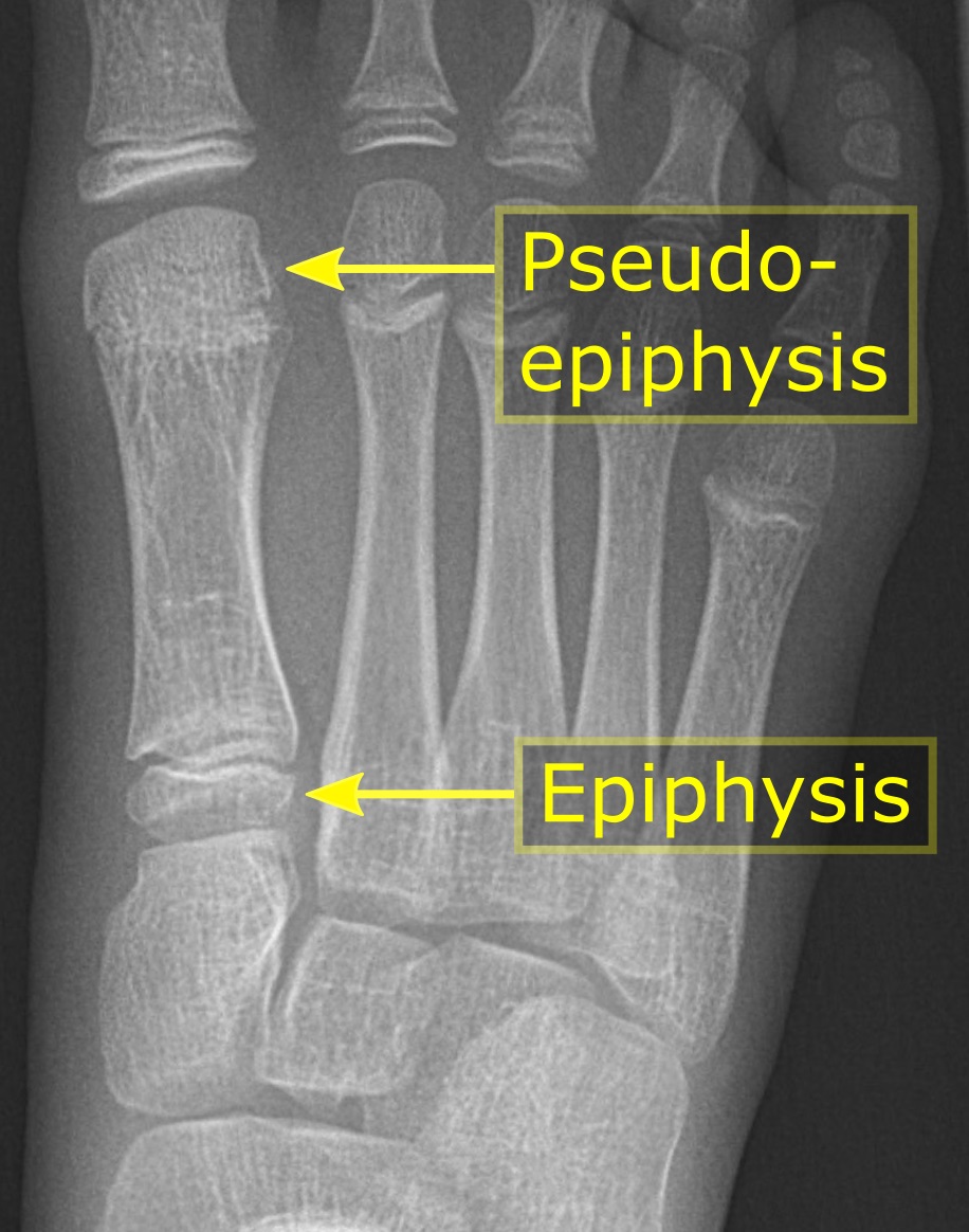
The epiphysis () is the rounded end of a long bone, at its joint with adjacent bone(s). Between the epiphysis and diaphysis (the long midsection of the long bone) lies the metaphysis, including the epiphyseal plate (growth plate). At the joint, the epiphysis is covered with articular cartilage; below that covering is a zone similar to the epiphyseal plate, known as subchondral bone. The epiphysis is filled with red bone marrow, which produces erythrocytes (red blood cells). Structure There are four types of epiphysis: # Pressure epiphysis: The region of the long bone that forms the joint is a pressure epiphysis (e.g. the head of the femur, part of the hip joint complex). Pressure epiphyses assist in transmitting the weight of the human body and are the regions of the bone that are under pressure during movement or locomotion. Another example of a pressure epiphysis is the head of the humerus which is part of the shoulder complex. condyles of femur and tibia also comes under ... [...More Info...] [...Related Items...] OR: [Wikipedia] [Google] [Baidu] |
Radiodensity
Radiodensity (or radiopacity) is opacity to the radio wave and X-ray portion of the electromagnetic spectrum: that is, the relative inability of those kinds of electromagnetic radiation to pass through a particular material. Radiolucency or hypodensity indicates greater passage (greater transradiancy) to X-ray photonsNovelline, Robert. ''Squire's Fundamentals of Radiology''. Harvard University Press. 5th edition. 1997. . and is the analogue of transparency and translucency with visible light. Materials that inhibit the passage of electromagnetic radiation are called radiodense or radiopaque, while those that allow radiation to pass more freely are referred to as radiolucent. Radiopaque volumes of material have white appearance on radiographs, compared with the relatively darker appearance of radiolucent volumes. For example, on typical radiographs, bones look white or light gray (radiopaque), whereas muscle and skin look black or dark gray, being mostly invisible (radiolucent). Th ... [...More Info...] [...Related Items...] OR: [Wikipedia] [Google] [Baidu] |
Femur
The femur (; ), or thigh bone, is the proximal bone of the hindlimb in tetrapod vertebrates. The head of the femur articulates with the acetabulum in the pelvic bone forming the hip joint, while the distal part of the femur articulates with the tibia (shinbone) and patella (kneecap), forming the knee joint. By most measures the two (left and right) femurs are the strongest bones of the body, and in humans, the largest and thickest. Structure The femur is the only bone in the upper leg. The two femurs converge medially toward the knees, where they articulate with the proximal ends of the tibiae. The angle of convergence of the femora is a major factor in determining the femoral-tibial angle. Human females have thicker pelvic bones, causing their femora to converge more than in males. In the condition ''genu valgum'' (knock knee) the femurs converge so much that the knees touch one another. The opposite extreme is ''genu varum'' (bow-leggedness). In the general populatio ... [...More Info...] [...Related Items...] OR: [Wikipedia] [Google] [Baidu] |
Humerus
The humerus (; ) is a long bone in the arm that runs from the shoulder to the elbow. It connects the scapula and the two bones of the lower arm, the radius and ulna, and consists of three sections. The humeral upper extremity consists of a rounded head, a narrow neck, and two short processes (tubercles, sometimes called tuberosities). The body is cylindrical in its upper portion, and more prismatic below. The lower extremity consists of 2 epicondyles, 2 processes (trochlea & capitulum), and 3 fossae (radial fossa, coronoid fossa, and olecranon fossa). As well as its true anatomical neck, the constriction below the greater and lesser tubercles of the humerus is referred to as its surgical neck due to its tendency to fracture, thus often becoming the focus of surgeons. Etymology The word "humerus" is derived from la, humerus, umerus meaning upper arm, shoulder, and is linguistically related to Gothic ''ams'' shoulder and Greek ''ōmos''. Structure Upper extremity The upper or pr ... [...More Info...] [...Related Items...] OR: [Wikipedia] [Google] [Baidu] |
Necrosis
Necrosis () is a form of cell injury which results in the premature death of cells in living tissue by autolysis. Necrosis is caused by factors external to the cell or tissue, such as infection, or trauma which result in the unregulated digestion of cell components. In contrast, apoptosis is a naturally occurring programmed and targeted cause of cellular death. While apoptosis often provides beneficial effects to the organism, necrosis is almost always detrimental and can be fatal. Cellular death due to necrosis does not follow the apoptotic signal transduction pathway, but rather various receptors are activated and result in the loss of cell membrane integrity and an uncontrolled release of products of cell death into the extracellular space. This initiates in the surrounding tissue an inflammatory response, which attracts leukocytes and nearby phagocytes which eliminate the dead cells by phagocytosis. However, microbial damaging substances released by leukocytes would crea ... [...More Info...] [...Related Items...] OR: [Wikipedia] [Google] [Baidu] |
Osteosclerosis
Osteosclerosis is a disorder that is characterized by abnormal hardening of bone and an elevation in bone density. It may predominantly affect the medullary portion and/or cortex of bone. Plain radiographs are a valuable tool for detecting and classifying osteosclerotic disorders. It can manifest in localized or generalized osteosclerosis. Localized osteosclerosis can be caused by Legg–Calvé–Perthes disease, sickle-cell disease and osteoarthritis among others. Osteosclerosis can be classified in accordance with the causative factor into acquired and hereditary. Types Acquired osteosclerosis * Osteogenic bone metastasis caused by carcinoma of prostate and breast * Paget's disease of bone * Myelofibrosis (primary disorder or secondary to intoxication or malignancy) * Osteosclerosing types of chronic osteomyelitis * Hypervitaminosis D * hyperparathyroidism * Schnitzler syndrome * Mastocytosis Skeletal fluorosis * Monoclonal IgM Kappa cryoglobulinemia * Hepatitis C. Heredita ... [...More Info...] [...Related Items...] OR: [Wikipedia] [Google] [Baidu] |
Cancellous Bone
A bone is a rigid organ that constitutes part of the skeleton in most vertebrate animals. Bones protect the various other organs of the body, produce red and white blood cells, store minerals, provide structure and support for the body, and enable mobility. Bones come in a variety of shapes and sizes and have complex internal and external structures. They are lightweight yet strong and hard and serve multiple functions. Bone tissue (osseous tissue), which is also called bone in the uncountable sense of that word, is hard tissue, a type of specialized connective tissue. It has a honeycomb-like matrix internally, which helps to give the bone rigidity. Bone tissue is made up of different types of bone cells. Osteoblasts and osteocytes are involved in the formation and mineralization of bone; osteoclasts are involved in the resorption of bone tissue. Modified (flattened) osteoblasts become the lining cells that form a protective layer on the bone surface. The mineralized matri ... [...More Info...] [...Related Items...] OR: [Wikipedia] [Google] [Baidu] |


