|
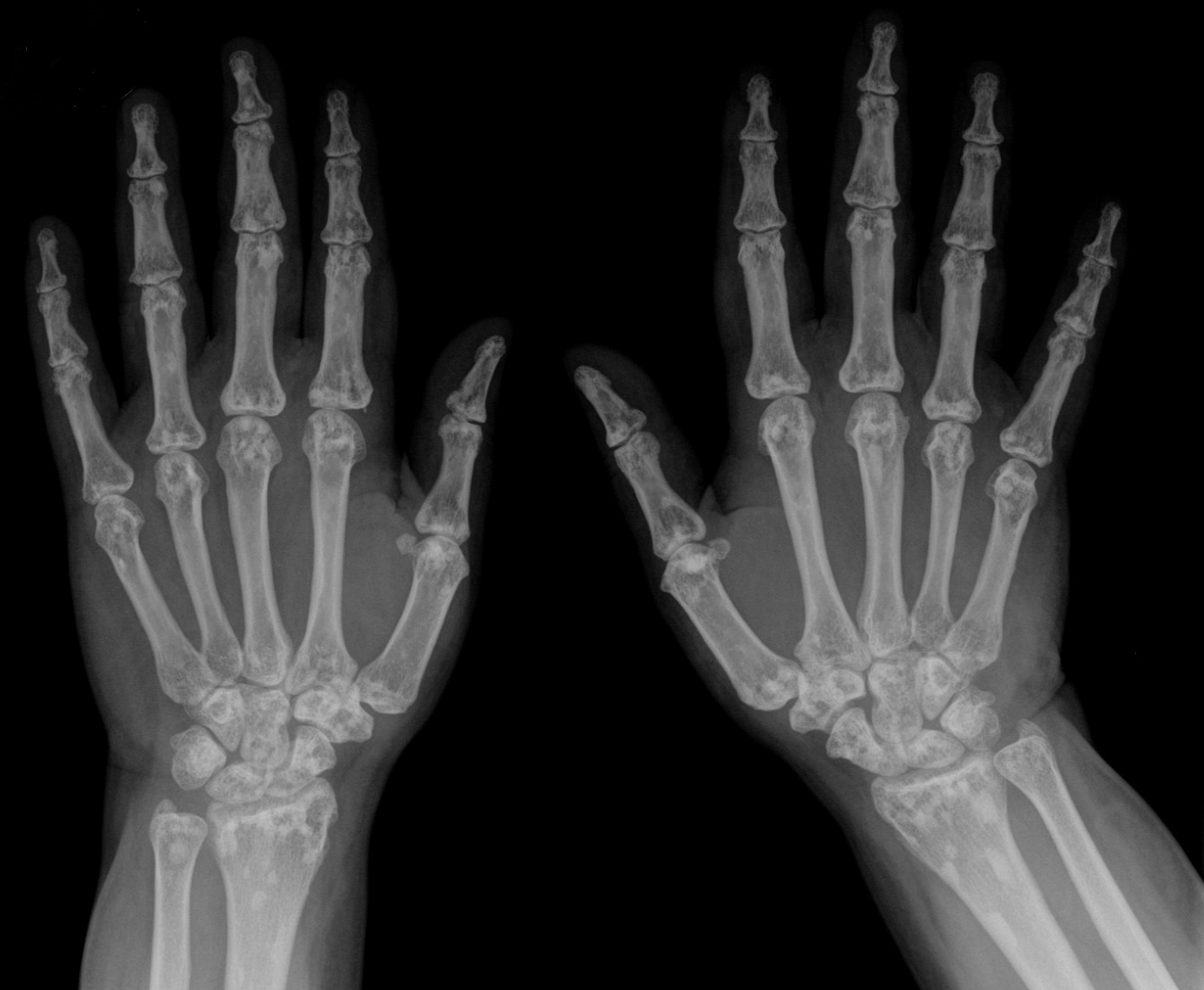
Osteosclerosis
Osteosclerosis is a disorder that is characterized by abnormal hardening of bone and an elevation in bone density. It may predominantly affect the medullary portion and/or cortex of bone. Plain radiographs are a valuable tool for detecting and classifying osteosclerotic disorders. It can manifest in localized or generalized osteosclerosis. Localized osteosclerosis can be caused by Legg–Calvé–Perthes disease, sickle-cell disease and osteoarthritis among others. Osteosclerosis can be classified in accordance with the causative factor into acquired and hereditary. Types Acquired osteosclerosis * Osteogenic bone metastasis caused by carcinoma of prostate and breast * Paget's disease of bone * Myelofibrosis (primary disorder or secondary to intoxication or malignancy) * Osteosclerosing types of chronic osteomyelitis * Hypervitaminosis D * hyperparathyroidism * Schnitzler syndrome * Mastocytosis Skeletal fluorosis * Monoclonal IgM Kappa cryoglobulinemia * Hepatitis C. Heredita ... [...More Info...] [...Related Items...] OR: [Wikipedia] [Google] [Baidu] |
Osteopetrosis
Osteopetrosis, literally "stone bone", also known as marble bone disease or Albers-Schönberg disease, is an extremely rare Biological inheritance, inherited disease, disorder whereby the bones harden, becoming Density, denser, in contrast to more prevalent conditions like osteoporosis, in which the bones become less dense and more brittle, or osteomalacia, in which the bones soften. Osteopetrosis can cause bones to dissolve and break. It is one of the hereditary causes of osteosclerosis. It is considered to be the prototype of osteosclerosing dysplasias. The cause of the disease is understood to be malfunctioning osteoclasts and their inability to resorb bone. Although human osteopetrosis is a heterogeneous disorder encompassing different molecular lesions and a range of clinical features, all forms share a single pathogenic nexus in the osteoclast. The exact molecular defects or location of the mutations taking place are unknown. Osteopetrosis was first described in 1903, by Ger ... [...More Info...] [...Related Items...] OR: [Wikipedia] [Google] [Baidu] |
Malignant Infantile Osteopetrosis
Malignant infantile osteopetrosis is a rare osteosclerosing type of skeletal dysplasia that typically presents in infancy and is characterized by a unique radiographic appearance of generalized hyperostosis (excessive growth of bone). The generalized increase in bone density has a special predilection to involve the medullary portion with relative sparing of the cortices.EL-Sobky TA, Elsobky E, Sadek I, Elsayed SM, Khattab MF (2016)"A case of infantile osteopetrosis: The radioclinical features with literature update' ''Bone Rep''. 4:11-16doi:10.1016/j.bonr.2015.11.002 Obliteration of bone marrow spaces and subsequent depression of the cellular function can result in serious hematologic complications. Optic atrophy and cranial nerve damage secondary to bony expansion can result in marked morbidity. The prognosis is extremely poor in ... [...More Info...] [...Related Items...] OR: [Wikipedia] [Google] [Baidu] |
Skeletal Fluorosis
Skeletal fluorosis is a bone disease caused by excessive accumulation of fluoride leading to weakened bones. In advanced cases, skeletal fluorosis causes painful damage to bones and joints. Symptoms Symptoms are mainly promoted in the bone structure. Due to a high fluoride concentration in the body, the bone is hardened and thus less elastic, resulting in an increased frequency of fractures. Other symptoms include thickening of the bone structure and accumulation of bone tissue, which both contribute to impaired joint mobility. Ligaments and cartilage can become ossified. Most patients with skeletal fluorosis show side effects from the high fluoride dose such as ruptures of the stomach lining and nausea. Fluoride can also damage the parathyroid glands, leading to hyperparathyroidism, the uncontrolled secretion of parathyroid hormones. These hormones regulate calcium concentration in the body. An elevated parathyroid hormone concentration results in a depletion of calcium in bone s ... [...More Info...] [...Related Items...] OR: [Wikipedia] [Google] [Baidu] |
Mastocytosis
Mastocytosis, a type of mast cell disease, is a rare disorder affecting both children and adults caused by the accumulation of functionally defective mast cells (also called ''mastocytes'') and CD34+ mast cell precursors. People affected by mastocytosis are susceptible to a variety of symptoms, including itching, hives, and anaphylactic shock, caused by the release of histamine and other pro-inflammatory substances from mast cells. Signs and symptoms When mast cells undergo degranulation, the substances that are released can cause a number of symptoms that can vary over time and can range in intensity from mild to severe. Because mast cells play a role in allergic reactions, the symptoms of mastocytosis often are similar to the symptoms of an allergic reaction. They may include, but are not limited to * Fatigue * Skin lesions (''urticaria pigmentosa''), itching, and dermatographic ''urticaria'' (skin writing) * "Darier's Sign", a reaction to stroking or scratching of urticar ... [...More Info...] [...Related Items...] OR: [Wikipedia] [Google] [Baidu] |
Osteomyelitis
Osteomyelitis (OM) is an infection of bone. Symptoms may include pain in a specific bone with overlying redness, fever, and weakness. The long bones of the arms and legs are most commonly involved in children e.g. the femur and humerus, while the feet, spine, and hips are most commonly involved in adults. The cause is usually a bacterial infection, but rarely can be a fungal infection. It may occur by spread from the blood or from surrounding tissue. Risks for developing osteomyelitis include diabetes, intravenous drug use, prior removal of the spleen, and trauma to the area. Diagnosis is typically suspected based on symptoms and basic laboratory tests as C-reactive protein (CRP) and erythrocyte sedimentation rate (ESR).This is because plain radiographs are unremarkable in the first few days following acute infection. Diagnosis is further confirmed by blood tests, medical imaging, or bone biopsy. Treatment of bacterial osteomyelitis often involves both antimicrobials and sur ... [...More Info...] [...Related Items...] OR: [Wikipedia] [Google] [Baidu] |
Myelofibrosis
Primary myelofibrosis (PMF) is a rare bone marrow blood cancer. It is classified by the World Health Organization (WHO) as a type of myeloproliferative neoplasm, a group of cancers in which there is growth of abnormal cells in the bone marrow. This is most often associated with a somatic mutation in the JAK2, CALR, or MPL gene markers. In PMF, the healthy marrow is replaced by scar tissue (fibrosis), resulting in a lack of production of normal blood cells. Symptoms include anemia, increased infection and an enlarged spleen ( splenomegaly). In 2016, prefibrotic primary myelofibrosis was formally classified as a distinct condition that progresses to overt PMF in many patients, the primary diagnostic difference being the grade of fibrosis. Signs and symptoms The primary feature of primary myelofibrosis is bone marrow fibrosis, but it is often accompanied by: * Abdominal fullness related to an enlarged spleen (splenomegaly). * Bone pain * Bruising and easy bleeding due to inad ... [...More Info...] [...Related Items...] OR: [Wikipedia] [Google] [Baidu] |
Buschke–Ollendorff Syndrome
Buschke–Ollendorff syndrome (BOS) is a rare genetic disorder associated with LEMD3. It is believed to be inherited in an autosomal dominant manner. It is named for Abraham Buschke and Helene Ollendorff Curth, who described it in a 45-year-old woman. Its frequency is almost 1 case per every 20,000 people, and it is equally found in both males and females. Signs and symptoms The signs and symptoms of this condition are consistent with the following (possible complications include aortic stenosis and hearing loss): :::::::*Osteopoikilosis :::::::*Bone pain :::::::* Connective tissue nevi :::::::*Metaphysis abnormality Pathogenesis Buschke–Ollendorff syndrome is caused by one important factor: mutations in the LEMD3 gene (12q14), located on chromosome 12. Among the important aspects of Buschke–Ollendorff syndrome condition, genetically speaking are: :::::::::* LEMD3 (protein) referred also as MAN1, is an important protein in inner nuclear membrane. :::::::::* LEMD3 gene g ... [...More Info...] [...Related Items...] OR: [Wikipedia] [Google] [Baidu] |
Osteopoikilosis
Osteopoikilosis is a benign, autosomal dominant sclerosing dysplasia of bone characterized by the presence of numerous bone islands in the skeleton. Presentation The radiographic appearance of osteopoikilosis on an X-ray is characterized by a pattern of numerous white densities of similar size spread throughout all the bones. This is a systemic condition. It must be differentiated from blastic metastasis, which can also present radiographically as white densities interspersed throughout bone. Blastic metastasis tends to present with larger and more irregular densities in less of a uniform pattern. Another differentiating factor is age, with blastic metastasis mostly affecting older people, and osteopoikilosis being found in people 20 years of age and younger. The distribution is variable, though it does not tend to affect the ribs, spine, or skull. Cause Epidemiology Men and women are affected in equal number, reflecting the fact that osteopoikilosis attacks indiscriminately. Ad ... [...More Info...] [...Related Items...] OR: [Wikipedia] [Google] [Baidu] |
Osteopathia Striata
Osteopathia striata is a rare entity characterized by fine linear striations about 2- to 3-mm-thick, visible by radiographic examination, in the metaphyses and diaphyses of long or flat bones. It is often asymptomatic, and is often discovered incidentally. See also * List of radiographic findings associated with cutaneous conditions Many conditions of or affecting the human integumentary system have associated features that may be found by performing an x-ray or CT scan of the affected person. See also * List of cutaneous conditions * List of contact allergens * List of ... References Radiologic signs {{med-sign-stub ... [...More Info...] [...Related Items...] OR: [Wikipedia] [Google] [Baidu] |
Diaphysis
The diaphysis is the main or midsection (shaft) of a long bone. It is made up of cortical bone and usually contains bone marrow and adipose tissue (fat). It is a middle tubular part composed of compact bone which surrounds a central marrow cavity which contains red or yellow marrow. In diaphysis, primary ossification occurs. Ewing sarcoma tends to occur at the diaphysis.Physical Medicine and Rehabilitation Board Review, Cuccurullo Additional images Illu long bone.jpg File:EpiMetaDiaphyse.jpg, Long bone See also *Epiphysis *Metaphysis The metaphysis is the neck portion of a long bone between the epiphysis and the diaphysis. It contains the growth plate, the part of the bone that grows during childhood, and as it grows it ossifies near the diaphysis and the epiphyses. The metap ... References Skeletal system Long bones {{musculoskeletal-stub ... [...More Info...] [...Related Items...] OR: [Wikipedia] [Google] [Baidu] |
Pyknodysostosis
Pycnodysostosis (from Greek: πυκνός (puknos) meaning "dense", ''dys'' ("defective"), and ''ostosis'' ("condition of the bone")), is a lysosomal storage disease of the bone caused by a mutation in the gene that codes the enzyme cathepsin K. It is also known as PKND and PYCD. History The disease was first described by Maroteaux and Lamy in 1962 at which time it was defined by the following characteristics: dwarfism; osteopetrosis; partial agenesis of the terminal digits of the hands and feet; cranial anomalies, such as persistence of fontanelles and failure of closure of cranial sutures; frontal and occipital bossing; and hypoplasia of the angle of the mandible. The defective gene responsible for the disease was discovered in 1996. The French painter Henri de Toulouse Lautrec (1864-1901) is believed to have had the disease. Signs and symptoms Pycnodysostosis causes the bones to be abnormally dense; the last bones of the fingers (the distal phalanges) to be unusually short; ... [...More Info...] [...Related Items...] OR: [Wikipedia] [Google] [Baidu] |
Dominance (genetics)
In genetics, dominance is the phenomenon of one variant (allele) of a gene on a chromosome masking or overriding the effect of a different variant of the same gene on the other copy of the chromosome. The first variant is termed dominant and the second recessive. This state of having two different variants of the same gene on each chromosome is originally caused by a mutation in one of the genes, either new (''de novo'') or inherited. The terms autosomal dominant or autosomal recessive are used to describe gene variants on non-sex chromosomes ( autosomes) and their associated traits, while those on sex chromosomes (allosomes) are termed X-linked dominant, X-linked recessive or Y-linked; these have an inheritance and presentation pattern that depends on the sex of both the parent and the child (see Sex linkage). Since there is only one copy of the Y chromosome, Y-linked traits cannot be dominant or recessive. Additionally, there are other forms of dominance such as incomplete d ... [...More Info...] [...Related Items...] OR: [Wikipedia] [Google] [Baidu] |


