|
Congenital Fibrosis Of The Extraocular Muscles
Congenital fibrosis of the extraocular muscles is a class of rare genetic disorders affecting one or more of the muscles that move the human eyeball, eyeballs. Individuals with congenital fibrosis of the extraocular muscles (CFEOM) have varying degrees of Ophthalmoparesis, ophthalmoplegia (an inability to move the eyes in one or more directions) and Ptosis (eyelid), ptosis. The condition is present from birth, non-progressive, runs in families, and usually affects both eyes similarly. In the most common form, the superior rectus muscle, superior recti are dysfunctional and the inferior rectus muscle, inferior recti, lacking proper opposition, pull the eyes down, forcing the head to be tilted upward in order to see straight ahead. There are three types of CFEOM, numbered 1-3. CFEOM1, the most common type, is now known to be caused by one of several mutations in the KIF21A gene, while CFEOM2 is caused by mutations in the PHOX2A gene.Engle, E.C., Genetic Basis of Congenital Strabis ... [...More Info...] [...Related Items...] OR: [Wikipedia] [Google] [Baidu] |
Fibrosis
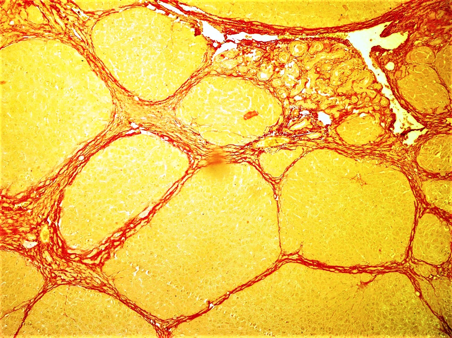
Fibrosis, also known as fibrotic scarring, is a pathological wound healing in which connective tissue replaces normal parenchymal tissue to the extent that it goes unchecked, leading to considerable tissue remodelling and the formation of permanent scar tissue. Repeated injuries, chronic inflammation and repair are susceptible to fibrosis where an accidental excessive accumulation of extracellular matrix components, such as the collagen is produced by fibroblasts, leading to the formation of a permanent fibrotic scar. In response to injury, this is called scarring, and if fibrosis arises from a single cell line, this is called a fibroma. Physiologically, fibrosis acts to deposit connective tissue, which can interfere with or totally inhibit the normal architecture and function of the underlying organ or tissue. Fibrosis can be used to describe the pathological state of excess deposition of fibrous tissue, as well as the process of connective tissue deposition in healing. Define ... [...More Info...] [...Related Items...] OR: [Wikipedia] [Google] [Baidu] |
Human Eyeball
The human eye is a sensory organ, part of the sensory nervous system, that reacts to visible light and allows humans to use visual information for various purposes including seeing things, keeping balance, and maintaining circadian rhythm. The eye can be considered as a living optical device. It is approximately spherical in shape, with its outer layers, such as the outermost, white part of the eye (the sclera) and one of its inner layers (the pigmented choroid) keeping the eye essentially light tight except on the eye's optic axis. In order, along the optic axis, the optical components consist of a first lens (the cornea—the clear part of the eye) that accomplishes most of the focussing of light from the outside world; then an aperture (the pupil) in a diaphragm (the iris—the coloured part of the eye) that controls the amount of light entering the interior of the eye; then another lens (the crystalline lens) that accomplishes the remaining focussing of light into ... [...More Info...] [...Related Items...] OR: [Wikipedia] [Google] [Baidu] |
Extraocular Muscles
The extraocular muscles (extrinsic ocular muscles), are the seven extrinsic muscles of the human eye. Six of the extraocular muscles, the four recti muscles, and the superior and inferior oblique muscles, control movement of the eye and the other muscle, the levator palpebrae superioris, controls eyelid elevation. The actions of the six muscles responsible for eye movement depend on the position of the eye at the time of muscle contraction. Structure Since only a small part of the eye called the fovea provides sharp vision, the eye must move to follow a target. Eye movements must be precise and fast. This is seen in scenarios like reading, where the reader must shift gaze constantly. Although under voluntary control, most eye movement is accomplished without conscious effort. Precisely how the integration between voluntary and involuntary control of the eye occurs is a subject of continuing research."eye, human."Encyclopædia Britannica from Encyclopædia Britannica 2006 Ulti ... [...More Info...] [...Related Items...] OR: [Wikipedia] [Google] [Baidu] |
Ophthalmoparesis
Ophthalmoparesis refers to weakness (-paresis) or paralysis (-plegia) of one or more extraocular muscles which are responsible for eye movements. It is a physical finding in certain neurologic, ophthalmologic, and endocrine disease. Internal ophthalmoplegia means involvement limited to the pupillary sphincter and ciliary muscle. External ophthalmoplegia refers to involvement of only the extraocular muscles. Complete ophthalmoplegia indicates involvement of both. Causes Ophthalmoparesis can result from disorders of various parts of the eye and nervous system: * Infection around the eye. Ophthalmoplegia is an important finding in orbital cellulitis. * The orbit of the eye, including mechanical restrictions of eye movement, as in Graves' disease. * The muscle, as in progressive external ophthalmoplegia or Kearns–Sayre syndrome. * The neuromuscular junction, as in myasthenia gravis. * The relevant cranial nerves (specifically the oculomotor, trochlear, and abducens), as in caver ... [...More Info...] [...Related Items...] OR: [Wikipedia] [Google] [Baidu] |
Ptosis (eyelid)
Ptosis, also known as blepharoptosis, is a drooping or falling of the upper eyelid. The drooping may be worse after being awake longer when the individual's muscles are tired. This condition is sometimes called "lazy eye", but that term normally refers to the condition amblyopia. If severe enough and left untreated, the drooping eyelid can cause other conditions, such as amblyopia or astigmatism. This is why it is especially important for this disorder to be treated in children at a young age, before it can interfere with vision development. The term is from Greek 'fall, falling'. Signs and symptoms Signs and symptoms typically seen in this condition include: * The eyelid(s) may appear to droop. * Droopy eyelids can give the face a false appearance of being fatigued, disinterested, or even sinister. * The eyelid may not protect the eye as effectively, allowing it to dry out. * Sagging upper eyelids can partially block the person's field of view. * Obstructed vision may cause ... [...More Info...] [...Related Items...] OR: [Wikipedia] [Google] [Baidu] |
Superior Rectus Muscle
The superior rectus muscle is a muscle in the orbit. It is one of the extraocular muscles. It is innervated by the superior division of the oculomotor nerve (III). In the primary position (looking straight ahead), its primary function is elevation, although it also contributes to intorsion and adduction. It is associated with a number of medical conditions, and may be weak, paralysed, overreactive, or even congenitally absent in some people. Structure The superior rectus muscle originates from the annulus of Zinn. It inserts into the anterosuperior surface of the eye. This insertion has a width of around 11 mm. It is around 8 mm from the corneal limbus. Nerve supply The superior rectus muscle is supplied by the superior division of the oculomotor nerve (III). Relations The superior rectus muscle is related to the other extraocular muscles, particularly to the medial rectus muscle and the lateral rectus muscle. The insertion of the superior rectus muscle is around 7.5 mm ... [...More Info...] [...Related Items...] OR: [Wikipedia] [Google] [Baidu] |
Inferior Rectus Muscle
The inferior rectus muscle is a muscle in the orbit near the eye. It is one of the four recti muscles in the group of extraocular muscles. It originates from the common tendinous ring, and inserts into the anteroinferior surface of the eye. It depresses the eye (downwards). Structure The inferior rectus muscle originates from the common tendinous ring (annulus of Zinn). It inserts into the anteroinferior surface of the eye. This insertion has a width of around 10.5 mm. It is around 7 mm from the corneal limbus. Blood supply The inferior rectus muscle is supplied by an inferior muscular branch of the ophthalmic artery. It may also be supplied by a branch of the infraorbital artery. It is drained by the corresponding veins: the inferior muscular branch of the ophthalmic vein, and sometimes a branch of the infraorbital vein. Nerve supply The inferior rectus muscle is supplied by the inferior division of the oculomotor nerve (III). Development The inferior rectus muscle devel ... [...More Info...] [...Related Items...] OR: [Wikipedia] [Google] [Baidu] |
KIF21A
Kinesin-like protein KIF21A is a protein that in humans is encoded by the ''KIF21A'' gene. KIF21A belongs to a family of plus end-directed kinesin (see MIM 600025) motor proteins. Neurons use kinesin and dynein (see MIM 600112) microtubule-dependent motor proteins to transport essential cellular components along axonal and dendritic microtubules Microtubules are polymers of tubulin that form part of the cytoskeleton and provide structure and shape to eukaryotic cells. Microtubules can be as long as 50 micrometres, as wide as 23 to 27 nm and have an inner diameter between 11 an .... upplied by OMIMref name="entrez" /> References Further reading * * * * * * * * * * * * * * * * * External links Engle Laboratory CFEOM page [...More Info...] [...Related Items...] OR: [Wikipedia] [Google] [Baidu] |
PHOX2A
Paired mesoderm homeobox protein 2A is a protein that in humans is encoded by the ''PHOX2A'' gene. Function The protein encoded by this gene contains a paired-like homeodomain most similar to that of the Drosophila aristaless gene product. This protein is expressed specifically in noradrenergic cell types. It regulates the expression of tyrosine hydroxylase and dopamine beta-hydroxylase, two catecholaminergic biosynthetic enzymes essential for the differentiation and maintenance of noradrenergic phenotype. Mutations in this gene have been associated with autosomal recessive congenital fibrosis of the extraocular muscles (CFEOM2). Interactions PHOX2A has been shown to interact with HAND2 Heart- and neural crest derivatives-expressed protein 2 is a protein that in humans is encoded by the ''HAND2'' gene. Function The protein encoded by this gene belongs to the basic helix-loop-helix family of transcription factors. This gene p .... References Further reading * ... [...More Info...] [...Related Items...] OR: [Wikipedia] [Google] [Baidu] |
Footnotes
A note is a string of text placed at the bottom of a page in a book or document or at the end of a chapter, volume, or the whole text. The note can provide an author's comments on the main text or citations of a reference work in support of the text. Footnotes are notes at the foot of the page while endnotes are collected under a separate heading at the end of a chapter, volume, or entire work. Unlike footnotes, endnotes have the advantage of not affecting the layout of the main text, but may cause inconvenience to readers who have to move back and forth between the main text and the endnotes. In some editions of the Bible, notes are placed in a narrow column in the middle of each page between two columns of biblical text. Numbering and symbols In English, a footnote or endnote is normally flagged by a superscripted number immediately following that portion of the text the note references, each such footnote being numbered sequentially. Occasionally, a number between brack ... [...More Info...] [...Related Items...] OR: [Wikipedia] [Google] [Baidu] |


