|
Bicornuate
A bicornuate uterus or bicornate uterus (from the Latin ''cornū'', meaning "horn"), is a type of mullerian anomaly in the human uterus, where there is a deep indentation at the fundus (top) of the uterus. Pathophysiology A bicornuate uterus develops during embryogenesis. It occurs when the proximal (upper) portions of the paramesonephric ducts do not fuse, but the distal portions that develops into the lower uterine segment, cervix, and upper vagina fuse normally. Diagnosis Diagnosis of bicornuate uterus typically involves imaging of the uterus with 2D or 3D ultrasound, hysterosalpingography, or magnetic resonance imaging (MRI). On imaging, a bicornuate uterus can be distinguished from a septate uterus by the angle between the cornua (intercornual angle): less than 75 degrees in a septate uterus, and greater than 105 degrees in a bicornuate uterus. Measuring the depth of the cleft between the cornua (fundal cleft) may also assist in diagnosis; a cleft of over is indicativ ... [...More Info...] [...Related Items...] OR: [Wikipedia] [Google] [Baidu] |
Bicornuate Uterus With Pregnancy
A bicornuate uterus or bicornate uterus (from the Latin ''cornū'', meaning "horn"), is a type of mullerian anomaly in the human uterus, where there is a deep indentation at the fundus (top) of the uterus. Pathophysiology A bicornuate uterus develops during embryogenesis. It occurs when the proximal (upper) portions of the paramesonephric ducts do not fuse, but the distal portions that develops into the lower uterine segment, cervix, and upper vagina fuse normally. Diagnosis Diagnosis of bicornuate uterus typically involves imaging of the uterus with 2D or 3D ultrasound, hysterosalpingography, or magnetic resonance imaging (MRI). On imaging, a bicornuate uterus can be distinguished from a septate uterus by the angle between the cornua (intercornual angle): less than 75 degrees in a septate uterus, and greater than 105 degrees in a bicornuate uterus. Measuring the depth of the cleft between the cornua (fundal cleft) may also assist in diagnosis; a cleft of over is indicativ ... [...More Info...] [...Related Items...] OR: [Wikipedia] [Google] [Baidu] |
Mullerian Anomalies
Müllerian duct anomalies are those structural anomalies caused by errors in müllerian duct development during embryonic morphogenesis. Factors that precipitate include genetics, and maternal exposure to teratogens. Genetic causes of müllerian duct anomalies are complicated and uncommon. Inheritance patterns can be autosomal dominant, autosomal recessive, and X-linked disorders. Müllerian anomalies can be part of a multiple malformation syndrome. Mullerian anomalies occur as a congenital malformation of the mullerian ducts during embryogenesis. The mullerian ducts are also referred to as paramesonephric ducts, referring to ducts next to (para) the mesonephric (Wolffian) duct during foetal development. Paramesonephric ducts are paired ducts derived from the embryo, and for females develop into the uterus, uterine tubes, cervix and upper two-thirds of the vagina. Embryogenesis of the mullerian ducts play important roles in ensuring normal development of the female reproductive tr ... [...More Info...] [...Related Items...] OR: [Wikipedia] [Google] [Baidu] |
Uterus
The uterus (from Latin ''uterus'', plural ''uteri'') or womb () is the organ in the reproductive system of most female mammals, including humans that accommodates the embryonic and fetal development of one or more embryos until birth. The uterus is a hormone-responsive sex organ that contains glands in its lining that secrete uterine milk for embryonic nourishment. In the human, the lower end of the uterus, is a narrow part known as the isthmus that connects to the cervix, leading to the vagina. The upper end, the body of the uterus, is connected to the fallopian tubes, at the uterine horns, and the rounded part above the openings to the fallopian tubes is the fundus. The connection of the uterine cavity with a fallopian tube is called the uterotubal junction. The fertilized egg is carried to the uterus along the fallopian tube. It will have divided on its journey to form a blastocyst that will implant itself into the lining of the uterus – the endometrium, where it will ... [...More Info...] [...Related Items...] OR: [Wikipedia] [Google] [Baidu] |
Uterine Malformation
A uterine malformation is a type of female genital malformation resulting from an abnormal development of the Müllerian duct(s) during embryogenesis. Symptoms range from amenorrhea, infertility, recurrent pregnancy loss, and pain, to normal functioning depending on the nature of the defect. Types The American Fertility Society (now American Society of Reproductive Medicine) Classification distinguishes: ; Class I—Müllerian agenesis (absent uterus). : This condition is represented by the hypoplasia or the agenesis (total absence) of the different parts of the uterus: :* Vaginal hypoplasia or agenesis :* Cervical hypoplasia or agenesis :* Fundal hypoplasia or agenesis (absence or hypoplasia of the fundus of the uterus) :* Tubal hypoplasia or agenesis (absence or hypoplasia of the Fallopian tubes) :* Combined hypoplasia the agenesis of different part of the uterus :This condition is also called Mayer-Rokitansky-Kuster-Hauser syndrome. The patient with MRKH syndrome will hav ... [...More Info...] [...Related Items...] OR: [Wikipedia] [Google] [Baidu] |
IUD With Progestogen
A hormonal intrauterine device (IUD), also known as a intrauterine system (IUS) with progestogen and sold under the brand name Mirena among others, is an intrauterine device that releases a progestogenic hormonal agent such as levonorgestrel into the uterus. It is used for birth control, heavy menstrual periods, and to prevent excessive build of the lining of the uterus in those on estrogen replacement therapy. It is one of the most effective forms of birth control with a one-year failure rate around 0.2%. The device is placed in the uterus and lasts three to eight years. Fertility often returns quickly following removal. Side effects include irregular periods, benign ovarian cysts, pelvic pain, and depression. Rarely uterine perforation may occur. Use is not recommended during pregnancy but is safe with breastfeeding. The IUD with progestogen is a type of long-acting reversible birth control. It works by thickening the mucus at the opening of the cervix, stopping the buildup ... [...More Info...] [...Related Items...] OR: [Wikipedia] [Google] [Baidu] |
Metroplasty
Metroplasty (also called Strassman metroplasty, uteroplasty or hysteroplasty) is a reconstructive surgery used to repair congenital anomalies of the uterus, including septate uterus and bicornuate uterus A bicornuate uterus or bicornate uterus (from the Latin ''cornū'', meaning "horn"), is a type of mullerian anomaly in the human uterus, where there is a deep indentation at the fundus (top) of the uterus. Pathophysiology A bicornuate uterus .... The surgery entails removing the abnormal tissue that separates the cornua of the uterus, then using several layers of stitches to create a normal shape. References {{Medicine Gynecological surgery ... [...More Info...] [...Related Items...] OR: [Wikipedia] [Google] [Baidu] |
Postpartum Bleeding
Postpartum bleeding or postpartum hemorrhage (PPH) is often defined as the loss of more than 500 ml or 1,000 ml of blood following childbirth. Some have added the requirement that there also be signs or symptoms of low blood volume for the condition to exist. Signs and symptoms may initially include: an increased heart rate, feeling faint upon standing, and an increased breathing rate. As more blood is lost, the patient may feel cold, blood pressure may drop, and they may become restless or unconscious. The condition can occur up to six weeks following delivery. The most common cause is poor contraction of the uterus following childbirth. Not all of the placenta being delivered, a tear of the uterus, or poor blood clotting are other possible causes. It occurs more commonly in those who: already have a low amount of red blood, are Asian, with bigger or more than one baby, are obese or are older than 40 years of age. It also occurs more commonly following caesarean s ... [...More Info...] [...Related Items...] OR: [Wikipedia] [Google] [Baidu] |
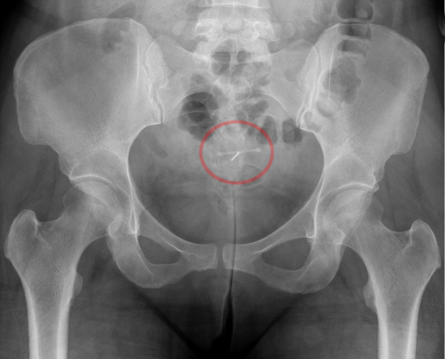
Intrauterine Device With Copper
Intrauterine device (IUD) with copper, also known as intrauterine coil or copper coil, is a type of intrauterine device which contains copper. It is used for birth control and emergency contraception within five days of unprotected sex. It is one of the most effective forms of birth control with a one-year failure rate around 0.7%. The device is placed in the uterus and lasts up to twelve years. It may be used by women of all ages regardless of whether or not they have had children. Following removal, fertility quickly returns. Side effects may be heavy menstrual periods, and/or rarely the device may come out. It is less recommended for people at high risk of sexually transmitted infections as it may increase the risk of pelvic inflammatory disease in the first three weeks after insertion. It is recommended for people who don't tolerate or hardly tolerate hormonal contraceptives. If a woman becomes pregnant with an IUD in place removal is recommended. Very rarely, uterine per ... [...More Info...] [...Related Items...] OR: [Wikipedia] [Google] [Baidu] |
Obstructed Labour
Obstructed labour, also known as labour dystocia, is the baby not exiting the pelvis because it is physically block during childbirth although the uterus contracts normally. Complications for the baby include not getting enough oxygen which may result in death. It increases the risk of the mother getting an infection, having uterine rupture, or having post-partum bleeding. Long-term complications for the mother include obstetrical fistula. Obstructed labour is said to result in prolonged labour, when the active phase of labour is longer than 12 hours. The main causes of obstructed labour include a large or abnormally positioned baby, a small pelvis, and problems with the birth canal. Abnormal positioning includes shoulder dystocia where the anterior shoulder does not pass easily below the pubic bone. Risk factors for a small pelvis include malnutrition and a lack of exposure to sunlight causing vitamin D deficiency. It is also more common in adolescence as the pelvis may not ha ... [...More Info...] [...Related Items...] OR: [Wikipedia] [Google] [Baidu] |
Shoulder Presentation
A shoulder presentation is a malpresentation at childbirth where the baby is in a transverse lie (its vertebral column is perpendicular to that of the mother), thus the leading part (the part that first enters the birth canal) is an arm, a shoulder, or the trunk. While a baby can be delivered vaginally when either the head or the feet/buttocks are the leading part, it usually cannot be expected to be delivered successfully with a shoulder presentation unless a cesarean section (C/S) is performed. Frequency and causes Shoulder presentations are uncommon (about 0.5% of births) since, usually, toward the end of gestation, either the head or the buttocks start to enter the upper part of the pelvis, anchoring the fetus in a longitudinal lie. It is not known in all cases of shoulder presentation why the longitudinal lie is not reached, but possible causes include bony abnormalities of the pelvis, uterine abnormalities such as malformations or tumors (fibroids), or other tumors in the pel ... [...More Info...] [...Related Items...] OR: [Wikipedia] [Google] [Baidu] |
Breech Birth
A breech birth is when a baby is born bottom first instead of head first, as is normal. Around 3–5% of pregnant women at term (37–40 weeks pregnant) have a breech baby. Due to their higher than average rate of possible complications for the baby, breech births are generally considered higher risk. Breech births also occur in many other mammals such as dogs and horses, see veterinary obstetrics. Most babies in the breech position are delivered via caesarean section because it is seen as safer than being born vaginally. Doctors and midwives in the developing world often lack many of the skills required to safely assist women giving birth to a breech baby vaginally. Also, delivering all breech babies by caesarean section in developing countries is difficult to implement as there are not always resources available to provide this service. OB-GYNs do not recommend home births if a breech birth is expected, even when attended by a medical professional. Cause With regard to the ... [...More Info...] [...Related Items...] OR: [Wikipedia] [Google] [Baidu] |


