|
Air Trapping
Air trapping, also called gas trapping, is an abnormal retention of air in the lungs where it is difficult to exhale completely. It is observed in obstructive lung diseases such as asthma, bronchiolitis obliterans syndrome and chronic obstructive pulmonary diseases such as emphysema and chronic bronchitis. Air trapping is not a diagnosis but is a presentation of an illness, and can be a guide to the appropriate differential. __TOC__ Imaging On high resolution CT air trapping has a typical imaging appearance, although often evaluation with both maximum inhalation and exhalation, or inspiratory and expiratory views, are needed for a more specific diagnosis. One of its typical imaging patterns is mosaic attenuation. In the classic presentation, the lung will appear normal at inspiration, but on exhalation, the diseased portions of the lung which have lost connective tissue recoil will remain lucent while the healthy portions of the lung will become more dense due to atelectasis. Th ... [...More Info...] [...Related Items...] OR: [Wikipedia] [Google] [Baidu] |
Lungs
The lungs are the primary organs of the respiratory system in humans and most other animals, including some snails and a small number of fish. In mammals and most other vertebrates, two lungs are located near the backbone on either side of the heart. Their function in the respiratory system is to extract oxygen from the air and transfer it into the bloodstream, and to release carbon dioxide from the bloodstream into the atmosphere, in a process of gas exchange. Respiration is driven by different muscular systems in different species. Mammals, reptiles and birds use their different muscles to support and foster breathing. In earlier tetrapods, air was driven into the lungs by the pharyngeal muscles via buccal pumping, a mechanism still seen in amphibians. In humans, the main muscle of respiration that drives breathing is the diaphragm. The lungs also provide airflow that makes vocal sounds including human speech possible. Humans have two lungs, one on the left and one on the ... [...More Info...] [...Related Items...] OR: [Wikipedia] [Google] [Baidu] |
Atelectasis
Atelectasis is the collapse or closure of a lung resulting in reduced or absent gas exchange. It is usually unilateral, affecting part or all of one lung. It is a condition where the alveoli are deflated down to little or no volume, as distinct from pulmonary consolidation, in which they are filled with liquid. It is often called a ''collapsed lung'', although that term may also refer to pneumothorax. It is a very common finding in chest X-rays and other radiological studies, and may be caused by normal exhalation or by various medical conditions. Although frequently described as a ''collapse of lung tissue'', atelectasis is not synonymous with a pneumothorax, which is a more specific condition that can cause atelectasis. Acute atelectasis may occur as a post-operative complication or as a result of surfactant deficiency. In premature babies, this leads to infant respiratory distress syndrome. The term uses combining forms of ''atel-'' + ''ectasis'', from el, ἀτελής, ... [...More Info...] [...Related Items...] OR: [Wikipedia] [Google] [Baidu] |
Computed Tomography
A computed tomography scan (CT scan; formerly called computed axial tomography scan or CAT scan) is a medical imaging technique used to obtain detailed internal images of the body. The personnel that perform CT scans are called radiographers or radiology technologists. CT scanners use a rotating X-ray tube and a row of detectors placed in a gantry to measure X-ray attenuations by different tissues inside the body. The multiple X-ray measurements taken from different angles are then processed on a computer using tomographic reconstruction algorithms to produce tomographic (cross-sectional) images (virtual "slices") of a body. CT scans can be used in patients with metallic implants or pacemakers, for whom magnetic resonance imaging (MRI) is contraindicated. Since its development in the 1970s, CT scanning has proven to be a versatile imaging technique. While CT is most prominently used in medical diagnosis, it can also be used to form images of non-living objects. The 1979 Nob ... [...More Info...] [...Related Items...] OR: [Wikipedia] [Google] [Baidu] |
Lung Volumes
Lung volumes and lung capacities refer to the volume of air in the lungs at different phases of the respiratory cycle. The average total lung capacity of an adult human male is about 6 litres of air. Tidal breathing is normal, resting breathing; the tidal volume is the volume of air that is inhaled or exhaled in only a single such breath. The average human respiratory rate is 30–60 breaths per minute at birth, decreasing to 12–20 breaths per minute in adults. Factors affecting volumes Several factors affect lung volumes; some can be controlled, and some cannot be controlled. Lung volumes vary with different people as follows: A person who is born and lives at sea level will develop a slightly smaller lung capacity than a person who spends their life at a high altitude. This is because the partial pressure of oxygen is lower at higher altitude which, as a result means that oxygen less readily diffuses into the bloodstream. In response to higher altitude, the body's diffu ... [...More Info...] [...Related Items...] OR: [Wikipedia] [Google] [Baidu] |
Spirometry
Spirometry (meaning ''the measuring of breath'') is the most common of the pulmonary function tests (PFTs). It measures lung function, specifically the amount (volume) and/or speed (flow) of air that can be inhaled and exhaled. Spirometry is helpful in assessing breathing patterns that identify conditions such as asthma, pulmonary fibrosis, cystic fibrosis, and COPD. It is also helpful as part of a system of health surveillance, in which breathing patterns are measured over time. Spirometry generates pneumotachographs, which are charts that plot the volume and flow of air coming in and out of the lungs from one inhalation and one exhalation. Indications Spirometry is indicated for the following reasons: * to diagnose or manage asthma * to detect respiratory disease in patients presenting with symptoms of breathlessness, and to distinguish respiratory from cardiac disease as the cause * to measure bronchial responsiveness in patients suspected of having asthma * to diagnose and ... [...More Info...] [...Related Items...] OR: [Wikipedia] [Google] [Baidu] |
Interstitial Lung Disease
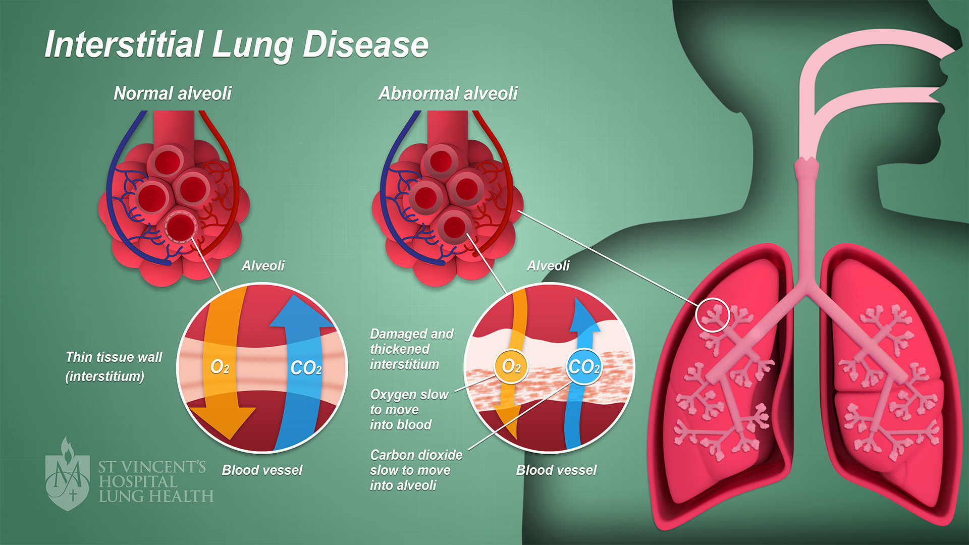
Interstitial lung disease (ILD), or diffuse parenchymal lung disease (DPLD), is a group of respiratory diseases affecting the interstitium (the tissue and space around the alveoli (air sacs)) of the lungs. It concerns alveolar epithelium, pulmonary capillary endothelium, basement membrane, and perivascular and perilymphatic tissues. It may occur when an injury to the lungs triggers an abnormal healing response. Ordinarily, the body generates just the right amount of tissue to repair damage, but in interstitial lung disease, the repair process is disrupted, and the tissue around the air sacs (alveoli) becomes scarred and thickened. This makes it more difficult for oxygen to pass into the bloodstream. The disease presents itself with the following symptoms: shortness of breath, nonproductive coughing, fatigue, and weight loss, which tend to develop slowly, over several months. The average rate of survival for someone with this disease is between three and five years. The term ILD i ... [...More Info...] [...Related Items...] OR: [Wikipedia] [Google] [Baidu] |
Usual Interstitial Pneumonitis
Usual may refer to: *Common *Normal *Standard Standard may refer to: Symbols * Colours, standards and guidons, kinds of military signs * Standard (emblem), a type of a large symbol or emblem used for identification Norms, conventions or requirements * Standard (metrology), an object th ... {{disambig ... [...More Info...] [...Related Items...] OR: [Wikipedia] [Google] [Baidu] |
Nonspecific Interstitial Pneumonia
Non-specific interstitial pneumonia (NSIP) is a form of idiopathic interstitial pneumonia. Symptoms Symptoms include cough, difficulty breathing, and fatigue. Causes It has been suggested that idiopathic nonspecific interstitial pneumonia has an autoimmune mechanism, and is a possible complication of undifferentiated connective tissue disease; however, not enough research has been done at this time to find a cause. Patients with NSIP will often have other unrelated lung diseases like COPD or emphysema, along with other auto-immune disorders. Diagnosis A full clinical diagnosis can only be made from a lung biopsy of the tissue, fully best performed by a VATS, done by a cardio-thoracic surgeon. Some pulmonologists may first attempt a bronchoscopy; however, this frequently fails to give a full or correct diagnosis. Lung biopsies performed on patients with NSIP reveal two different disease patterns – cellular and fibrosing – which are associated with different prognoses. T ... [...More Info...] [...Related Items...] OR: [Wikipedia] [Google] [Baidu] |
Mosaic Attenuation
A mosaic is a pattern or image made of small regular or irregular pieces of colored stone, glass or ceramic, held in place by plaster/mortar, and covering a surface. Mosaics are often used as floor and wall decoration, and were particularly popular in the Ancient Rome, Ancient Roman world. Mosaic today includes not just murals and pavements, but also artwork, hobby crafts, and industrial and construction forms. Mosaics have a long history, starting in Mesopotamia in the 3rd millennium BC. Pebble mosaics were made in Tiryns in Mycenean civilisation, Mycenean Greece; mosaics with patterns and pictures became widespread in classical times, both in Ancient Greece and Ancient Rome. Early Christian basilicas from the 4th century onwards were decorated with wall and ceiling mosaics. Mosaic art flourished in the Byzantine Empire from the 6th to the 15th centuries; that tradition was adopted by the Norman dynasty, Norman Kingdom of Sicily in the 12th century, by the eastern-influenced R ... [...More Info...] [...Related Items...] OR: [Wikipedia] [Google] [Baidu] |
Obstructive Lung Disease
Obstructive lung disease is a category of respiratory disease characterized by airway obstruction. Many obstructive diseases of the lung result from narrowing (obstruction) of the smaller bronchi and larger bronchioles, often because of excessive contraction of the smooth muscle itself. It is generally characterized by inflamed and easily collapsible airways, obstruction to airflow, problems exhaling, and frequent medical clinic visits and hospitalizations. Types of obstructive lung disease include; asthma, bronchiectasis, bronchitis and chronic obstructive pulmonary disease (COPD). Although COPD shares similar characteristics with all other obstructive lung diseases, such as the signs of coughing and wheezing, they are distinct conditions in terms of disease onset, frequency of symptoms, and reversibility of airway obstruction. Cystic fibrosis is also sometimes included in obstructive pulmonary disease. Types Asthma Asthma is an obstructive lung disease where the bronchial t ... [...More Info...] [...Related Items...] OR: [Wikipedia] [Google] [Baidu] |
High Resolution CT
High-resolution computed tomography (HRCT) is a type of computed tomography (CT) with specific techniques to enhance image resolution. It is used in the diagnosis of various health problems, though most commonly for lung disease, by assessing the lung parenchyma. On the other hand, HRCT of the temporal bone is used to diagnose various middle ear diseases such as otitis media, cholesteatoma, and evaluations after ear operations. Technique HRCT is performed using a conventional CT scanner. However, imaging parameters are chosen so as to maximize spatial resolution: a narrow slice width is used (usually 1–2 mm), a high spatial resolution image reconstruction algorithm is used, field of view is minimized, so as to minimize the size of each pixel, and other scan factors (e.g. focal spot) may be optimized for resolution at the expense of scan speed. Depending on the suspected diagnosis, the scan may be performed in both inspiration and expiration. In inspiration images are ... [...More Info...] [...Related Items...] OR: [Wikipedia] [Google] [Baidu] |
Differential Diagnosis
In healthcare, a differential diagnosis (abbreviated DDx) is a method of analysis of a patient's history and physical examination to arrive at the correct diagnosis. It involves distinguishing a particular disease or condition from others that present with similar clinical features. Differential diagnostic procedures are used by clinicians to diagnose the specific disease in a patient, or, at least, to consider any imminently life-threatening conditions. Often, each individual option of a possible disease is called a differential diagnosis (e.g., acute bronchitis could be a differential diagnosis in the evaluation of a cough, even if the final diagnosis is common cold). More generally, a differential diagnostic procedure is a systematic diagnostic method used to identify the presence of a disease entity where multiple alternatives are possible. This method may employ algorithms, akin to the process of elimination, or at least a process of obtaining information that shrinks the "p ... [...More Info...] [...Related Items...] OR: [Wikipedia] [Google] [Baidu] |


.jpg)