|
Acyanotic Heart Defect
An acyanotic heart defect, is a class of congenital heart defects. In these, blood is shunted (flows) from the left side of the heart to the right side of the heart, most often due to a structural defect (hole) in the interventricular septum. People often retain normal levels of oxyhemoglobin saturation in systemic circulation. This term is outdated, because a person with an acyanotic heart defect may show cyanosis (turn blue due to insufficient oxygen in the blood). Signs and symptoms Presentation is the following: * Shortness of breath * Congested cough * Diaphoresis * Fatigue * Frequent respiratory infections * Machine-like heart murmur * Tachycardia * Tachypnea * Respiratory distress * Mild cyanosis (in right sided heart failure) * Poor growth and development (from increased energy spent on breathing) Complications This condition can cause congestive heart failure. Diagnosis Types Left to right shunting heart defects include: * Ventricular septal defect (VSD) (30% o ... [...More Info...] [...Related Items...] OR: [Wikipedia] [Google] [Baidu] |
Congenital Heart Defect
A congenital heart defect (CHD), also known as a congenital heart anomaly and congenital heart disease, is a defect in the structure of the heart or great vessels that is present at birth. A congenital heart defect is classed as a cardiovascular disease. Signs and symptoms depend on the specific type of defect. Symptoms can vary from none to life-threatening. When present, symptoms may include rapid breathing, bluish skin (cyanosis), poor weight gain, and feeling tired. CHD does not cause chest pain. Most congenital heart defects are not associated with other diseases. A complication of CHD is heart failure. The cause of a congenital heart defect is often unknown. Risk factors include certain infections during pregnancy such as rubella, use of certain medications or drugs such as alcohol or tobacco, parents being closely related, or poor nutritional status or obesity in the mother. Having a parent with a congenital heart defect is also a risk factor. A number of genetic conditio ... [...More Info...] [...Related Items...] OR: [Wikipedia] [Google] [Baidu] |
Patent Ductus Arteriosus
''Patent ductus arteriosus'' (PDA) is a medical condition in which the ''ductus arteriosus'' fails to close after birth: this allows a portion of oxygenated blood from the left heart to flow back to the lungs by flowing from the aorta, which has a higher pressure, to the pulmonary artery. Symptoms are uncommon at birth and shortly thereafter, but later in the first year of life there is often the onset of an increased work of breathing and failure to gain weight at a normal rate. With time, an uncorrected PDA usually leads to pulmonary hypertension followed by right-sided heart failure. The ''ductus arteriosus'' is a fetal blood vessel that normally closes soon after birth. In a PDA, the vessel does not close, but remains ''patent'' (open), resulting in an abnormal transmission of blood from the aorta to the pulmonary artery. PDA is common in newborns with persistent respiratory problems such as hypoxia, and has a high occurrence in premature newborns. Premature newborns are ... [...More Info...] [...Related Items...] OR: [Wikipedia] [Google] [Baidu] |
Furosemide
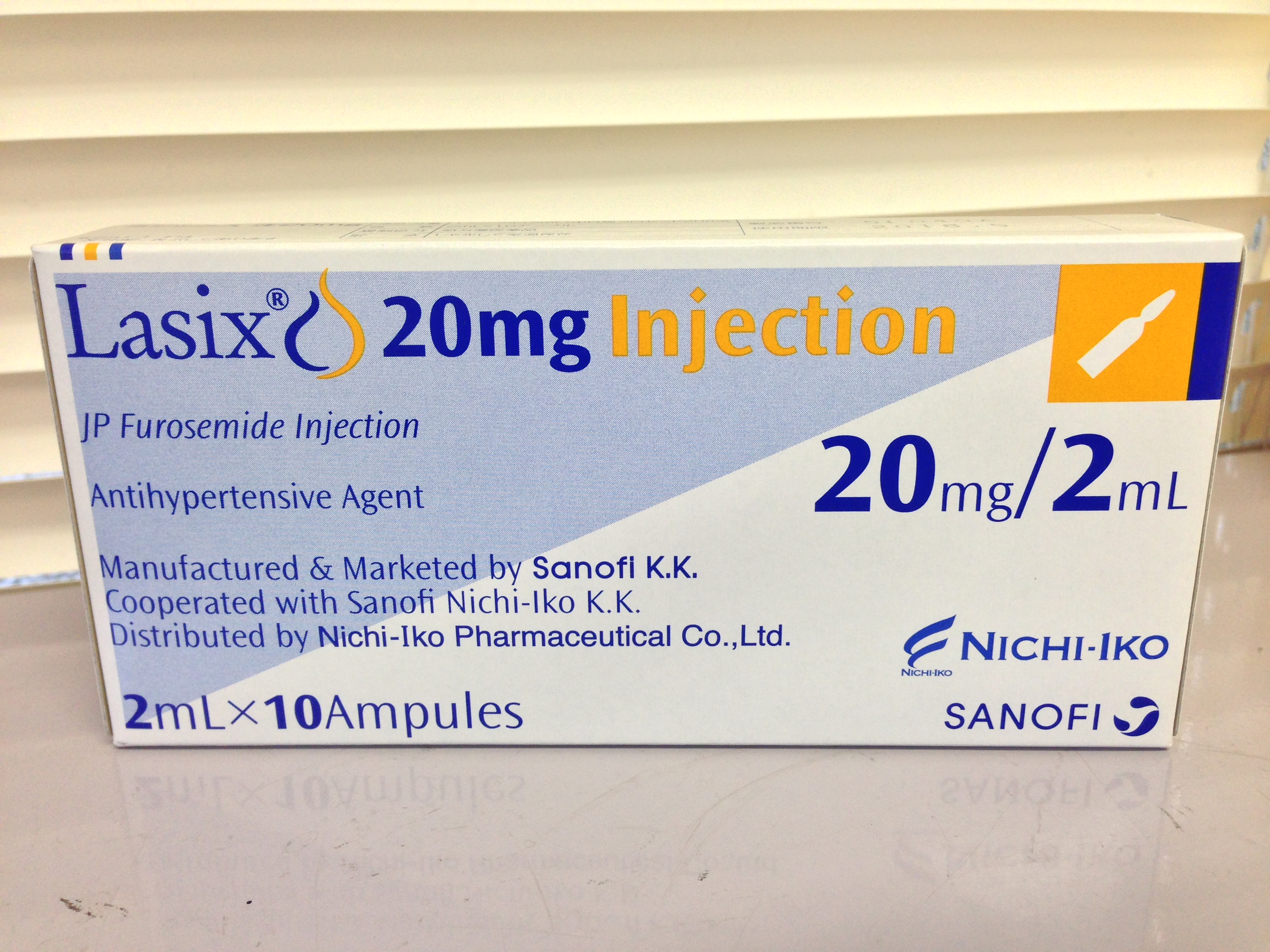
Furosemide is a loop diuretic medication used to treat fluid build-up due to heart failure, liver scarring, or kidney disease. It may also be used for the treatment of high blood pressure. It can be taken by injection into a vein or by mouth. When taken by mouth, it typically begins working within an hour, while intravenously, it typically begins working within five minutes. Common side effects include feeling lightheaded while standing, ringing in the ears, and sensitivity to light. Potentially serious side effects include electrolyte abnormalities, low blood pressure, and hearing loss. Blood tests are recommended regularly for those on treatment. Furosemide is a type of loop diuretic that works by decreasing the reabsorption of sodium by the kidneys. Common side effects of furosemide injection include hypokalemia (low potassium level), hypotension (low blood pressure), and dizziness. Furosemide was patented in 1959 and approved for medical use in 1964. It is on the Wo ... [...More Info...] [...Related Items...] OR: [Wikipedia] [Google] [Baidu] |
Digoxin
Digoxin (better known as Digitalis), sold under the brand name Lanoxin among others, is a medication used to treat various heart conditions. Most frequently it is used for atrial fibrillation, atrial flutter, and heart failure. Digoxin is one of the oldest medications used in the field of cardiology. It works by increasing myocardial contractility, increasing stroke volume and blood pressure, reducing heart rate, and somewhat extending the time frame of the contraction. Digoxin is taken by mouth or by injection into a vein. Digoxin has a half life of approximately 36 hours given at average doses in patients with normal renal function. It is excreted mostly unchanged in the urine. Common side effects include breast enlargement with other side effects generally due to an excessive dose. These side effects may include loss of appetite, nausea, trouble seeing, confusion, and an irregular heartbeat. Greater care is required in older people and those with poor kidney function. It ... [...More Info...] [...Related Items...] OR: [Wikipedia] [Google] [Baidu] |
Coarctation Of The Aorta
Coarctation of the aorta (CoA or CoAo), also called aortic narrowing, is a congenital condition whereby the aorta is narrow, usually in the area where the ductus arteriosus (ligamentum arteriosum after regression) inserts. The word ''coarctation'' means "pressing or drawing together; narrowing". Coarctations are most common in the aortic arch. The arch may be small in babies with coarctations. Other heart defects may also occur when coarctation is present, typically occurring on the left side of the heart. When a patient has a coarctation, the left ventricle has to work harder. Since the aorta is narrowed, the left ventricle must generate a much higher pressure than normal in order to force enough blood through the aorta to deliver blood to the lower part of the body. If the narrowing is severe enough, the left ventricle may not be strong enough to push blood through the coarctation, thus resulting in a lack of blood to the lower half of the body. Physiologically its complete form ... [...More Info...] [...Related Items...] OR: [Wikipedia] [Google] [Baidu] |
Aortic Stenosis
Aortic stenosis (AS or AoS) is the narrowing of the exit of the left ventricle of the heart (where the aorta begins), such that problems result. It may occur at the aortic valve as well as above and below this level. It typically gets worse over time. Symptoms often come on gradually with a decreased ability to exercise often occurring first. If heart failure, loss of consciousness, or heart related chest pain occur due to AS the outcomes are worse. Loss of consciousness typically occurs with standing or exercising. Signs of heart failure include shortness of breath especially when lying down, at night, or with exercise, and swelling of the legs. Thickening of the valve without narrowing is known as aortic sclerosis. Causes include being born with a bicuspid aortic valve, and rheumatic fever; a normal valve may also harden over the decades. A bicuspid aortic valve affects about one to two percent of the population. As of 2014 rheumatic heart disease mostly occurs in the dev ... [...More Info...] [...Related Items...] OR: [Wikipedia] [Google] [Baidu] |
Pulmonary Valve
The pulmonary valve (sometimes referred to as the pulmonic valve) is a valve of the heart that lies between the right ventricle and the pulmonary artery and has three cusps. It is one of the four valves of the heart and one of the two semilunar valves, the other being the aortic valve. Similar to the aortic valve, the pulmonary valve opens in ventricular systole, when the pressure in the right ventricle rises above the pressure in the pulmonary artery. At the end of ventricular systole, when the pressure in the right ventricle falls rapidly, the pressure in the pulmonary artery will close the pulmonary valve. The closure of the pulmonary valve contributes the P2 component of the second heart sound (S2). The right heart is a low-pressure system, so the P2 component of the second heart sound is usually softer than the A2 component of the second heart sound. However, it is physiologically normal in some young people to hear both components separated during inhalation. Description * ... [...More Info...] [...Related Items...] OR: [Wikipedia] [Google] [Baidu] |
Pulmonary Stenosis
Pulmonic stenosis, is a dynamic or fixed obstruction of flow from the right ventricle of the heart to the pulmonary artery. It is usually first diagnosed in childhood. Signs and symptoms Cause Pulmonic stenosis is usually due to isolated valvular obstruction (pulmonary valve stenosis), but it may be due to subvalvular or supravalvular obstruction, such as infundibular stenosis. It may occur in association with other congenital heart defects as part of more complicated syndromes (for example, tetralogy of Fallot). Pathophysiology When pulmonic stenosis (PS) is present, resistance to blood flow causes right ventricular hypertrophy. If right ventricular failure develops, right atrial pressure will increase, and this may result in a persistent opening of the foramen ovale, shunting of unoxygenated blood from the right atrium into the left atrium, and systemic cyanosis. If pulmonary stenosis is severe, congestive heart failure occurs, and systemic venous engorgement will be noted. A ... [...More Info...] [...Related Items...] OR: [Wikipedia] [Google] [Baidu] |
Levo-Transposition Of The Great Arteries
Levo-Transposition of the great arteries is an acyanotic congenital heart defect in which the primary arteries (the aorta and the pulmonary artery) are transposed, with the aorta anterior and to the left of the pulmonary artery; the morphological left and right ventricles with their corresponding atrioventricular valves are also transposed. Use of the term "corrected" has been disputed by many due to the frequent occurrence of other abnormalities and or acquired disorders in l-TGA patients. In segmental analysis, this condition is described as atrioventricular discordance (ventricular inversion) with ventriculoarterial discordance. l-TGA is often referred to simply as transposition of the great arteries (TGA); however, TGA is a more general term which may also refer to dextro-transposition of the great arteries (d-TGA). Signs and symptoms Simple l-TGA does not immediately produce any visually identifiable symptoms, but since each ventricle is intended to handle different blood ... [...More Info...] [...Related Items...] OR: [Wikipedia] [Google] [Baidu] |
Atrioventricular Septal Defect
Atrioventricular septal defect (AVSD) or atrioventricular canal defect (AVCD), also known as "common atrioventricular canal" (CAVC) or "endocardial cushion defect" (ECD), is characterized by a deficiency of the atrioventricular septum of the heart that creates connections between all four of its chambers. It is caused by an abnormal or inadequate fusion of the superior and inferior endocardial cushions with the mid portion of the atrial septum and the muscular portion of the ventricular septum. Symptoms and signs Symptoms include difficulty breathing (dyspnea) and bluish discoloration on skin, fingernails, and lips (cyanosis). A newborn baby will show signs of heart failure such as edema, fatigue, wheezing, sweating and irregular heartbeat. Complications Normally, the four chambers of the heart divide oxygenated and de-oxygenated blood into separate pools. When holes form between the chambers, as in AVSD, the pools can mix. Consequently, arterial blood supplies become less oxygena ... [...More Info...] [...Related Items...] OR: [Wikipedia] [Google] [Baidu] |
Interventricular Septum
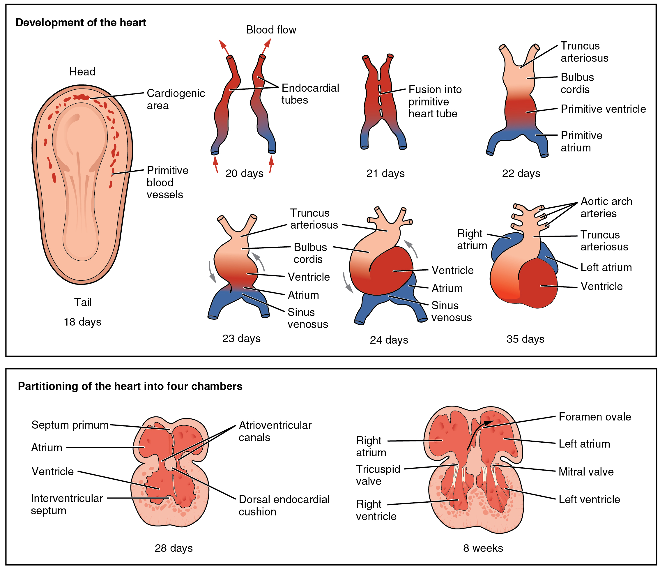
The interventricular septum (IVS, or ventricular septum, or during development septum inferius) is the stout wall separating the ventricles, the lower chambers of the heart, from one another. The ventricular septum is directed obliquely backward to the right and curved with the convexity toward the right ventricle; its margins correspond with the anterior and posterior interventricular sulci. The lower part of the septum, which is the major part, is thick and muscular, and its much smaller upper part is thin and membraneous. During each cardiac cycle the interventricular septum contracts by shortening longitudinally and becoming thicker. Structure The interventricular septum is the stout wall separating the ventricles, the lower chambers of the heart, from one another. The ventricular septum is directed obliquely backward to the right and curved with the convexity toward the right ventricle; its margins correspond with the anterior and posterior longitudinal sulci. The gr ... [...More Info...] [...Related Items...] OR: [Wikipedia] [Google] [Baidu] |
Atrial Septal Defect
Atrial septal defect (ASD) is a congenital heart defect in which blood flows between the atria (upper chambers) of the heart. Some flow is a normal condition both pre-birth and immediately post-birth via the foramen ovale; however, when this does not naturally close after birth it is referred to as a patent (open) foramen ovale (PFO). It is common in patients with a congenital atrial septal aneurysm (ASA). After PFO closure the atria normally are separated by a dividing wall, the interatrial septum. If this septum is defective or absent, then oxygen-rich blood can flow directly from the left side of the heart to mix with the oxygen-poor blood in the right side of the heart; or the opposite, depending on whether the left or right atrium has the higher blood pressure. In the absence of other heart defects, the left atrium has the higher pressure. This can lead to lower-than-normal oxygen levels in the arterial blood that supplies the brain, organs, and tissues. However, an ASD m ... [...More Info...] [...Related Items...] OR: [Wikipedia] [Google] [Baidu] |


