|
Acute Interstitial Pneumonitis
Acute interstitial pneumonitis is a rare, severe lung disease that usually affects otherwise healthy individuals. There is no known cause or cure. Acute interstitial pneumonitis is often categorized as both an interstitial lung disease and a form of acute respiratory distress syndrome (ARDS) but it is distinguished from the ''chronic'' forms of interstitial pneumonia such as idiopathic pulmonary fibrosis. Symptoms and signs The most common symptoms of acute interstitial pneumonitis are highly productive cough with expectoration of thick mucus, fever, and difficulties breathing. These often occur over a period of one to two weeks before medical attention is sought. The presence of fluid means the person experiences a feeling similar to 'drowning'. Difficulties breathing can quickly progress to an inability to breathe without support (respiratory failure). Acute interstitial pneumonitis typically progresses rapidly, with hospitalization and mechanical ventilation often required ... [...More Info...] [...Related Items...] OR: [Wikipedia] [Google] [Baidu] |
Micrograph
A micrograph or photomicrograph is a photograph or digital image taken through a microscope or similar device to show a magnify, magnified image of an object. This is opposed to a macrograph or photomacrograph, an image which is also taken on a microscope but is only slightly magnified, usually less than 10 times. Micrography is the practice or art of using microscopes to make photographs. A micrograph contains extensive details of microstructure. A wealth of information can be obtained from a simple micrograph like behavior of the material under different conditions, the phases found in the system, failure analysis, grain size estimation, elemental analysis and so on. Micrographs are widely used in all fields of microscopy. Types Photomicrograph A light micrograph or photomicrograph is a micrograph prepared using an optical microscope, a process referred to as ''photomicroscopy''. At a basic level, photomicroscopy may be performed simply by connecting a camera to a micros ... [...More Info...] [...Related Items...] OR: [Wikipedia] [Google] [Baidu] |
Biopsy
A biopsy is a medical test commonly performed by a surgeon, interventional radiologist, or an interventional cardiologist. The process involves extraction of sample cells or tissues for examination to determine the presence or extent of a disease. The tissue is then fixed, dehydrated, embedded, sectioned, stained and mounted before it is generally examined under a microscope by a pathologist; it may also be analyzed chemically. When an entire lump or suspicious area is removed, the procedure is called an excisional biopsy. An incisional biopsy or core biopsy samples a portion of the abnormal tissue without attempting to remove the entire lesion or tumor. When a sample of tissue or fluid is removed with a needle in such a way that cells are removed without preserving the histological architecture of the tissue cells, the procedure is called a needle aspiration biopsy. Biopsies are most commonly performed for insight into possible cancerous or inflammatory conditions. H ... [...More Info...] [...Related Items...] OR: [Wikipedia] [Google] [Baidu] |
Respiratory Diseases Principally Affecting The Interstitium
The respiratory system (also respiratory apparatus, ventilatory system) is a biological system consisting of specific organs and structures used for gas exchange in animals and plants. The anatomy and physiology that make this happen varies greatly, depending on the size of the organism, the environment in which it lives and its evolutionary history. In land animals the respiratory surface is internalized as linings of the lungs. Gas exchange in the lungs occurs in millions of small air sacs; in mammals and reptiles these are called alveoli, and in birds they are known as atria. These microscopic air sacs have a very rich blood supply, thus bringing the air into close contact with the blood. These air sacs communicate with the external environment via a system of airways, or hollow tubes, of which the largest is the trachea, which branches in the middle of the chest into the two main bronchi. These enter the lungs where they branch into progressively narrower secondary and te ... [...More Info...] [...Related Items...] OR: [Wikipedia] [Google] [Baidu] |
Arnold Rice Rich
Arnold Rice Rich (March 28, 1893 – April 17, 1968) was an American pathologist. Career Born March 28, 1893, in Birmingham, Alabama, Rich attended the University of Virginia, majoring in biology, and then the Johns Hopkins Medical School in Baltimore, Maryland, from which he received his M.D. degree in 1919. He remained associated with Hopkins the rest of his career. He was appointed Chairman of the Department of Pathology and pathologist-in-chief of the Johns Hopkins Hospital in 1944, until he retired in 1958. Work Rich had broad interests in medicine. Among his many contributions, he classified jaundice, helped understand the formation of bile pigment, studied the relationship between hypersensitivity and immunity, especially in tuberculosis (on which he was one of the reigning experts) and discovered the phagocytic function of the Gaucher cell, the hallmark of Gaucher's disease. A number of diseases or conditions are named after Rich, including: * Hamman-Rich syndrome a ... [...More Info...] [...Related Items...] OR: [Wikipedia] [Google] [Baidu] |
Louis Hamman
Louis Virgil Hamman (December 21, 1877 – April 28, 1946) was recognized as one of the great clinicians in his time. Early life Louis Virgil Hamman was born on December 21, 1877, in Baltimore, Maryland, to Agatha (née Haseneyer) and John A. Hamman. Hamman graduated from Calvert Hall College High School. He then graduated with a Bachelor of Science from Rock Hill College in 1895. He was graduated M.D. from Johns Hopkins University School of Medicine in 1901. Career Hamman interned at New York Hospital from 1901 to 1902 and served as a resident physician until 1903. Hamman returned in 1903 to his alma mater to practice medicine. Hamman served as assistant in medicine at Johns Hopkins School of Medicine from 1903 to 1906. He then served as instructor from 1906 to 1908 and associate in medicine from 1908 to 1915. He was associate professor of clinical medicine from 1915 to 1932. In 1932, he was appointed associate professor of medicine. He become head of the new Phipps Tubercul ... [...More Info...] [...Related Items...] OR: [Wikipedia] [Google] [Baidu] |
Tobacco Smoking
Tobacco smoking is the practice of burning tobacco and ingesting the resulting smoke. The smoke may be inhaled, as is done with cigarettes, or simply released from the mouth, as is generally done with pipes and cigars. The practice is believed to have begun as early as 5000–3000 BC in Mesoamerica and South America. Tobacco was introduced to Eurasia in the late 17th century by European colonists, where it followed common trade routes. The practice encountered criticism from its first import into the Western world onwards but embedded itself in certain strata of a number of societies before becoming widespread upon the introduction of automated cigarette-rolling apparatus. Smoking is the most common method of consuming tobacco, and tobacco is the most common substance smoked. The agricultural product is often mixed with additives and then combusted. The resulting smoke is then inhaled and the active substances absorbed through the alveoli in the lungs or the oral mucosa. ... [...More Info...] [...Related Items...] OR: [Wikipedia] [Google] [Baidu] |
Lung Transplant
Lung transplantation, or pulmonary transplantation, is a surgical procedure in which one or both lungs are replaced by lungs from a donor. Donor lungs can be retrieved from a living or deceased donor. A living donor can only donate one lung lobe. With some lung diseases, a recipient may only need to receive a single lung. With other lung diseases such as cystic fibrosis, it is imperative that a recipient receive two lungs. While lung transplants carry certain associated risks, they can also extend life expectancy and enhance the quality of life for those with end stage pulmonary disease. Qualifying conditions Lung transplantation is the therapeutic measure of last resort for patients with end-stage lung disease who have exhausted all other available treatments without improvement. A variety of conditions may make such surgery necessary. As of 2005, the most common reasons for lung transplantation in the United States were: * 27% chronic obstructive pulmonary disease (COPD), in ... [...More Info...] [...Related Items...] OR: [Wikipedia] [Google] [Baidu] |
Corticosteroids
Corticosteroids are a class of steroid hormones that are produced in the adrenal cortex of vertebrates, as well as the synthetic analogues of these hormones. Two main classes of corticosteroids, glucocorticoids and mineralocorticoids, are involved in a wide range of physiological processes, including stress response, immune response, and regulation of inflammation, carbohydrate metabolism, protein catabolism, blood electrolyte levels, and behavior. Some common naturally occurring steroid hormones are cortisol (), corticosterone (), cortisone () and aldosterone (). (Note that cortisone and aldosterone are isomers.) The main corticosteroids produced by the adrenal cortex are cortisol and aldosterone. Classes * Glucocorticoids such as cortisol affect carbohydrate, fat, and protein metabolism, and have anti-inflammatory, immunosuppressive, anti-proliferative, and vasoconstrictive effects. Anti-inflammatory effects are mediated by blocking the action of infl ... [...More Info...] [...Related Items...] OR: [Wikipedia] [Google] [Baidu] |
Mechanical Ventilation
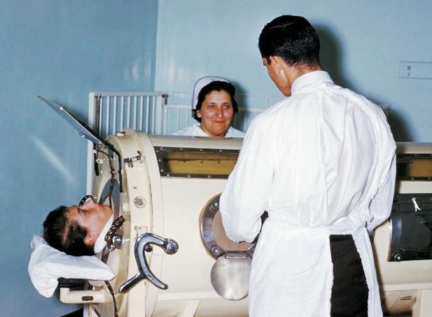
Mechanical ventilation, assisted ventilation or intermittent mandatory ventilation (IMV), is the medical term for using a machine called a ventilator to fully or partially provide artificial ventilation. Mechanical ventilation helps move air into and out of the lungs, with the main goal of helping the delivery of oxygen and removal of carbon dioxide. Mechanical ventilation is used for many reasons, including to protect the airway due to mechanical or neurologic cause, to ensure adequate oxygenation, or to remove excess carbon dioxide from the lungs. Various healthcare providers are involved with the use of mechanical ventilation and people who require ventilators are typically monitored in an intensive care unit. Mechanical ventilation is termed invasive if it involves an instrument to create an airway that is placed inside the trachea. This is done through an endotracheal tube or nasotracheal tube. For non-invasive ventilation in people who are conscious, face or nasal ... [...More Info...] [...Related Items...] OR: [Wikipedia] [Google] [Baidu] |
Intensive Care Unit
220px, Intensive care unit An intensive care unit (ICU), also known as an intensive therapy unit or intensive treatment unit (ITU) or critical care unit (CCU), is a special department of a hospital or health care facility that provides intensive care medicine. Intensive care units cater to patients with severe or life-threatening illnesses and injuries, which require constant care, close supervision from life support equipment and medication in order to ensure normal bodily functions. They are staffed by highly trained physicians, nurses and respiratory therapists who specialize in caring for critically ill patients. ICUs are also distinguished from general hospital wards by a higher staff-to-patient ratio and access to advanced medical resources and equipment that is not routinely available elsewhere. Common conditions that are treated within ICUs include acute respiratory distress syndrome, septic shock, and other life-threatening conditions. Patients may be referred di ... [...More Info...] [...Related Items...] OR: [Wikipedia] [Google] [Baidu] |
Bronchoalveolar Lavage
Bronchoalveolar lavage (BAL) (also known as bronchoalveolar washing) is a diagnostic method of the lower respiratory system in which a bronchoscope is passed through the mouth or nose into an appropriate airway in the lungs, with a measured amount of fluid introduced and then collected for examination. This method is typically performed to diagnose pathogenic infections of the lower respiratory airways (leading to, for example pneumonia and COVID-19), though it also has been shown to have utility in diagnosing interstitial lung disease. Bronchoalveolar lavage can be a more sensitive method of detection than nasal swabs in respiratory molecular diagnostics, as has been the case with SARS-CoV-2 where bronchoalveolar lavage samples detect copies of viral RNA after negative nasal swab testing. In particular, bronchoalveolar lavage is commonly used to diagnose infections in people with immune system problems, pneumonia in people on ventilators, and acute respiratory distress syndrom ... [...More Info...] [...Related Items...] OR: [Wikipedia] [Google] [Baidu] |
Pulmonary Alveolus
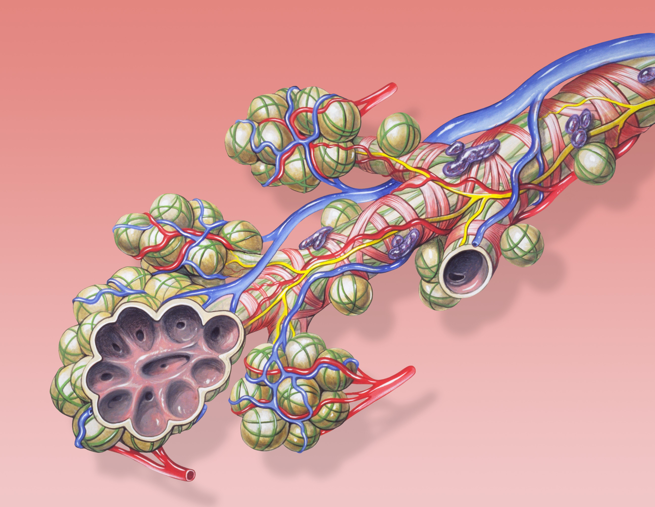
A pulmonary alveolus (plural: alveoli, from Latin ''alveolus'', "little cavity"), also known as an air sac or air space, is one of millions of hollow, distensible cup-shaped cavities in the lungs where oxygen Molecular diffusion#Biology, is exchanged for carbon dioxide. Alveoli make up the functional tissue of the mammalian lungs known as the lung parenchyma, which takes up 90 percent of the total lung volume. Alveoli are first located in the respiratory bronchioles that mark the beginning of the respiratory zone. They are located sparsely in these bronchioles, line the walls of the alveolar ducts, and are more numerous in the blind-ended alveolar sacs. The Pulmonary acinus, acini are the basic units of respiration, with gas exchange taking place in all the alveoli present. The alveolar membrane is the gas exchange surface, surrounded by a network of capillary, capillaries. Across the membrane oxygen is Diffusion#Diffusion vs. bulk flow, diffused into the capillaries and carbon ... [...More Info...] [...Related Items...] OR: [Wikipedia] [Google] [Baidu] |


.jpg)


