|
Acrania
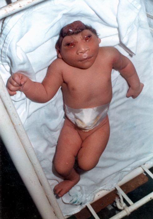
Acrania is a rare congenital disorder that occurs in the human fetus in which the flat bones in the cranial vault are either completely or partially absent. The cerebral hemispheres develop completely but abnormally. The condition is frequently, though not always, associated with anencephaly. The fetus is said to have acrania if it meets the following criteria: the fetus should have a perfectly normal facial bone, a normal cervical column but without the fetal skull and a volume of brain tissue equivalent to at least one-third of the normal brain size. Causes Genetics There are no known family ties in acrania and recurrence rates are extremely low. Not much is known about the exact mechanism involved in acrania. It is hypothesized that, like other developmental malformations, there are multiple origins for acrania. Recent work has identified mutations in the hedgehog acyltransferase (HHAT) gene that have caused acrania along with holoprosencephaly and agnathia. The mutation ... [...More Info...] [...Related Items...] OR: [Wikipedia] [Google] [Baidu] |
Human Cranium
The skull is a bone protective cavity for the brain. The skull is composed of four types of bone i.e., cranial bones, facial bones, ear ossicles and hyoid bone. However two parts are more prominent: the cranium and the mandible. In humans, these two parts are the neurocranium and the viscerocranium (facial skeleton) that includes the mandible as its largest bone. The skull forms the anterior-most portion of the skeleton and is a product of cephalisation—housing the brain, and several sensory structures such as the eyes, ears, nose, and mouth. In humans these sensory structures are part of the facial skeleton. Functions of the skull include protection of the brain, fixing the distance between the eyes to allow stereoscopic vision, and fixing the position of the ears to enable sound localisation of the direction and distance of sounds. In some animals, such as horned ungulates (mammals with hooves), the skull also has a defensive function by providing the mount (on the fronta ... [...More Info...] [...Related Items...] OR: [Wikipedia] [Google] [Baidu] |
Anencephaly
Anencephaly is the absence of a major portion of the brain, skull, and scalp that occurs during embryonic development. It is a cephalic disorder that results from a neural tube defect that occurs when the rostral (head) end of the neural tube fails to close, usually between the 23rd and 26th day following conception. Strictly speaking, the Greek term translates as "without a brain" (or totally lacking the inside part of the head), but it is accepted that children born with this disorder usually only lack a telencephalon, the largest part of the brain consisting mainly of the cerebral hemispheres, including the neocortex, which is responsible for cognition. The remaining structure is usually covered only by a thin layer of membrane—skin, bone, meninges, etc., are all lacking. With very few exceptions, infants with this disorder do not survive longer than a few hours or days after birth. Signs and symptoms The National Institute of Neurological Disorders and Stroke (NINDS) descr ... [...More Info...] [...Related Items...] OR: [Wikipedia] [Google] [Baidu] |
Medical Genetics
Medical genetics is the branch tics in that human genetics is a field of scientific research that may or may not apply to medicine, while medical genetics refers to the application of genetics to medical care. For example, research on the causes and inheritance of genetic disorders would be considered within both human genetics and medical genetics, while the diagnosis, management, and counselling people with genetic disorders would be considered part of medical genetics. In contrast, the study of typically non-medical phenotypes such as the genetics of eye color would be considered part of human genetics, but not necessarily relevant to medical genetics (except in situations such as albinism). ''Genetic medicine'' is a newer term for medical genetics and incorporates areas such as gene therapy, personalized medicine, and the rapidly emerging new medical specialty, predictive medicine. Scope Medical genetics encompasses many different areas, including clinical practice of ... [...More Info...] [...Related Items...] OR: [Wikipedia] [Google] [Baidu] |
Fibroblast Growth Factor
Fibroblast growth factors (FGF) are a family of cell signalling proteins produced by macrophages; they are involved in a wide variety of processes, most notably as crucial elements for normal development in animal cells. Any irregularities in their function lead to a range of developmental defects. These growth factors typically act as systemic or locally circulating molecules of extracellular origin that activate cell surface receptors. A defining property of FGFs is that they bind to heparin and to heparan sulfate. Thus, some are sequestered in the extracellular matrix of tissues that contains heparan sulfate proteoglycans and are released locally upon injury or tissue remodeling. Families In humans, 23 members of the FGF family have been identified, all of which are ''structurally'' related signaling molecules: * Members FGF1 through FGF10 all bind fibroblast growth factor receptors (FGFRs). FGF1 is also known as ''acidic fibroblast growth factor'', and FGF2 is also known a ... [...More Info...] [...Related Items...] OR: [Wikipedia] [Google] [Baidu] |
Mesenchyme
Mesenchyme () is a type of loosely organized animal embryonic connective tissue of undifferentiated cells that give rise to most tissues, such as skin, blood or bone. The interactions between mesenchyme and epithelium help to form nearly every organ in the developing embryo. Vertebrates Structure Mesenchyme is characterized morphologically by a prominent ground substance matrix containing a loose aggregate of reticular fibers and unspecialized mesenchymal stem cells. Mesenchymal cells can migrate easily (in contrast to epithelial cells, which lack mobility), are organized into closely adherent sheets, and are polarized in an apical-basal orientation. Development The mesenchyme originates from the mesoderm. From the mesoderm, the mesenchyme appears as an embryologically primitive "soup". This "soup" exists as a combination of the mesenchymal cells plus serous fluid plus the many different tissue proteins. Serous fluid is typically stocked with the many serous elements, such a ... [...More Info...] [...Related Items...] OR: [Wikipedia] [Google] [Baidu] |
Diencephalon
The diencephalon (or interbrain) is a division of the forebrain (embryonic ''prosencephalon''). It is situated between the telencephalon and the midbrain (embryonic ''mesencephalon''). The diencephalon has also been known as the 'tweenbrain in older literature. It consists of structures that are on either side of the third ventricle, including the thalamus, the hypothalamus, the epithalamus and the subthalamus. The diencephalon is one of the main vesicles of the brain formed during embryogenesis. During the third week of development a neural tube is created from the ectoderm, one of the three primary germ layers. The tube forms three main vesicles during the third week of development: the prosencephalon, the mesencephalon and the rhombencephalon. The prosencephalon gradually divides into the telencephalon and the diencephalon. Structure The diencephalon consists of the following structures: *Thalamus *Hypothalamus including the posterior pituitary *Epithalamus which consists of: ... [...More Info...] [...Related Items...] OR: [Wikipedia] [Google] [Baidu] |
Neurocranium
In human anatomy, the neurocranium, also known as the braincase, brainpan, or brain-pan is the upper and back part of the skull, which forms a protective case around the brain. In the human skull, the neurocranium includes the calvaria (skull), calvaria or skullcap. The remainder of the skull is the facial skeleton. In comparative anatomy, neurocranium is sometimes used synonymously with endocranium or chondrocranium. Structure The neurocranium is divided into two portions: * the membranous part, consisting of flat bones, which surround the brain; and * the cartilaginous part, or chondrocranium, which forms bones of the base of the skull. In humans, the neurocranium is usually considered to include the following eight bones: * 1 ethmoid bone * 1 frontal bone * 1 occipital bone * 2 parietal bones * 1 sphenoid bone * 2 temporal bones The ossicles (three on each side) are usually not included as bones of the neurocranium. There may variably also be extra sutural bones present. B ... [...More Info...] [...Related Items...] OR: [Wikipedia] [Google] [Baidu] |
Ectoderm
The ectoderm is one of the three primary germ layers formed in early embryonic development. It is the outermost layer, and is superficial to the mesoderm (the middle layer) and endoderm (the innermost layer). It emerges and originates from the outer layer of germ cells. The word ectoderm comes from the Greek ''ektos'' meaning "outside", and ''derma'' meaning "skin".Gilbert, Scott F. Developmental Biology. 9th ed. Sunderland, MA: Sinauer Associates, 2010: 333-370. Print. Generally speaking, the ectoderm differentiates to form epithelial and neural tissues (spinal cord, peripheral nerves and brain). This includes the skin, linings of the mouth, anus, nostrils, sweat glands, hair and nails, and tooth enamel. Other types of epithelium are derived from the endoderm. In vertebrate embryos, the ectoderm can be divided into two parts: the dorsal surface ectoderm also known as the external ectoderm, and the neural plate, which invaginates to form the neural tube and neural crest. Th ... [...More Info...] [...Related Items...] OR: [Wikipedia] [Google] [Baidu] |
Mesenchyme
Mesenchyme () is a type of loosely organized animal embryonic connective tissue of undifferentiated cells that give rise to most tissues, such as skin, blood or bone. The interactions between mesenchyme and epithelium help to form nearly every organ in the developing embryo. Vertebrates Structure Mesenchyme is characterized morphologically by a prominent ground substance matrix containing a loose aggregate of reticular fibers and unspecialized mesenchymal stem cells. Mesenchymal cells can migrate easily (in contrast to epithelial cells, which lack mobility), are organized into closely adherent sheets, and are polarized in an apical-basal orientation. Development The mesenchyme originates from the mesoderm. From the mesoderm, the mesenchyme appears as an embryologically primitive "soup". This "soup" exists as a combination of the mesenchymal cells plus serous fluid plus the many different tissue proteins. Serous fluid is typically stocked with the many serous elements, such a ... [...More Info...] [...Related Items...] OR: [Wikipedia] [Google] [Baidu] |
Neuropore
Neurulation refers to the folding process in vertebrate embryos, which includes the transformation of the neural plate into the neural tube. The embryo at this stage is termed the neurula. The process begins when the notochord induces the formation of the central nervous system (CNS) by signaling the ectoderm germ layer above it to form the thick and flat neural plate. The neural plate folds in upon itself to form the neural tube, which will later differentiate into the spinal cord and the brain, eventually forming the central nervous system. Computer simulations found that cell wedging and differential proliferation are sufficient for mammalian neurulation. Different portions of the neural tube form by two different processes, called primary and secondary neurulation, in different species. * In primary neurulation, the neural plate creases inward until the edges come in contact and fuse. * In secondary neurulation, the tube forms by hollowing out of the interior of a solid precur ... [...More Info...] [...Related Items...] OR: [Wikipedia] [Google] [Baidu] |
Amniotic Band Syndrome
Constriction ring syndrome (CRS) is a congenital disorder with unknown cause. Because of the unknown cause there are many different, and sometimes incorrect names. It is a malformation due to intrauterine bands or rings that give deep grooves in, most commonly, distal extremities like fingers and toes. In rare cases the constriction ring can form around other parts of the fetus and cause amputation or even intrauterine death. The anatomy proximal to the site of constriction (or amputation) is developmentally normal. CRS can be associated with other malformations with club foot being most common. The precise configuration of the bands, lymphedema, and character of the amputations are not predictable and vary with each individual patient. Also more than one extremity is usually affected, and it is rare for only one ring to present as an isolated malformation with no other manifestation of this syndrome. Signs and symptoms The constriction of appendages by amniotic bands may result in ... [...More Info...] [...Related Items...] OR: [Wikipedia] [Google] [Baidu] |
Acalvaria
Acalvaria is a rare malformation consisting of absence of the calvarial bones, dura mater and associated muscles in the presence of a normal skull base and normal facial bones. The central nervous system is usually unaffected. The presumed pathogenesis of acalvaria is faulty migration of the membranous neurocranium with normal placement of the embryonic ectoderm, resulting in absence of the calvaria but an intact layer of skin over the brain parenchyma. In other words, instead of having a skull cap protecting the brain, there is only skin covering it."Acalvaria." Right Diagnosis. Health Grades Inc., n.d. Web. 27 Nov. 2012. < ...
|


