|
Abdominal Aortic Plexus
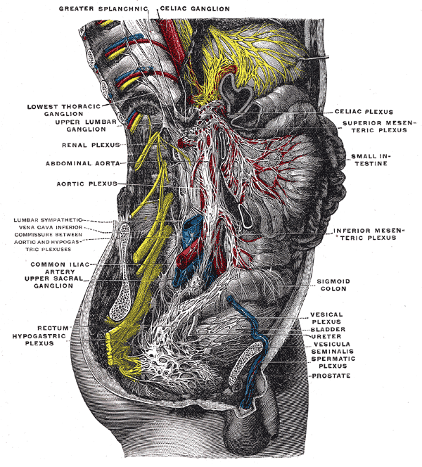
The abdominal aortic plexus (not to be confused with the thoracic aortic plexus) is formed by branches derived, on either side, from the celiac plexus and ganglia, and receives filaments from some of the lumbar ganglia. It is situated upon the sides and front of the aorta, between the origins of the superior and inferior mesenteric arteries. From this plexus arise part of the spermatic, the inferior mesenteric, and the hypogastric plexuses; it also distributes filaments to the inferior vena cava The inferior vena cava is a large vein that carries the deoxygenated blood from the lower and middle body into the right atrium of the heart. It is formed by the joining of the right and the left common iliac veins, usually at the level of the .... The abdominal aortic plexus contains the spermatic ganglia, the inferior mesenteric ganglion, and thprehypogastricganglion. Additional images Image:Gray838.png, The right sympathetic chain and its connections with the thoracic, abdomi ... [...More Info...] [...Related Items...] OR: [Wikipedia] [Google] [Baidu] |
Celiac Ganglia
The celiac ganglia or coeliac ganglia are two large irregularly shaped masses of nerve tissue in the upper abdomen. Part of the sympathetic subdivision of the autonomic nervous system (ANS), the two celiac ganglia are the largest ganglia in the ANS, and they innervate most of the digestive tract. They have the appearance of lymph glands and are placed on either side of the midline in front of the crura of the diaphragm, close to the suprarenal glands (also called adrenal glands). The ganglion on the right side is placed behind the inferior vena cava. They are sometimes referred to as the semilunar ganglia or the solar ganglia. Neurotransmission The celiac ganglion is part of the sympathetic prevertebral chain possessing a great variety of specific receptors and neurotransmitters such as catecholamines, neuropeptides, and nitric oxide and constitutes a modulation center in the pathway of the afferent and efferent fibers between the central nervous system and the ovary. T ... [...More Info...] [...Related Items...] OR: [Wikipedia] [Google] [Baidu] |
Celiac Plexus
The celiac plexus, also known as the solar plexus because of its radiating nerve fibers, is a complex network of nerves located in the abdomen, near where the celiac trunk, superior mesenteric artery, and renal arteries branch from the abdominal aorta. It is behind the stomach and the omental bursa, and in front of the crura of the diaphragm, on the level of the first lumbar vertebra. The plexus is formed in part by the greater and lesser splanchnic nerves of both sides, and fibers from the anterior and posterior vagal trunks. The celiac plexus proper consists of the celiac ganglia with a network of interconnecting fibers. The aorticorenal ganglia are often considered to be part of the celiac ganglia, and thus, part of the plexus. Structure The celiac plexus includes a number of smaller plexuses: Other plexuses that are derived from the celiac plexus: Terminology The celiac plexus is often popularly referred to as the solar plexus. In the context of sparring or in ... [...More Info...] [...Related Items...] OR: [Wikipedia] [Google] [Baidu] |
Superior Mesenteric Plexus
The superior mesenteric plexus is a continuation of the lower part of the celiac plexus, receiving a branch from the junction of the right vagus nerve with the plexus. It surrounds the superior mesenteric artery, accompanies it into the mesentery, and divides into a number of secondary plexuses, which are distributed to all the parts supplied by the artery, viz., pancreatic branches to the pancreas; intestinal branches to the small intestine; and ileocolic, right colic, and middle colic branches, which supply the corresponding parts of the great intestine. The nerves composing this plexus are white in color and firm in texture; in the upper part of the plexus close to the origin of the superior mesenteric artery is the superior mesenteric ganglion. Additional images File:Gray838.png, The right sympathetic chain and its connections with the thoracic, abdominal, and pelvic plexuses. File:Gray839.png, Diagram of efferent sympathetic nervous system. File:Gray849.png, Lower half ... [...More Info...] [...Related Items...] OR: [Wikipedia] [Google] [Baidu] |
Superior Hypogastric Plexus
The superior hypogastric plexus (in older texts, hypogastric plexus or presacral nerve) is a plexus of nerves situated on the vertebral bodies anterior to the bifurcation of the abdominal aorta. Structure From the plexus, sympathetic fibers are carried into the pelvis as two main trunks- the right and left hypogastric nerves- each lying medial to the internal iliac artery and its branches. The right and left hypogastric nerves continues as Inferior hypogastric plexus; these hypogastric nerves send sympathetic fibers to the ovarian and ureteric plexuses, which originate within the renal and abdominal aortic sympathetic plexuses. The superior hypogastric plexus receives contributions from the two lower lumbar splanchnic nerves (L3-L4), which are branches of the chain ganglia. They also contain parasympathetic fibers which arise from pelvic splanchnic nerve (S2-S4) and ascend from Inferior hypogastric plexus; it is more usual for these parasympathetic fibers to ascend to the left-hand ... [...More Info...] [...Related Items...] OR: [Wikipedia] [Google] [Baidu] |
Thoracic Aortic Plexus
The thoracic aortic plexus is a sympathetic plexus in the region of the thoracic aorta The descending thoracic aorta is a part of the aorta located in the thorax. It is a continuation of the aortic arch. It is located within the posterior mediastinal cavity, but frequently bulges into the left pleural cavity. The descending thoracic .... References External links * Nerve plexus Nerves of the torso {{Neuroanatomy-stub ... [...More Info...] [...Related Items...] OR: [Wikipedia] [Google] [Baidu] |
Celiac Plexus
The celiac plexus, also known as the solar plexus because of its radiating nerve fibers, is a complex network of nerves located in the abdomen, near where the celiac trunk, superior mesenteric artery, and renal arteries branch from the abdominal aorta. It is behind the stomach and the omental bursa, and in front of the crura of the diaphragm, on the level of the first lumbar vertebra. The plexus is formed in part by the greater and lesser splanchnic nerves of both sides, and fibers from the anterior and posterior vagal trunks. The celiac plexus proper consists of the celiac ganglia with a network of interconnecting fibers. The aorticorenal ganglia are often considered to be part of the celiac ganglia, and thus, part of the plexus. Structure The celiac plexus includes a number of smaller plexuses: Other plexuses that are derived from the celiac plexus: Terminology The celiac plexus is often popularly referred to as the solar plexus. In the context of sparring or in ... [...More Info...] [...Related Items...] OR: [Wikipedia] [Google] [Baidu] |
Ganglia
A ganglion is a group of neuron cell bodies in the peripheral nervous system. In the somatic nervous system this includes dorsal root ganglia and trigeminal ganglia among a few others. In the autonomic nervous system there are both sympathetic and parasympathetic ganglia which contain the cell bodies of postganglionic sympathetic and parasympathetic neurons respectively. A pseudoganglion looks like a ganglion, but only has nerve fibers and has no nerve cell bodies. Structure Ganglia are primarily made up of somata and dendritic structures which are bundled or connected. Ganglia often interconnect with other ganglia to form a complex system of ganglia known as a plexus. Ganglia provide relay points and intermediary connections between different neurological structures in the body, such as the peripheral and central nervous systems. Among vertebrates there are three major groups of ganglia: *Dorsal root ganglia (also known as the spinal ganglia) contain the cell bodies of se ... [...More Info...] [...Related Items...] OR: [Wikipedia] [Google] [Baidu] |
Lumbar Ganglia
The lumbar ganglia are paravertebral ganglia located in the inferior portion of the sympathetic trunk. The lumbar portion of the sympathetic trunk typically has 4 lumbar ganglia. The lumbar splanchnic nerves arise from the ganglia here, and contribute sympathetic efferent fibers to the nearby plexuses. The first two lumbar ganglia have both white and gray rami communicates. Function Paravertebral ganglia are divided into cervical, thoracic, lumbar, and sacral ganglia. Each controls different glands and muscle groups since each muscle and gland receives input from postganglionic neurons that originated from different levels of paravertebral ganglia. The lumbar part of the sympathetic trunk contains four interconnected ganglia. Superiorly, it is continuous with thoracic sympathetic ganglion and inferiorly continuous with sacral sympathetic ganglion. Presynaptic neurons traveling from the spinal cord terminate in the paravertebral ganglia (cervical, thoracic, lumbar, sacral) or the ... [...More Info...] [...Related Items...] OR: [Wikipedia] [Google] [Baidu] |
Aorta
The aorta ( ) is the main and largest artery in the human body, originating from the left ventricle of the heart and extending down to the abdomen, where it splits into two smaller arteries (the common iliac arteries). The aorta distributes oxygenated blood to all parts of the body through the systemic circulation. Structure Sections In anatomical sources, the aorta is usually divided into sections. One way of classifying a part of the aorta is by anatomical compartment, where the thoracic aorta (or thoracic portion of the aorta) runs from the heart to the diaphragm. The aorta then continues downward as the abdominal aorta (or abdominal portion of the aorta) from the diaphragm to the aortic bifurcation. Another system divides the aorta with respect to its course and the direction of blood flow. In this system, the aorta starts as the ascending aorta, travels superiorly from the heart, and then makes a hairpin turn known as the aortic arch. Following the aortic arch ... [...More Info...] [...Related Items...] OR: [Wikipedia] [Google] [Baidu] |
Inferior Mesenteric Artery
In human anatomy, the inferior mesenteric artery, often abbreviated as IMA, is the third main branch of the abdominal aorta and arises at the level of L3, supplying the large intestine from the distal transverse colon to the upper part of the anal canal. The regions supplied by the IMA are the descending colon, the sigmoid colon, and part of the rectum. Structure Proximally, its territory of distribution overlaps (forms a watershed) with the middle colic artery, and therefore the superior mesenteric artery. The SMA and IMA anastomose via the marginal artery of the colon (artery of Drummond) and via Riolan's arcade (also called the "meandering artery", an arterial connection between the left colic artery and the middle colic artery). The territory of distribution of the IMA is more or less equivalent to the embryonic hindgut. Branches The IMA branches off the anterior surface of the abdominal aorta below the renal artery branch points, 3-4 cm above the aortic bifurcation (into t ... [...More Info...] [...Related Items...] OR: [Wikipedia] [Google] [Baidu] |
Spermatic
The spermatic plexus (or testicular plexus) is derived from the renal plexus, receiving branches from the aortic plexus. It accompanies the internal spermatic artery to the testis A testicle or testis (plural testes) is the male reproductive gland or gonad in all bilaterians, including humans. It is homologous to the female ovary. The functions of the testes are to produce both sperm and androgens, primarily testostero .... Additional images File:Gray849.png, Lower half of right sympathetic cord. File:Gray1144.png, The scrotum. References External links Nerve plexus Nerves of the torso {{Neuroanatomy-stub ... [...More Info...] [...Related Items...] OR: [Wikipedia] [Google] [Baidu] |
Hypogastrium
The hypogastrium (also called the hypogastric region or suprapubic region) is a region of the abdomen located below the umbilical region. Etymology The roots of the word ''hypogastrium'' mean "below the stomach"; the roots of ''suprapubic'' mean "above the pubic bone". Boundaries The upper limit is the umbilicus while the pubis bone constitutes its lower limit. The lateral boundaries are formed are drawing straight lines through the midway between the anterior superior iliac spine and symphisis pubis The pubic symphysis is a secondary cartilaginous joint between the left and right superior rami of the pubis of the hip bones. It is in front of and below the urinary bladder. In males, the suspensory ligament of the penis attaches to the pubic .... References External links * Abdomen {{Anatomy-stub ... [...More Info...] [...Related Items...] OR: [Wikipedia] [Google] [Baidu] |


