|
Atrioventricular Nodal Branch
The atrioventricular nodal branch is a coronary artery that feeds the atrioventricular node, necessary for the excitation of the ventricles. Structure The atrioventricular nodal branch sees significant variation in origin: * proximal posterolateral branch from the right coronary artery in around 77%. * distal posterolateral branch from the right coronary artery in around 2%. * distal right coronary artery in around 10%. * right posterior interventricular artery in around 7%. * distal circumflex branch of left coronary artery in around 4%. The right coronary artery supplies the atrioventricular node in around 90% of people. In approximately 2% of people, the vascular supply to the atrioventricular node arises from both the right coronary artery and the left circumflex branch.Sow ML, Ndoye JM, Lo EA. The artery of the atrioventricular node: an anatomic study based on 38 injection-dissections. Surg Radiol Anat 1996;18:183–187 Function The atrioventricular nodal branch suppli ... [...More Info...] [...Related Items...] OR: [Wikipedia] [Google] [Baidu] |
Right Coronary Artery
In the blood supply of the heart, the right coronary artery (RCA) is an artery originating above the right cusp of the aortic valve, at the right aortic sinus in the heart. It travels down the right coronary sulcus, towards the crux of the heart. It supplies the right side of the heart, and the interventricular septum. Structure The right coronary artery originates above the right aortic sinus above the aortic valve. It passes through the right coronary sulcus (right atrioventricular groove), towards the crux of the heart. It gives off many branches, including the posterior interventricular artery, the right marginal artery, the conus artery, and the sinoatrial nodal artery. Segments * Proximal: starting at RCA origin, spanning half the distance to the acute margin * Middle: from proximal segment to the acute margin * Distal: from middle segment to origination point of the posterior interventricular artery, where the posterior interventricular sulcus meets the atrioven ... [...More Info...] [...Related Items...] OR: [Wikipedia] [Google] [Baidu] |
Great Cardiac Vein
The great cardiac vein (left coronary vein) begins at the apex of the heart and ascends along the anterior longitudinal sulcus to the base of the ventricles. It then curves around the left margin of the heart to reach the posterior surface. It merges with the oblique vein of the left atrium to form the coronary sinus, which drains into the right atrium. At the junction of the great cardiac vein and the coronary sinus, there is typically a valve present. This is the Vieussens valve of the coronary sinus. It receives tributaries from the left atrium and from both ventricles: one, the left marginal vein The great cardiac vein receives tributaries from the left atrium and from both ventricles: one, the left marginal vein, is of considerable size, and ascends along the left margin of the heart The heart is a muscular organ in most animals ..., is of considerable size, and ascends along the left margin of the heart. References External links * - "Heart: Cardiac veins" ... [...More Info...] [...Related Items...] OR: [Wikipedia] [Google] [Baidu] |
Ventricle (heart)
A ventricle is one of two large chambers toward the bottom of the heart that collect and expel blood towards the peripheral beds within the body and lungs. The blood pumped by a ventricle is supplied by an atrium, an adjacent chamber in the upper heart that is smaller than a ventricle. Interventricular means between the ventricles (for example the interventricular septum), while intraventricular means within one ventricle (for example an intraventricular block). In a four-chambered heart, such as that in humans, there are two ventricles that operate in a double circulatory system: the right ventricle pumps blood into the pulmonary circulation to the lungs, and the left ventricle pumps blood into the systemic circulation through the aorta. Structure Ventricles have thicker walls than atria and generate higher blood pressures. The physiological load on the ventricles requiring pumping of blood throughout the body and lungs is much greater than the pressure generated by the atria ... [...More Info...] [...Related Items...] OR: [Wikipedia] [Google] [Baidu] |
Muscle Contraction
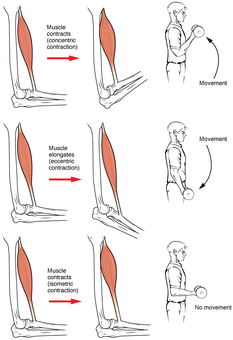
Muscle contraction is the activation of tension-generating sites within muscle cells. In physiology, muscle contraction does not necessarily mean muscle shortening because muscle tension can be produced without changes in muscle length, such as when holding something heavy in the same position. The termination of muscle contraction is followed by muscle relaxation, which is a return of the muscle fibers to their low tension-generating state. For the contractions to happen, the muscle cells must rely on the interaction of two types of filaments which are the thin and thick filaments. Thin filaments are two strands of actin coiled around each, and thick filaments consist of mostly elongated proteins called myosin. Together, these two filaments form myofibrils which are important organelles in the skeletal muscle system. Muscle contraction can also be described based on two variables: length and tension. A muscle contraction is described as isometric if the muscle tension changes ... [...More Info...] [...Related Items...] OR: [Wikipedia] [Google] [Baidu] |
Atrioventricular Node
The atrioventricular node or AV node electrically connects the heart's atria and ventricles to coordinate beating in the top of the heart; it is part of the electrical conduction system of the heart. The AV node lies at the lower back section of the interatrial septum near the opening of the coronary sinus, and conducts the normal electrical impulse from the atria to the ventricles. The AV node is quite compact (~1 x 3 x 5 mm).Full Size Picture triangle of-Koch.jpg Retrieved on 2008-12-22 Structure Location The AV node lies at the lower back section of the |
Coronary Arteries
The coronary arteries are the arterial blood vessels of coronary circulation, which transport oxygenated blood to the heart muscle. The heart requires a continuous supply of oxygen to function and survive, much like any other tissue or organ of the body. The coronary arteries wrap around the entire heart. The two main branches are the left coronary artery and right coronary artery. The arteries can additionally be categorized based on the area of the heart for which they provide circulation. These categories are called ''epicardial'' (above the epicardium, or the outermost tissue of the heart) and ''microvascular'' (close to the endocardium, or the innermost tissue of the heart). Reduced function of the coronary arteries can lead to decreased flow of oxygen and nutrients to the heart. Not only does this affect supply to the heart muscle itself, but it also can affect the ability of the heart to pump blood throughout the body. Therefore, any disorder or disease of the coronary ar ... [...More Info...] [...Related Items...] OR: [Wikipedia] [Google] [Baidu] |
Coronary Sinus
In anatomy, the coronary sinus () is a collection of veins joined together to form a large vessel that collects blood from the heart muscle ( myocardium). It delivers deoxygenated blood to the right atrium, as do the superior and inferior venae cavae. It is present in all mammals, including humans. The coronary sinus drains into the right atrium, at the coronary sinus orifice, an opening between the inferior vena cava and the right atrioventricular orifice or tricuspid valve. It returns blood from the heart muscle, and is protected by a semicircular fold of the lining membrane of the auricle, the valve of coronary sinus (or valve of Thebesius). The sinus, before entering the atrium, is considerably dilated - nearly to the size of the end of the little finger. Its wall is partly muscular, and at its junction with the great cardiac vein is somewhat constricted and furnished with a valve, known as the valve of Vieussens consisting of two unequal segments. Structure The coron ... [...More Info...] [...Related Items...] OR: [Wikipedia] [Google] [Baidu] |
Middle Cardiac Vein
The middle cardiac vein commences at the apex of the heart; ascends in the posterior longitudinal sulcus, and ends in the coronary sinus In anatomy, the coronary sinus () is a collection of veins joined together to form a large vessel that collects blood from the heart muscle (myocardium). It delivers deoxygenated blood to the right atrium, as do the superior and inferior vena ... near its right extremity. Structure Variation The middle cardiac vein has a constant location on the surface of the ventricles. Clinical significance The middle cardiac vein is useful for epicardial access to the inferior side of the ventricles. References External links * - "Posterior view of the heart." Veins of the torso {{circulatory-stub ... [...More Info...] [...Related Items...] OR: [Wikipedia] [Google] [Baidu] |
Anterior Cardiac Veins
The anterior cardiac veins (or anterior veins of right ventricle) comprise a variable number of small vessels, usually between two and five, which collect blood from the front of the right ventricle and open into the right atrium; the right marginal vein frequently opens into the right atrium, and is therefore sometimes regarded as belonging to this group. Unlike most cardiac veins, they do not end in the coronary sinus. Instead, these veins drain directly into the anterior wall of the right atrium The atrium ( la, ātrium, , entry hall) is one of two upper chambers in the heart that receives blood from the circulatory system. The blood in the atria is pumped into the heart ventricles through the atrioventricular valves. There are two at .... References External links Veins of the torso {{circulatory-stub ... [...More Info...] [...Related Items...] OR: [Wikipedia] [Google] [Baidu] |
Atrial Branches Of Coronary Arteries
The atrial branches of right coronary artery derive from the right coronary artery and provide part of the blood supply to the right atrium and left atrium. Although named for the right coronary artery in Terminologia anatomica, a portion of the blood supply to the atria derives from the Circumflex branch of left coronary artery The circumflex branch of left coronary artery, or left circumflex artery or circumflex artery, is a branch of the left coronary artery. Description The left circumflex artery follows the left part of the coronary sulcus, running first to the l .... External links Image {{Authority control Arteries of the thorax ... [...More Info...] [...Related Items...] OR: [Wikipedia] [Google] [Baidu] |
Small Cardiac Vein
The small cardiac vein, also known as the right coronary vein, is a coronary vein that drains the right atrium and right ventricle of the heart. Despite its size, it is one of the major drainage vessels for the heart. Location The small cardiac vein runs in the coronary sulcus between the right atrium and right ventricle, and opens into the right extremity of the coronary sinus. Function The small cardiac vein receives blood from the posterior portion of the right atrium and ventricle. Variations The small cardiac vein may drain to the coronary sinus, right atrium, middle cardiac vein The middle cardiac vein commences at the apex of the heart; ascends in the posterior longitudinal sulcus, and ends in the coronary sinus In anatomy, the coronary sinus () is a collection of veins joined together to form a large vessel that c ..., or be absent. References External links * - "Anterior view of the heart." {{Authority control Veins of the torso ... [...More Info...] [...Related Items...] OR: [Wikipedia] [Google] [Baidu] |


