|
Anterior Segment Mesenchymal Dysgenesis
Anterior segment mesenchymal dysgenesis, or simply anterior segment dysgenesis (ASD), is a failure of the normal development of the tissues of the anterior segment of the eye. It leads to anomalies in the structure of the mature anterior segment, associated with an increased risk of glaucoma and corneal opacity. Peters' (frequently misspelled as Peter's) anomaly is a specific type of mesenchymal anterior segment dysgenesis, in which there is central corneal leukoma, adhesions of the iris and cornea and abnormalities of the posterior corneal stroma, Descemet's membrane, corneal endothelium, lens and anterior chamber. Pathophysiology Several gene mutations have been identified underlying these anomalies, with the majority of ASD genes encoding transcriptional regulators. In this review, the role of the ASD genes, ''PITX2'' and ''FOXC1'', is considered in relation to the embryology of the anterior segment, the biochemical function of these proteins, and their role in development and ... [...More Info...] [...Related Items...] OR: [Wikipedia] [Google] [Baidu] |
Autism Spectrum Disorder
The autism spectrum, often referred to as just autism or in the context of a professional diagnosis autism spectrum disorder (ASD) or autism spectrum condition (ASC), is a neurodevelopmental disorder, neurodevelopmental condition (or conditions) characterized by difficulties in Social relation, social interaction, verbal and nonverbal communication, and the presence of repetitive behavior and restricted interests. Other common signs include unusual responses to Multisensory integration, sensory stimuli. Autism is generally understood as a ''spectrum disorder'', which means that it can manifest differently in each person: any given autistic individual is likely to show some, but not all, of the characteristics associated with it, and the person may exhibit them to varying degrees. Some autistic people remain nonverbal autism, nonspeaking over the course of their lifespan, while others have relatively unimpaired spoken language. There is large variation in the level of support peop ... [...More Info...] [...Related Items...] OR: [Wikipedia] [Google] [Baidu] |
Lens (anatomy)
The lens, or crystalline lens, is a transparent biconvex structure in the eye that, along with the cornea, helps to refract light to be focused on the retina. By changing shape, it functions to change the focal length of the eye so that it can focus on objects at various distances, thus allowing a sharp real image of the object of interest to be formed on the retina. This adjustment of the lens is known as '' accommodation'' (see also below). Accommodation is similar to the focusing of a photographic camera via movement of its lenses. The lens is flatter on its anterior side than on its posterior side. In humans, the refractive power of the lens in its natural environment is approximately 18 dioptres, roughly one-third of the eye's total power. Structure The lens is part of the anterior segment of the human eye. In front of the lens is the iris, which regulates the amount of light entering into the eye. The lens is suspended in place by the suspensory ligament of the lens ... [...More Info...] [...Related Items...] OR: [Wikipedia] [Google] [Baidu] |
Gene Dosage
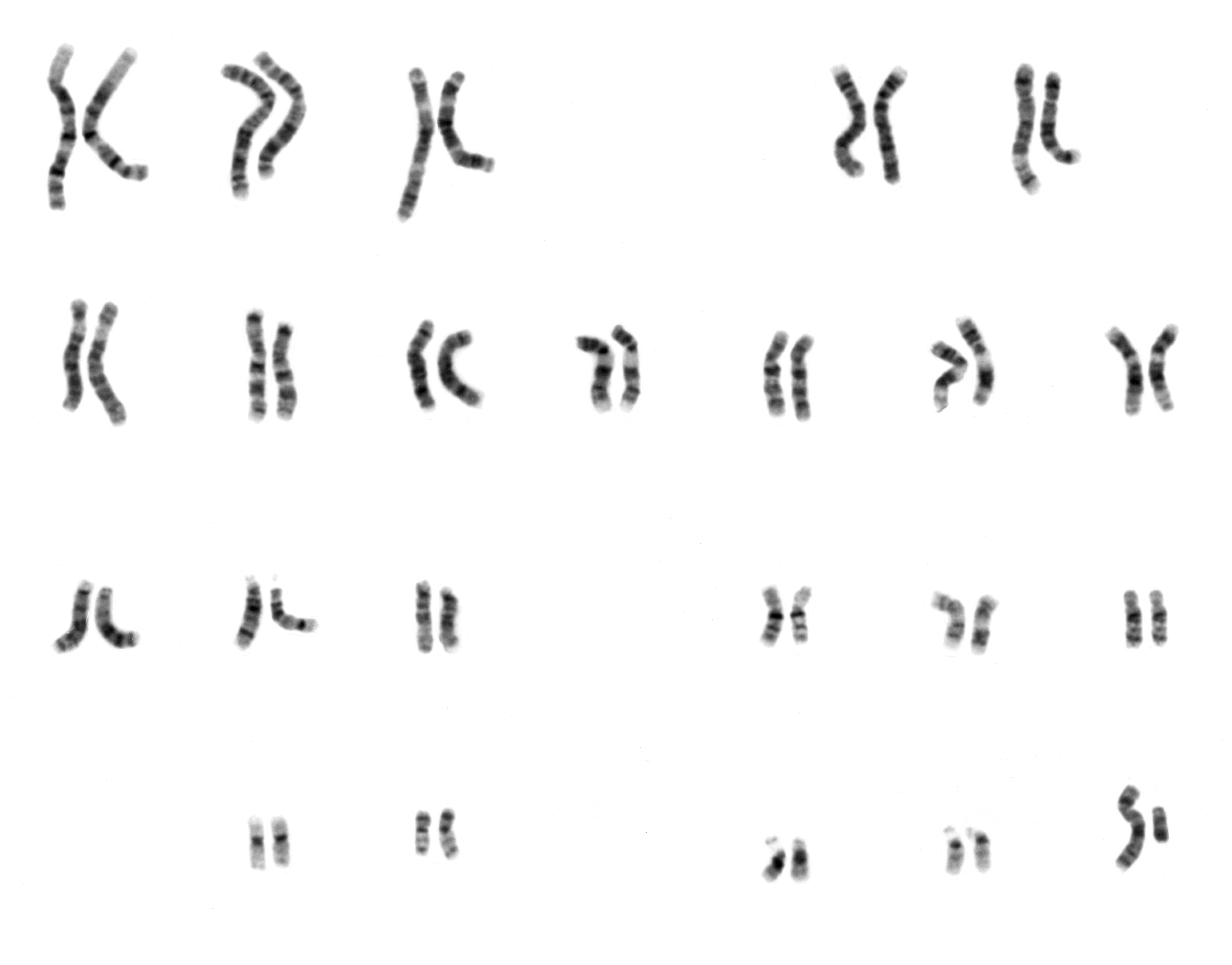
Gene dosage is the number of copies of a particular gene present in a genome. Gene dosage is related to the amount of gene product (proteins or functional RNAs) the cell is able to express. Since, a gene acts as a template, the number of templates in the cell contributes to the amount of gene product able to be produced. However, the amount of gene product produced in a cell is more commonly dependent on regulation of gene expression. The normal gene dosage is dependent on species, humans generally have two doses, one copy from the mother and one from the father. Changes in gene dosage can be a result of copy number variation (gene insertions or gene deletions), or aneuploidy (chromosome number abnormalities). These changes can have significant phenotypic consequences. Ploidy Ploidy refers to the number of complete sets of chromosomes in a cell. Humans typically have a gene dosage of two because they are diploid, they have two sets of 23 different chromosomes. The number of copie ... [...More Info...] [...Related Items...] OR: [Wikipedia] [Google] [Baidu] |
Mesenchymal Stem Cell
Mesenchymal stem cells (MSCs) also known as mesenchymal stromal cells or medicinal signaling cells are multipotent stromal cells that can differentiate into a variety of cell types, including osteoblasts (bone cells), chondrocytes (cartilage cells), myocytes (muscle cells) and adipocytes (fat cells which give rise to marrow adipose tissue). Structure Definition While the terms ''mesenchymal stem cell'' (MSC) and ''marrow stromal cell'' have been used interchangeably for many years, neither term is sufficiently descriptive: * Mesenchyme is embryonic connective tissue that is derived from the mesoderm and that differentiates into hematopoietic and connective tissue, whereas MSCs do not differentiate into hematopoietic cells. * Stromal cells are connective tissue cells that form the supportive structure in which the functional cells of the tissue reside. While this is an accurate description for one function of MSCs, the term fails to convey the relatively recently discove ... [...More Info...] [...Related Items...] OR: [Wikipedia] [Google] [Baidu] |
Optic Cup (embryology)
During embryonic development of the eye, the outer wall of the bulb of the optic vesicles becomes thickened and invaginated, and the bulb is thus converted into a cup, the optic cup (or ophthalmic cup), consisting of two strata of cells. These two strata are continuous with each other at the cup margin, which ultimately overlaps the front of the lens and reaches as far forward as the future aperture of the pupil. The optic cup is part of the diencephalon and gives rise to the retina The retina (from la, rete "net") is the innermost, light-sensitive layer of tissue of the eye of most vertebrates and some molluscs. The optics of the eye create a focused two-dimensional image of the visual world on the retina, which then ... of the eye. References External links Overview at temple.edu Embryology of nervous system Eye {{eye-stub ... [...More Info...] [...Related Items...] OR: [Wikipedia] [Google] [Baidu] |
Neural Crest
Neural crest cells are a temporary group of cells unique to vertebrates that arise from the embryonic ectoderm germ layer, and in turn give rise to a diverse cell lineage—including melanocytes, craniofacial cartilage and bone, smooth muscle, peripheral and enteric neurons and glia. After gastrulation, neural crest cells are specified at the border of the neural plate and the non-neural ectoderm. During neurulation, the borders of the neural plate, also known as the neural folds, converge at the dorsal midline to form the neural tube. Subsequently, neural crest cells from the roof plate of the neural tube undergo an epithelial to mesenchymal transition, delaminating from the neuroepithelium and migrating through the periphery where they differentiate into varied cell types. The emergence of neural crest was important in vertebrate evolution because many of its structural derivatives are defining features of the vertebrate clade. Underlying the development of neural crest is ... [...More Info...] [...Related Items...] OR: [Wikipedia] [Google] [Baidu] |
FOXC1
Forkhead box C1, also known as FOXC1, is a protein which in humans is encoded by the ''FOXC1'' gene. Function This gene belongs to the forkhead family of transcription factors which is characterized by a distinct DNA-binding fork head domain. The specific function of this gene has not yet been determined; however, it has been shown to play a role in the regulation of embryonic and ocular development. Heart development and somitogenesis FOXC1 and its close relative, FOXC2 are both critical components in the development of the heart and blood vessels, as well as the segmentation of the paraxial mesoderm and the formation of somites. Expression of the Fox proteins range from low levels in the posterior pre-somitic mesoderm (PSM) to the highest levels in the anterior PSM. Homozygous mutant embryos for both Fox proteins failed to form somites 1-8, which indicates the importance of these proteins early on in somite development. In cardiac morphogenesis, FOXC1 and FOXC2 are requir ... [...More Info...] [...Related Items...] OR: [Wikipedia] [Google] [Baidu] |
PITX2
Paired-like homeodomain transcription factor 2 also known as pituitary homeobox 2 is a protein that in humans is encoded by the ''PITX2'' gene. Function This gene encodes a member of the RIEG/PITX homeobox family, which is in the bicoid class of homeodomain proteins. This protein acts as a transcription factor and regulates procollagen lysyl hydroxylase gene expression. This protein is involved in the development of the eye, tooth and abdominal organs. This protein acts as a transcriptional regulator involved in basal and hormone-regulated activity of prolactin. A similar protein in other vertebrates is involved in the determination of left-right asymmetry during development. Three transcript variants encoding distinct isoforms have been identified for this gene. Pitx2 is responsible for the establishment of the left-right axis, the asymmetrical development of the heart, lungs, and spleen, twisting of the gut and stomach, as well as the development of the eyes. Once activated ... [...More Info...] [...Related Items...] OR: [Wikipedia] [Google] [Baidu] |
Regulation Of Gene Expression
Regulation of gene expression, or gene regulation, includes a wide range of mechanisms that are used by cells to increase or decrease the production of specific gene products (protein or RNA). Sophisticated programs of gene expression are widely observed in biology, for example to trigger developmental pathways, respond to environmental stimuli, or adapt to new food sources. Virtually any step of gene expression can be modulated, from Transcriptional regulation, transcriptional initiation, to RNA processing, and to the post-translational modification of a protein. Often, one gene regulator controls another, and so on, in a gene regulatory network. Gene regulation is essential for viruses, prokaryotes and eukaryotes as it increases the versatility and adaptability of an organism by allowing the cell to express protein when needed. Although as early as 1951, Barbara McClintock showed interaction between two genetic loci, Activator (''Ac'') and Dissociator (''Ds''), in the color f ... [...More Info...] [...Related Items...] OR: [Wikipedia] [Google] [Baidu] |
Anterior Chamber
The anterior chamber ( AC) is the aqueous humor-filled space inside the eye between the iris and the cornea's innermost surface, the endothelium. Hyphema, anterior uveitis and glaucoma are three main pathologies in this area. In hyphema, blood fills the anterior chamber as a result of a hemorrhage, most commonly after a blunt eye injury. Anterior uveitis is an inflammatory process affecting the iris and ciliary body, with resulting inflammatory signs in the anterior chamber. In glaucoma, blockage of the trabecular meshwork prevents the normal outflow of aqueous humour, resulting in increased intraocular pressure, progressive damage to the optic nerve head, and eventually blindness. The depth of the anterior chamber of the eye varies between 1.5 and 4.0 mm, averaging 3.0 mm. It tends to become shallower at older age and in eyes with hypermetropia (far sightedness). As depth decreases below 2.5 mm, the risk for angle closure glaucoma increases. Clinical significance ... [...More Info...] [...Related Items...] OR: [Wikipedia] [Google] [Baidu] |
Corneal Endothelium
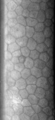
The corneal endothelium is a single layer of endothelial cells on the inner surface of the cornea. It faces the chamber formed between the cornea and the iris. The corneal endothelium are specialized, flattened, mitochondria-rich cells that line the posterior surface of the cornea and face the anterior chamber of the eye. The corneal endothelium governs fluid and solute transport across the posterior surface of the cornea and maintains the cornea in the slightly dehydrated state that is required for optical transparency. Embryology and anatomy The corneal endothelium is embryologically derived from the neural crest. The postnatal total endothelial cellularity of the cornea (approximately 300,000 cells per cornea) is achieved as early as the second trimester of gestation. Thereafter the endothelial cell density (but not the absolute number of cells) rapidly declines, as the fetal cornea grows in surface area, achieving a final adult density of approximately 2400 - 3200 cells ... [...More Info...] [...Related Items...] OR: [Wikipedia] [Google] [Baidu] |
Autosomal Dominant
In genetics, dominance is the phenomenon of one variant (allele) of a gene on a chromosome masking or overriding the effect of a different variant of the same gene on the other copy of the chromosome. The first variant is termed dominant and the second recessive. This state of having two different variants of the same gene on each chromosome is originally caused by a mutation in one of the genes, either new (''de novo'') or inherited. The terms autosomal dominant or autosomal recessive are used to describe gene variants on non-sex chromosomes ( autosomes) and their associated traits, while those on sex chromosomes (allosomes) are termed X-linked dominant, X-linked recessive or Y-linked; these have an inheritance and presentation pattern that depends on the sex of both the parent and the child (see Sex linkage). Since there is only one copy of the Y chromosome, Y-linked traits cannot be dominant or recessive. Additionally, there are other forms of dominance such as incomplete d ... [...More Info...] [...Related Items...] OR: [Wikipedia] [Google] [Baidu] |


.jpg)

