|
X-type Histiocytosis
X-type histiocytoses are a clinically well-defined group of cutaneous syndromes characterized by infiltrates of Langerhans cells, as opposed to Non-X histiocytosis in which the infiltrates contain monocytes/macrophages. Conditions included in this group are: :* Congenital self-healing reticulohistiocytosis :* Langerhans cell histiocytosis See also * Non-X histiocytosis * Histiocytosis In medicine, histiocytosis is an excessive number of histiocytes (tissue macrophages), and the term is also often used to refer to a group of rare diseases which share this sign as a characteristic. Occasionally and confusingly, the term "histioc ... References Monocyte- and macrophage-related cutaneous conditions Histiocytosis {{Cutaneous-condition-stub ... [...More Info...] [...Related Items...] OR: [Wikipedia] [Google] [Baidu] |
Langerhans Cell
A Langerhans cell (LC) is a tissue-resident macrophage of the skin. These cells contain organelles called Birbeck granules. They are present in all layers of the epidermis and are most prominent in the stratum spinosum. They also occur in the papillary dermis, particularly around blood vessels, as well as in the mucosa of the mouth, foreskin, and vaginal epithelium. They can be found in other tissues, such as lymph nodes, particularly in association with the condition Langerhans cell histiocytosis (LCH). Function In skin infections, the local Langerhans cells take up and process microbial antigens to become fully functional antigen-presenting cells. Generally, tissue-resident macrophages are involved in immune homeostasis and the uptake of apoptotic bodies. However, Langerhans cells can also take on a dendritic cell-like phenotype and migrate to lymph nodes to interact with naive T-cells. Langerhans cells derive from primitive erythro-myeloid progenitors that arise in the ... [...More Info...] [...Related Items...] OR: [Wikipedia] [Google] [Baidu] |
Non-X Histiocytosis
Non-X histiocytoses are a clinically well-defined group of cutaneous syndromes characterized by infiltrates of monocytes/macrophages, as opposed to X-type histiocytoses in which the infiltrates contain Langerhans cells. Conditions included in this group are: :* Juvenile xanthogranuloma :* Benign cephalic histiocytosis :* Generalized eruptive histiocytoma :* Xanthoma disseminatum :* Progressive nodular histiocytosis :* Papular xanthoma :* Hereditary progressive mucinous histiocytosis :* Reticulohistiocytosis :* Indeterminate cell histiocytosis :* Sea-blue histiocytosis :* Erdheim–Chester disease See also * X-type histiocytosis * Histiocytosis In medicine, histiocytosis is an excessive number of histiocytes (tissue macrophages), and the term is also often used to refer to a group of rare diseases which share this sign as a characteristic. Occasionally and confusingly, the term "histioc ... References Monocyte- and macrophage-related cutaneous conditions Histiocytosi ... [...More Info...] [...Related Items...] OR: [Wikipedia] [Google] [Baidu] |
Monocytes
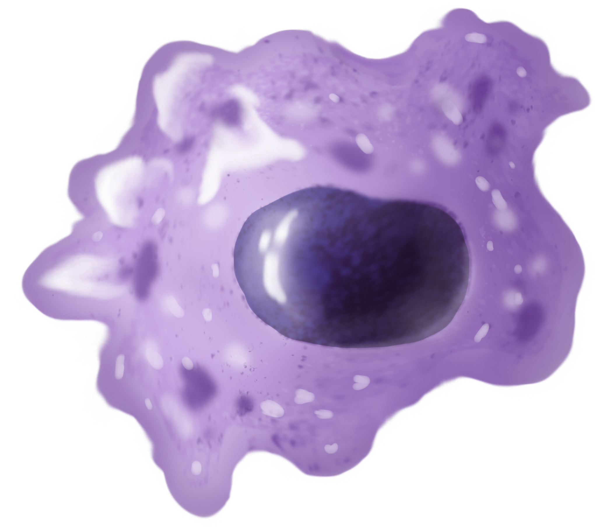
Monocytes are a type of leukocyte or white blood cell. They are the largest type of leukocyte in blood and can differentiate into macrophages and conventional dendritic cells. As a part of the vertebrate innate immune system monocytes also influence adaptive immune responses and exert tissue repair functions. There are at least three subclasses of monocytes in human blood based on their phenotypic receptors. Structure Monocytes are amoeboid in appearance, and have nongranulated cytoplasm. Thus they are classified as agranulocytes, although they might occasionally display some azurophil granules and/or vacuoles. With a diameter of 15–22 μm, monocytes are the largest cell type in peripheral blood. Monocytes are mononuclear cells and the ellipsoidal nucleus is often lobulated/indented, causing a bean-shaped or kidney-shaped appearance. Monocytes compose 2% to 10% of all leukocytes in the human body. Development Monocytes are produced by the bone marrow from precursors cal ... [...More Info...] [...Related Items...] OR: [Wikipedia] [Google] [Baidu] |
Macrophages
Macrophages (abbreviated as M φ, MΦ or MP) ( el, large eaters, from Greek ''μακρός'' (') = large, ''φαγεῖν'' (') = to eat) are a type of white blood cell of the immune system that engulfs and digests pathogens, such as cancer cells, microbes, cellular debris, and foreign substances, which do not have proteins that are specific to healthy body cells on their surface. The process is called phagocytosis, which acts to defend the host against infection and injury. These large phagocytes are found in essentially all tissues, where they patrol for potential pathogens by amoeboid movement. They take various forms (with various names) throughout the body (e.g., histiocytes, Kupffer cells, alveolar macrophages, microglia, and others), but all are part of the mononuclear phagocyte system. Besides phagocytosis, they play a critical role in nonspecific defense (innate immunity) and also help initiate specific defense mechanisms (adaptive immunity) by recruiting other immun ... [...More Info...] [...Related Items...] OR: [Wikipedia] [Google] [Baidu] |
Congenital Self-healing Reticulohistiocytosis
Congenital self-healing reticulohistiocytosis is a condition that is a self-limited form of Langerhans cell histiocytosis. Symptoms Non-specific inflammatory response, which includes fever, lethargy, and weight loss. This is suspected of being a genetic disorder, and as the name implies, is self healing. *Skin: Commonly seen are a rash which varies from scaly erythematous lesions to red papules pronounced in intertriginous areas. Up to 80% of patients have extensive eruptions on the scalp. *Lymph node: Enlargement of the lymph nodes in 50% of Histiocytosis cases. Diagnosis Treatment History It was first described by Ken Hashimoto and M. S. Pritzkar in 1973. See also * List of cutaneous conditions * X-type histiocytosis X-type histiocytoses are a clinically well-defined group of cutaneous syndromes characterized by infiltrates of Langerhans cells, as opposed to Non-X histiocytosis in which the infiltrates contain monocytes/macrophages. Conditions included in this ... Re ... [...More Info...] [...Related Items...] OR: [Wikipedia] [Google] [Baidu] |
Langerhans Cell Histiocytosis
Langerhans cell histiocytosis (LCH) is an abnormal clonal proliferation of Langerhans cells, abnormal cells deriving from bone marrow and capable of migrating from skin to lymph nodes. Symptoms range from isolated bone lesions to multisystem disease. LCH is part of a group of syndromes called histiocytoses, which are characterized by an abnormal proliferation of histiocytes (an archaic term for activated dendritic cells and macrophages). These diseases are related to other forms of abnormal proliferation of white blood cells, such as leukemias and lymphomas. The disease has gone by several names, including Hand–Schüller–Christian disease, Abt-Letterer-Siwe disease, Hashimoto-Pritzker disease (a very rare self-limiting variant seen at birth) and histiocytosis X, until it was renamed in 1985 by the Histiocyte Society. Classification The disease spectrum results from clonal accumulation and proliferation of cells resembling the epidermal dendritic cells called Langerhan ... [...More Info...] [...Related Items...] OR: [Wikipedia] [Google] [Baidu] |
Histiocytosis
In medicine, histiocytosis is an excessive number of histiocytes (tissue macrophages), and the term is also often used to refer to a group of rare diseases which share this sign as a characteristic. Occasionally and confusingly, the term "histiocytosis" is sometimes used to refer to individual diseases. According to the Histiocytosis Association of America, 1 in 200,000 children in the United States are born with histiocytosis each year. HAA also states that most of the people diagnosed with histiocytosis are children under the age of 10, although the disease can afflict adults. The disease usually occurs from birth to age 15. Histiocytosis (and malignant histiocytosis) are both important in veterinary as well as human pathology. Diagnosis Histiocytosis is a rare disease, thus its diagnosis may be challenging. A variety of tests may be used, including: * Imaging ** CT scans of various organs such as lung, heart and kidneys. ** MRI of the brain, pituitary gland, heart, among ... [...More Info...] [...Related Items...] OR: [Wikipedia] [Google] [Baidu] |

.png)


.jpg)