|
White Pulp
White pulp is a histological designation for regions of the spleen (named because it appears whiter than the surrounding red pulp on gross section), that encompasses approximately 25% of splenic tissue. White pulp consists entirely of lymphoid tissue. Specifically, the white pulp encompasses several areas with distinct functions: * The periarteriolar lymphoid sheaths (PALS) are typically associated with the arteriole supply of the spleen; they contain T lymphocytes. * Lymph follicles with dividing B lymphocytes are located between the PALS and the marginal zone bordering on the red pulp. IgM and IgG2 are produced in this zone. These molecules play a role in opsonization of extracellular organisms, encapsulated bacteria in particular. * The marginal zone exists between the white pulp and red pulp. It is located farther away from the central arteriole, in proximity to the red pulp. It contains antigen-presenting cells (APCs), such as dendritic cells and macrophages. Some of the wh ... [...More Info...] [...Related Items...] OR: [Wikipedia] [Google] [Baidu] |
Spleen
The spleen is an organ found in almost all vertebrates. Similar in structure to a large lymph node, it acts primarily as a blood filter. The word spleen comes .σπλήν Henry George Liddell, Robert Scott, ''A Greek-English Lexicon'', on Perseus Digital Library The spleen plays very important roles in regard to s (erythrocytes) and the . It removes old red blood cells and holds a reserve of blood, which can be valuable in case of |
B Cell
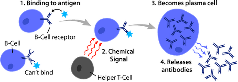
B cells, also known as B lymphocytes, are a type of white blood cell of the lymphocyte subtype. They function in the humoral immunity component of the adaptive immune system. B cells produce antibody molecules which may be either secreted or inserted into the plasma membrane where they serve as a part of B-cell receptors. When a naïve or memory B cell is activated by an antigen, it proliferates and differentiates into an antibody-secreting effector cell, known as a plasmablast or plasma cell. Additionally, B cells present antigens (they are also classified as professional antigen-presenting cells (APCs)) and secrete cytokines. In mammals, B cells mature in the bone marrow, which is at the core of most bones. In birds, B cells mature in the bursa of Fabricius, a lymphoid organ where they were first discovered by Chang and Glick, which is why the 'B' stands for bursa and not bone marrow as commonly believed. B cells, unlike the other two classes of lymphocytes, T cells and ... [...More Info...] [...Related Items...] OR: [Wikipedia] [Google] [Baidu] |
Mucosa-associated Lymphoid Tissue
The mucosa-associated lymphoid tissue (MALT), also called mucosa-associated lymphatic tissue, is a diffuse system of small concentrations of lymphoid tissue found in various submucosal membrane sites of the body, such as the gastrointestinal tract, nasopharynx, thyroid, breast, lung, salivary glands, eye, and skin. MALT is populated by lymphocytes such as T cells and B cells, as well as plasma cells and macrophages, each of which is well situated to encounter antigens passing through the mucosal epithelium. In the case of intestinal MALT, M cells are also present, which sample antigen from the lumen and deliver it to the lymphoid tissue. MALT constitute about 50% of the lymphoid tissue in human body. Immune responses that occur at mucous membranes are studied by mucosal immunology. Categorization The components of MALT are sometimes subdivided into the following: * GALT (gut-associated lymphoid tissue. Peyer's patches are a component of GALT found in the lining of the small in ... [...More Info...] [...Related Items...] OR: [Wikipedia] [Google] [Baidu] |
Marginal Zone
The marginal zone is the region at the interface between the non-lymphoid red pulp and the lymphoid white-pulp of the spleen. (Some sources consider it to be the part of red pulp which borders on the white pulp, while other sources consider it to be neither red pulp nor white pulp.) A marginal zone also exists in lymph nodes. Structure It is composed of cells derived primarily from the myeloid compartment of bone marrow differentiation. More recently, a population of Neutrophil#Subpopulations, neutrophil-killers has been described to populate peripheral areas of the marginal zone. At least three distinct cellular markers can be used to identify cells of the marginal zone, MOMA-1, ERTR-9 and MARCO. Blood flow The marginal zone (MZ) is a highly transited area that receives large amounts of blood from the general circulation. Remarkably, the splenic Microvessel, microvasculature shows striking differences in mice and humans. In humans, the spleen receives blood from the splenic ar ... [...More Info...] [...Related Items...] OR: [Wikipedia] [Google] [Baidu] |
Lymph Node
A lymph node, or lymph gland, is a kidney-shaped organ of the lymphatic system and the adaptive immune system. A large number of lymph nodes are linked throughout the body by the lymphatic vessels. They are major sites of lymphocytes that include B and T cells. Lymph nodes are important for the proper functioning of the immune system, acting as filters for foreign particles including cancer cells, but have no detoxification function. In the lymphatic system a lymph node is a secondary lymphoid organ. A lymph node is enclosed in a fibrous capsule and is made up of an outer cortex and an inner medulla. Lymph nodes become inflamed or enlarged in various diseases, which may range from trivial throat infections to life-threatening cancers. The condition of lymph nodes is very important in cancer staging, which decides the treatment to be used and determines the prognosis. Lymphadenopathy refers to glands that are enlarged or swollen. When inflamed or enlarged, lymph nodes can be ... [...More Info...] [...Related Items...] OR: [Wikipedia] [Google] [Baidu] |
Lymphocyte
A lymphocyte is a type of white blood cell (leukocyte) in the immune system of most vertebrates. Lymphocytes include natural killer cells (which function in cell-mediated, cytotoxic innate immunity), T cells (for cell-mediated, cytotoxic adaptive immunity), and B cells (for humoral, antibody-driven adaptive immunity). They are the main type of cell found in lymph, which prompted the name "lymphocyte". Lymphocytes make up between 18% and 42% of circulating white blood cells. Types The three major types of lymphocyte are T cells, B cells and natural killer (NK) cells. Lymphocytes can be identified by their large nucleus. T cells and B cells T cells (thymus cells) and B cells ( bone marrow- or bursa-derived cells) are the major cellular components of the adaptive immune response. T cells are involved in cell-mediated immunity, whereas B cells are primarily responsible for humoral immunity (relating to antibodies). The function of T cells and B cells is to recognize sp ... [...More Info...] [...Related Items...] OR: [Wikipedia] [Google] [Baidu] |
Antigen Presentation
Antigen presentation is a vital immune process that is essential for T cell immune response triggering. Because T cells recognize only fragmented antigens displayed on cell surfaces, antigen processing must occur before the antigen fragment, now bound to the major histocompatibility complex (MHC), is transported to the surface of the cell, a process known as presentation, where it can be recognized by a T-cell receptor. If there has been an infection with viruses or bacteria, the cell will present an endogenous or exogenous peptide fragment derived from the antigen by MHC molecules. There are two types of MHC molecules which differ in the behaviour of the antigens: MHC class I molecules (MHC-I) bind peptides from the cell cytosol, while peptides generated in the endocytic vesicles after internalisation are bound to MHC class II (MHC-II). Cellular membranes separate these two cellular environments - intracellular and extracellular. Each T cell can only recognize tens to hundreds ... [...More Info...] [...Related Items...] OR: [Wikipedia] [Google] [Baidu] |
Monocyte
Monocytes are a type of leukocyte or white blood cell. They are the largest type of leukocyte in blood and can differentiate into macrophages and conventional dendritic cells. As a part of the vertebrate innate immune system monocytes also influence adaptive immune responses and exert tissue repair functions. There are at least three subclasses of monocytes in human blood based on their phenotypic receptors. Structure Monocytes are amoeboid in appearance, and have nongranulated cytoplasm. Thus they are classified as agranulocytes, although they might occasionally display some azurophil granules and/or vacuoles. With a diameter of 15–22 μm, monocytes are the largest cell type in peripheral blood. Monocytes are mononuclear cells and the ellipsoidal nucleus is often lobulated/indented, causing a bean-shaped or kidney-shaped appearance. Monocytes compose 2% to 10% of all leukocytes in the human body. Development Monocytes are produced by the bone marrow from precursors ca ... [...More Info...] [...Related Items...] OR: [Wikipedia] [Google] [Baidu] |
Phosphatidylserine
Phosphatidylserine (abbreviated Ptd-L-Ser or PS) is a phospholipid and is a component of the cell membrane. It plays a key role in cell cycle signaling, specifically in relation to apoptosis. It is a key pathway for viruses to enter cells via apoptotic mimicry. Its exposure on the outer surface of a membrane marks the cell for destruction via apoptosis. Structure Phosphatidylserine is a phospholipid—more specifically a glycerophospholipid—which consists of two fatty acids attached in ester linkage to the first and second carbon of glycerol and serine attached through a phosphodiester linkage to the third carbon of the glycerol. Phosphatidylserine sourced from plants differs in fatty acid composition from that sourced from animals. It is commonly found in the inner (cytoplasmic) leaflet of biological membranes. It is almost entirely found in the inner monolayer of the membrane with only less than 10% of it in the outer monolayer. Introduction Phosphatidylserine (PS) is ... [...More Info...] [...Related Items...] OR: [Wikipedia] [Google] [Baidu] |
Tingible Body Macrophage
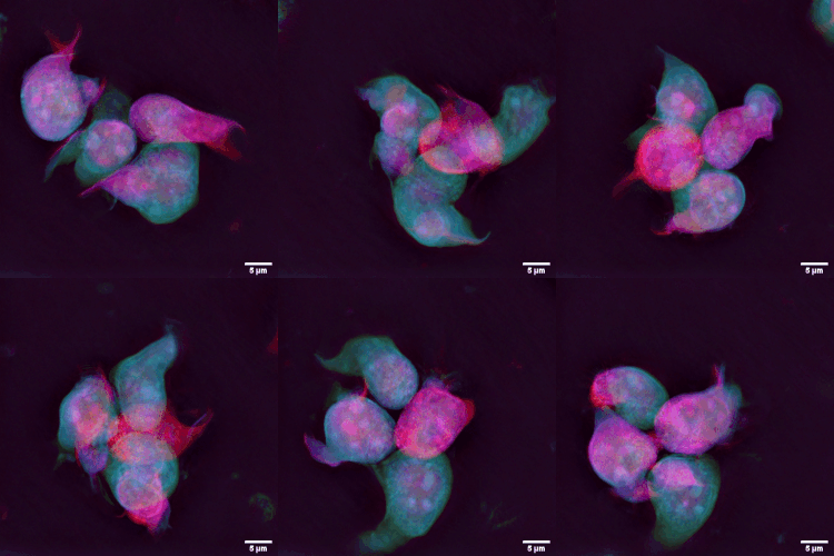
A tingible body macrophage (TBMs) is a type of macrophage predominantly found in germinal centers, containing many phagocytized, apoptotic cells in various states of degradation, referred to as tingible bodies (tingible meaning stainable). Tingible body macrophages contain condensed chromatin fragments. TBMs are licensed (empowered) for phagocytosis by follicular dendritic cells (FDCs). FDCs provide TBMs with Milk fat globule-EGF factor 8 protein (Mfge8), which is a phosphatidylserine-binding "eat me" signal for removal of apoptotic germinal center B cells. It is thought that they may play a role in downregulating the germinal center reaction by the release of prostaglandins and hence a reduced B-cell induction of IL-2. Macrophages that contain debris from ingested lymphocyte A lymphocyte is a type of white blood cell (leukocyte) in the immune system of most vertebrates. Lymphocytes include natural killer cells (which function in cell-mediated, cytotoxic innate immuni ... [...More Info...] [...Related Items...] OR: [Wikipedia] [Google] [Baidu] |
Apoptosis
Apoptosis (from grc, ἀπόπτωσις, apóptōsis, 'falling off') is a form of programmed cell death that occurs in multicellular organisms. Biochemical events lead to characteristic cell changes (morphology) and death. These changes include blebbing, cell shrinkage, nuclear fragmentation, chromatin condensation, DNA fragmentation, and mRNA decay. The average adult human loses between 50 and 70 billion cells each day due to apoptosis. For an average human child between eight and fourteen years old, approximately twenty to thirty billion cells die per day. In contrast to necrosis, which is a form of traumatic cell death that results from acute cellular injury, apoptosis is a highly regulated and controlled process that confers advantages during an organism's life cycle. For example, the separation of fingers and toes in a developing human embryo occurs because cells between the digits undergo apoptosis. Unlike necrosis, apoptosis produces cell fragments called apoptotic ... [...More Info...] [...Related Items...] OR: [Wikipedia] [Google] [Baidu] |
Isotype Switching
Immunoglobulin class switching, also known as isotype switching, isotypic commutation or class-switch recombination (CSR), is a biological mechanism that changes a B cell's production of immunoglobulin from one type to another, such as from the isotype IgM to the isotype IgG. During this process, the constant-region portion of the antibody heavy chain is changed, but the variable region of the heavy chain stays the same (the terms ''variable'' and ''constant'' refer to changes or lack thereof between antibodies that target different epitopes). Since the variable region does not change, class switching does not affect antigen specificity. Instead, the antibody retains affinity for the same antigens, but can interact with different effector molecules. Mechanism Class switching occurs after activation of a mature B cell via its membrane-bound antibody molecule (or B cell receptor) to generate the different classes of antibody, all with the same variable domains as the original ... [...More Info...] [...Related Items...] OR: [Wikipedia] [Google] [Baidu] |


.jpg)

.png)


