|
Westermark Sign
In chest radiography, the Westermark sign is a sign that represents a focus of oligemia (hypovolemia) (leading to collapse of vessel) seen distal to a pulmonary embolism (PE). While the chest x-ray is normal in the majority of PE cases, the Westermark sign is seen in 2% of patients. Essentially, this is a plain X-ray version of a filling defect as seen on computed tomography pulmonary arteriogram. The sign results from a combination of: # the dilation of the pulmonary arteries proximal to the embolus and # the collapse of the distal vasculature creating the appearance of a sharp cut off on chest radiography. Sensitivity and specificity The Westermark sign, like Hampton's hump (a wedge shaped, pleural based consolidation associated with pulmonary infarction), has a low sensitivity (11%) and high specificity (92%) for the diagnosis of pulmonary embolism. Put more simply, the Westermark sign is seldom seen in pulmonary embolism. When visible on a chest X-ray, the Positive Predict ... [...More Info...] [...Related Items...] OR: [Wikipedia] [Google] [Baidu] |
Chest Radiograph
A chest radiograph, called a chest X-ray (CXR), or chest film, is a projection radiograph of the chest used to diagnose conditions affecting the chest, its contents, and nearby structures. Chest radiographs are the most common film taken in medicine. Like all methods of radiography, chest radiography employs ionizing radiation in the form of X-rays to generate images of the chest. The mean radiation dose to an adult from a chest radiograph is around 0.02 mSv (2 mrem) for a front view (PA, or posteroanterior) and 0.08 mSv (8 mrem) for a side view (LL, or latero-lateral). Together, this corresponds to a background radiation equivalent time of about 10 days. Medical uses Conditions commonly identified by chest radiography * Pneumonia * Pneumothorax * Interstitial lung disease * Heart failure * Bone fracture * Hiatal hernia Chest radiographs are used to diagnose many conditions involving the chest wall, including its bones, and also structures contained within the thoracic ... [...More Info...] [...Related Items...] OR: [Wikipedia] [Google] [Baidu] |
Sign (medicine)
Signs and symptoms are the observed or detectable signs, and experienced symptoms of an illness, injury, or condition. A sign for example may be a higher or lower temperature than normal, raised or lowered blood pressure or an abnormality showing on a medical scan. A symptom is something out of the ordinary that is experienced by an individual such as feeling feverish, a headache or other pain or pains in the body. Signs and symptoms Signs A medical sign is an objective observable indication of a disease, injury, or abnormal physiological state that may be detected during a physical examination, examining the patient history, or diagnostic procedure. These signs are visible or otherwise detectable such as a rash or bruise. Medical signs, along with symptoms, assist in formulating diagnostic hypothesis. Examples of signs include elevated blood pressure, nail clubbing of the fingernails or toenails, staggering gait, and arcus senilis and arcus juvenilis of the eyes. Ind ... [...More Info...] [...Related Items...] OR: [Wikipedia] [Google] [Baidu] |
Oligemia
Hypovolemia, also known as volume depletion or volume contraction, is a state of abnormally low extracellular fluid in the body. This may be due to either a loss of both salt and water or a decrease in blood volume. Hypovolemia refers to the loss of extracellular fluid and should not be confused with dehydration. Hypovolemia is caused by a variety of events, but these can be simplified into two categories: those that are associated with kidney function and those that are not. The signs and symptoms of hypovolemia worsen as the amount of fluid lost increases. Immediately or shortly after mild fluid loss (from blood donation, diarrhea, vomiting, bleeding from trauma, etc.), one may experience headache, fatigue, weakness, dizziness, or thirst. Untreated hypovolemia or excessive and rapid losses of volume may lead to hypovolemic shock. Signs and symptoms of hypovolemic shock include increased heart rate, low blood pressure, pale or cold skin, and altered mental status. When these si ... [...More Info...] [...Related Items...] OR: [Wikipedia] [Google] [Baidu] |
Pulmonary Embolism
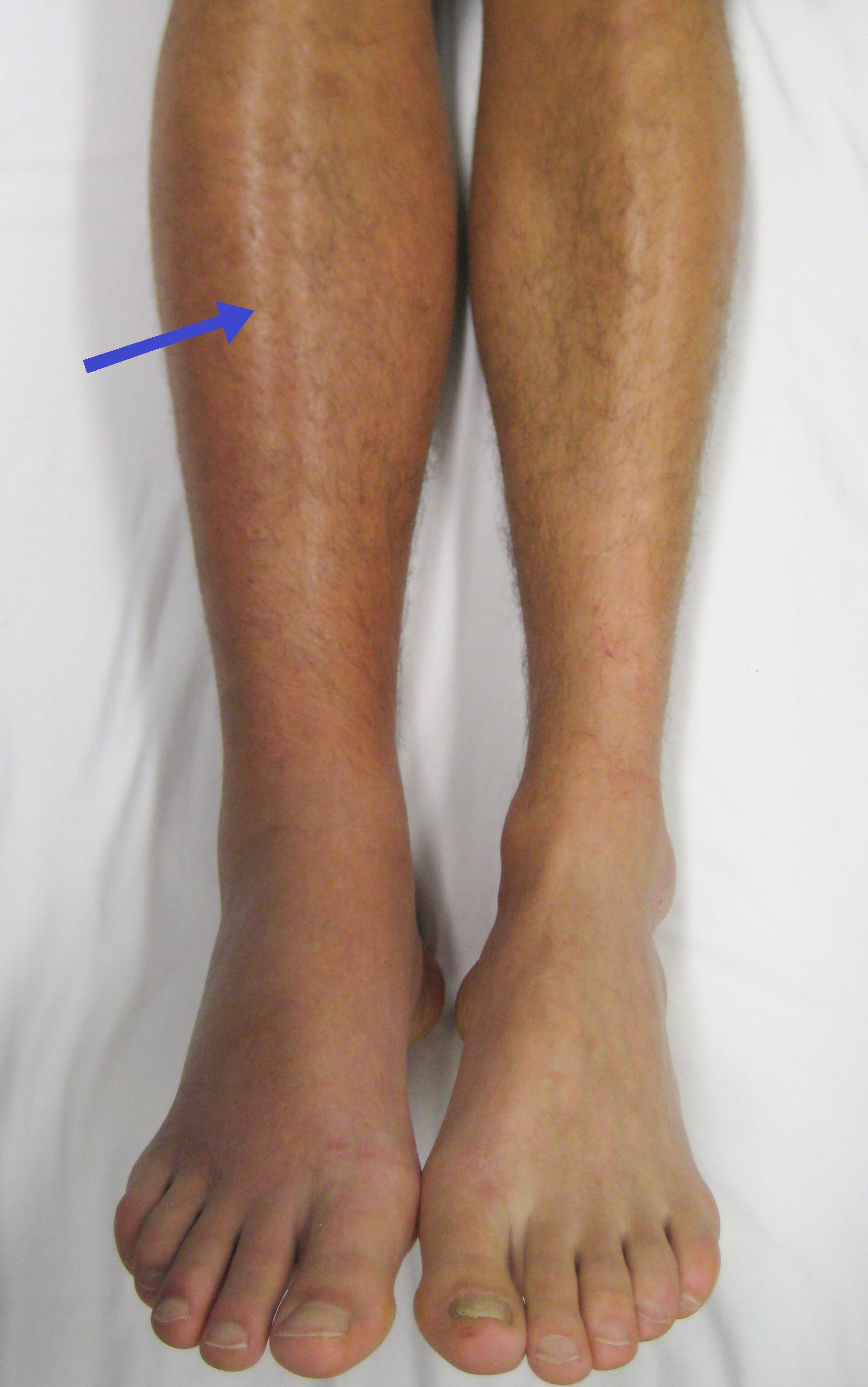
Pulmonary embolism (PE) is a blockage of an artery in the lungs by a substance that has moved from elsewhere in the body through the bloodstream (embolism). Symptoms of a PE may include shortness of breath, chest pain particularly upon breathing in, and coughing up blood. Symptoms of a blood clot in the leg may also be present, such as a red, warm, swollen, and painful leg. Signs of a PE include low blood oxygen levels, rapid breathing, rapid heart rate, and sometimes a mild fever. Severe cases can lead to passing out, abnormally low blood pressure, obstructive shock, and sudden death. PE usually results from a blood clot in the leg that travels to the lung. The risk of blood clots is increased by advanced age, cancer, prolonged bed rest and immobilization, smoking, stroke, long-haul travel over 4 hours, certain genetic conditions, estrogen-based medication, pregnancy, obesity, trauma or bone fracture, and after some types of surgery. A small proportion of cases are due ... [...More Info...] [...Related Items...] OR: [Wikipedia] [Google] [Baidu] |
Pulmonary Arteries
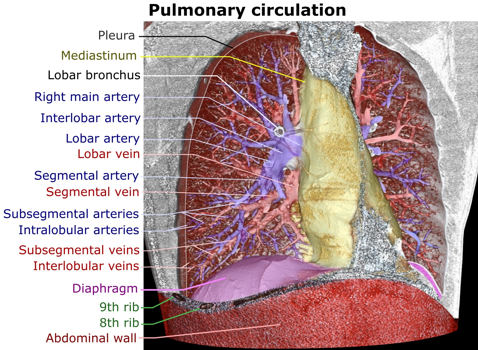
A pulmonary artery is an artery in the pulmonary circulation that carries deoxygenated blood from the right side of the heart to the lungs. The largest pulmonary artery is the ''main pulmonary artery'' or ''pulmonary trunk'' from the heart, and the smallest ones are the arterioles, which lead to the capillaries that surround the pulmonary alveoli. Structure The pulmonary arteries are blood vessels that carry systemic venous blood from the right ventricle of the heart to the microcirculation of the lungs. Unlike in other organs where arteries supply oxygenated blood, the blood carried by the pulmonary arteries is deoxygenated, as it is venous blood returning to the heart. The main pulmonary arteries emerge from the right side of the heart, and then split into smaller arteries that progressively divide and become arterioles, eventually narrowing into the capillary microcirculation of the lungs where gas exchange occurs. Pulmonary trunk In order of blood flow, the pulmonary a ... [...More Info...] [...Related Items...] OR: [Wikipedia] [Google] [Baidu] |
Anatomical Terms Of Location
Standard anatomical terms of location are used to unambiguously describe the anatomy of animals, including humans. The terms, typically derived from Latin or Greek roots, describe something in its standard anatomical position. This position provides a definition of what is at the front ("anterior"), behind ("posterior") and so on. As part of defining and describing terms, the body is described through the use of anatomical planes and anatomical axes. The meaning of terms that are used can change depending on whether an organism is bipedal or quadrupedal. Additionally, for some animals such as invertebrates, some terms may not have any meaning at all; for example, an animal that is radially symmetrical will have no anterior surface, but can still have a description that a part is close to the middle ("proximal") or further from the middle ("distal"). International organisations have determined vocabularies that are often used as standard vocabularies for subdisciplines o ... [...More Info...] [...Related Items...] OR: [Wikipedia] [Google] [Baidu] |
Hampton's Hump
Hampton's hump, also called Hampton hump, is a radiologic sign which consists of a shallow wedge-shaped opacity in the periphery of the lung with its base against the pleural surface. It is named after Aubrey Otis Hampton Aubrey Otis Hampton (September 10, 1900 in Copeville, Texas – July 17, 1955 in Weare, New Hampshire) was an American radiologist remembered for describing Hampton's hump and Hampton's line. He graduated from Baylor College of Medicine in 1925, ..., who first described it in 1940. Hampton's hump along with Westermark sign may aid in the diagnosis of pulmonary embolism, although they are rare and their sensitivities and interoperator reliabilities are low. If the sign is present in an image, there is a high chance that the person has a pulmonary embolism, but when the sign is absent a pulmonary embolism is not ruled out. References {{Radiologic signs Radiologic signs ... [...More Info...] [...Related Items...] OR: [Wikipedia] [Google] [Baidu] |
Sensitivity (test)
In medicine and statistics, sensitivity and specificity mathematically describe the accuracy of a test that reports the presence or absence of a medical condition. If individuals who have the condition are considered "positive" and those who do not are considered "negative", then sensitivity is a measure of how well a test can identify true positives and specificity is a measure of how well a test can identify true negatives: * Sensitivity (true positive rate) is the probability of a positive test result, conditioned on the individual truly being positive. * Specificity (true negative rate) is the probability of a negative test result, conditioned on the individual truly being negative. If the true status of the condition cannot be known, sensitivity and specificity can be defined relative to a " gold standard test" which is assumed correct. For all testing, both diagnoses and screening, there is usually a trade-off between sensitivity and specificity, such that higher sensit ... [...More Info...] [...Related Items...] OR: [Wikipedia] [Google] [Baidu] |
Nils Westermark
Nils Johan Hugo Westermark (September 9, 1892 – January 24, 1980) was a Swedish sailor who competed in the 1912 Summer Olympics. He later became a radiologist, and described the Westermark sign In chest radiography, the Westermark sign is a sign that represents a focus of oligemia (hypovolemia) (leading to collapse of vessel) seen distal to a pulmonary embolism (PE). While the chest x-ray is normal in the majority of PE cases, the Westerm .... He was a crew member of the Swedish boat ''Sans Atout'', which won the silver medal in the 8 metre class. References External linksprofile 1892 births 1980 deaths Swedish male sailors (sport) Sailors at the 1912 Summer Olympics – 8 Metre Olympic sailors for Sweden Olympic silver medalists for Sweden Olympic medalists in sailing Medalists at the 1912 Summer Olympics {{Sweden-yachtracing-bio-stub ... [...More Info...] [...Related Items...] OR: [Wikipedia] [Google] [Baidu] |


