|
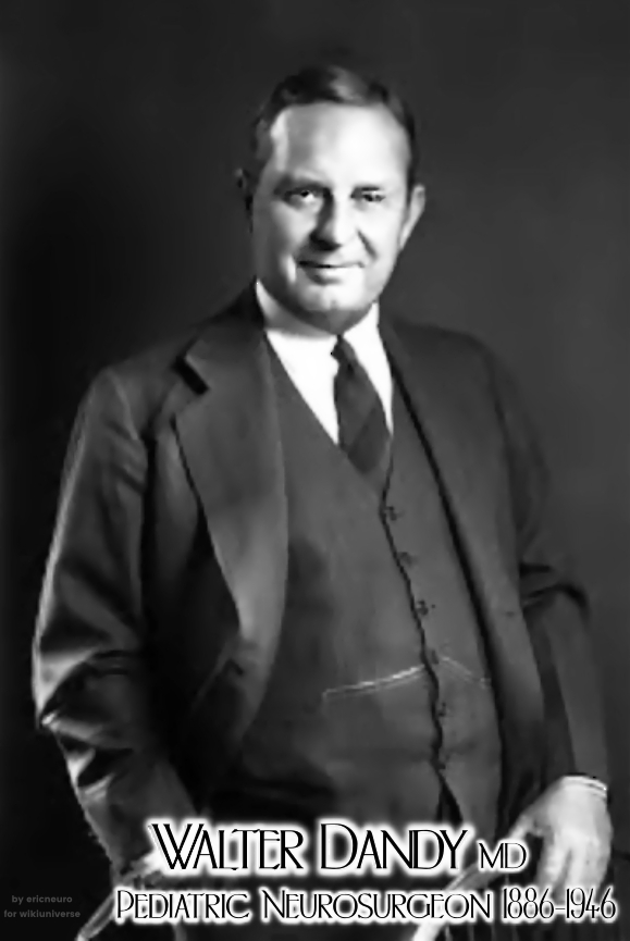
Walter Dandy
Walter Edward Dandy (April 6, 1886 – April 19, 1946) was an American neurosurgeon and scientist. He is considered one of the founding fathers of neurosurgery, along with Victor Horsley (1857–1916) and Harvey Cushing (1869–1939). Dandy is credited with numerous neurosurgical discoveries and innovations, including the description of the circulation of cerebrospinal fluid in the brain, surgical treatment of hydrocephalus, the invention of air ventriculography and pneumoencephalography, the description of brain endoscopy, the establishment of the first intensive care unit ( Fox 1984, p. 82), and the first clipping of an intracranial aneurysm, which marked the birth of cerebrovascular neurosurgery. During his 40-year medical career, Dandy published five books and more than 160 peer-reviewed articles while conducting a full-time, ground-breaking neurosurgical practice in which he performed during his peak years about 1000 operations per year ( Sherman et al. 2006). He was re ... [...More Info...] [...Related Items...] OR: [Wikipedia] [Google] [Baidu] |
Sedalia, Missouri
Sedalia is a city located approximately south of the Missouri River and, as the county seat of Pettis County, Missouri, United States, it is the principal city of the Sedalia Micropolitan Statistical Area. As of the 2010 census, the city had a total population of 21,387. Sedalia is also the location of the Missouri State Fair and the Scott Joplin Ragtime Festival. U.S. Routes 50 and 65 intersect in the city. History Indigenous peoples lived along the Missouri River and its tributaries for thousands of years before European contact. Historians believe the entire area around Sedalia was long occupied by the Osage (among historical American Indian tribes). When the land was first settled by European Americans, bands of Shawnee, who had migrated from east of the Mississippi River, lived in the vicinity of Sedalia. Until the city was incorporated in 1860 as Sedalia, it had existed only "on paper" from November 30, 1857, to October 16, 1860. According to local lore, the tow ... [...More Info...] [...Related Items...] OR: [Wikipedia] [Google] [Baidu] |
Aneurysm
An aneurysm is an outward bulging, likened to a bubble or balloon, caused by a localized, abnormal, weak spot on a blood vessel wall. Aneurysms may be a result of a hereditary condition or an acquired disease. Aneurysms can also be a nidus (starting point) for clot formation (thrombosis) and embolization. As an aneurysm increases in size, the risk of rupture, which leads to uncontrolled bleeding, increases. Although they may occur in any blood vessel, particularly lethal examples include aneurysms of the Circle of Willis in the brain, aortic aneurysms affecting the thoracic aorta, and abdominal aortic aneurysms. Aneurysms can arise in the heart itself following a heart attack, including both ventricular and atrial septal aneurysms. There are congenital atrial septal aneurysms, a rare heart defect. Etymology The word is from Greek: ἀνεύρυσμα, aneurysma, "dilation", from ἀνευρύνειν, aneurynein, "to dilate". Classification Aneurysms are classified by type, ... [...More Info...] [...Related Items...] OR: [Wikipedia] [Google] [Baidu] |
Ménière's Disease
Ménière's disease (MD) is a disease of the inner ear that is characterized by potentially severe and incapacitating episodes of vertigo, tinnitus, hearing loss, and a feeling of fullness in the ear. Typically, only one ear is affected initially, but over time, both ears may become involved. Episodes generally last from 20 minutes to a few hours. The time between episodes varies. The hearing loss and ringing in the ears can become constant over time. The cause of Ménière's disease is unclear, but likely involves both genetic and environmental factors. A number of theories exist for why it occurs, including constrictions in blood vessels, viral infections, and autoimmune reactions. About 10% of cases run in families. Symptoms are believed to occur as the result of increased fluid buildup in the labyrinth of the inner ear. Diagnosis is based on the symptoms and a hearing test. Other conditions that may produce similar symptoms include vestibular migraine and transient ischemi ... [...More Info...] [...Related Items...] OR: [Wikipedia] [Google] [Baidu] |
Trigeminal Neuralgia
Trigeminal neuralgia (TN or TGN), also called Fothergill disease, tic douloureux, or trifacial neuralgia is a long-term pain disorder that affects the trigeminal nerve, the nerve responsible for sensation in the face and motor functions such as biting and chewing. It is a form of neuropathic pain. There are two main types: typical and atypical trigeminal neuralgia. The typical form results in episodes of severe, sudden, shock-like pain in one side of the face that lasts for seconds to a few minutes. Groups of these episodes can occur over a few hours. The atypical form results in a constant burning pain that is less severe. Episodes may be triggered by any touch to the face. Both forms may occur in the same person. It is regarded as one of the most painful disorders known to medicine, and often results in depression. The exact cause is unknown, but believed to involve loss of the myelin of the trigeminal nerve. This might occur due to compression from a blood vessel as the nerv ... [...More Info...] [...Related Items...] OR: [Wikipedia] [Google] [Baidu] |
Trigeminal Nerve
In neuroanatomy, the trigeminal nerve ( lit. ''triplet'' nerve), also known as the fifth cranial nerve, cranial nerve V, or simply CN V, is a cranial nerve responsible for sensation in the face and motor functions such as biting and chewing; it is the most complex of the cranial nerves. Its name ("trigeminal", ) derives from each of the two nerves (one on each side of the pons) having three major branches: the ophthalmic nerve (V), the maxillary nerve (V), and the mandibular nerve (V). The ophthalmic and maxillary nerves are purely sensory, whereas the mandibular nerve supplies motor as well as sensory (or "cutaneous") functions. Adding to the complexity of this nerve is that autonomic nerve fibers as well as special sensory fibers (taste) are contained within it. The motor division of the trigeminal nerve derives from the basal plate of the embryonic pons, and the sensory division originates in the cranial neural crest. Sensory information from the face and body is proc ... [...More Info...] [...Related Items...] OR: [Wikipedia] [Google] [Baidu] |
Acoustic Neuroma
A vestibular schwannoma (VS), also called acoustic neuroma, is a benign tumor that develops on the vestibulocochlear nerve that passes from the inner ear to the brain. The tumor originates when Schwann cells that form the insulating myelin sheath on the nerve malfunction. Normally, Schwann cells function beneficially to protect the nerves which transmit balance and sound information to the brain. However, sometimes a mutation in the tumor suppressor gene, NF2, located on chromosome 22, results in abnormal production of the cell protein named ''Merlin'', and Schwann cells multiply to form a tumor. The tumor originates mostly on the vestibular division of the nerve rather than the cochlear division, but hearing as well as balance will be affected as the tumor enlarges. The great majority of these VSs (95%) are unilateral, in one ear only. They are called "sporadic" (i.e., by-chance, non-hereditary). Although non-cancerous, they can do harm or even become life-threatening if they grow ... [...More Info...] [...Related Items...] OR: [Wikipedia] [Google] [Baidu] |
Cerebellopontine Angle
The cerebellopontine angle (CPA) ( la, angulus cerebellopontinus) is located between the cerebellum and the pons. The cerebellopontine angle is the site of the cerebellopontine angle cistern one of the subarachnoid cisterns that contains cerebrospinal fluid, arachnoid tissue, cranial nerves, and associated vessels. The cerebellopontine angle is also the site of a set of neurological disorders known as the cerebellopontine angle syndrome. Structure The cerebellopontine angle is formed by the cerebellopontine fissure. This fissure is made when the cerebellum folds over to the pons, creating a sharply defined angle between them. The angle formed in turn creates a subarachnoid cistern, the cerebellopontine angle cistern. The pia mater follows the outline of the fissure and the arachnoid mater continues across the divide so that the subarachnoid space is dilated at this area, forming the cerebellopontine angle cistern. The anterior inferior cerebellar artery (AICA) is the principa ... [...More Info...] [...Related Items...] OR: [Wikipedia] [Google] [Baidu] |
CT Scan
A computed tomography scan (CT scan; formerly called computed axial tomography scan or CAT scan) is a medical imaging technique used to obtain detailed internal images of the body. The personnel that perform CT scans are called radiographers or radiology technologists. CT scanners use a rotating X-ray tube and a row of detectors placed in a gantry (medical), gantry to measure X-ray Attenuation#Radiography, attenuations by different tissues inside the body. The multiple X-ray measurements taken from different angles are then processed on a computer using tomographic reconstruction algorithms to produce Tomography, tomographic (cross-sectional) images (virtual "slices") of a body. CT scans can be used in patients with metallic implants or pacemakers, for whom magnetic resonance imaging (MRI) is Contraindication, contraindicated. Since its development in the 1970s, CT scanning has proven to be a versatile imaging technique. While CT is most prominently used in medical diagnosis, ... [...More Info...] [...Related Items...] OR: [Wikipedia] [Google] [Baidu] |
Pneumoencephalogram
Pneumoencephalography (sometimes abbreviated PEG; also referred to as an "air study") was a common medical procedure in which most of the cerebrospinal fluid (CSF) was drained from around the brain by means of a lumbar puncture and replaced with air, oxygen, or helium to allow the structure of the brain to show up more clearly on an X-ray image. It was derived from ventriculography, an earlier and more primitive method where the air is injected through holes drilled in the skull. The procedure was introduced in 1919 by the American neurosurgeon Walter Dandy and was performed extensively until the late 1970s, when it was replaced by more-sophisticated and less-invasive modern neuroimaging techniques. Procedure Though pneumoencephalography was the single most important way of localizing brain lesions of its time, it was, nevertheless, extremely painful and generally not well tolerated by conscious patients. Pneumoencephalography was associated with a wide range of side-effects, inclu ... [...More Info...] [...Related Items...] OR: [Wikipedia] [Google] [Baidu] |
Dandy–Walker Malformation
Dandy–Walker malformation (DWM), also known as Dandy–Walker syndrome (DWS), is a rare congenital brain malformation in which the part joining the two hemispheres of the cerebellum (the cerebellar vermis) does not fully form, and the fourth ventricle and space behind the cerebellum (the posterior fossa) are enlarged with cerebrospinal fluid. Most of those affected develop hydrocephalus within the first year of life, which can present as increasing head size, vomiting, excessive sleepiness, irritability, downward deviation of the eyes and seizures. Other, less common symptoms are generally associated with comorbid genetic conditions and can include congenital heart defects, eye abnormalities, intellectual disability, congenital tumours, other brain defects such as agenesis of the corpus callosum, skeletal abnormalities, an occipital encephalocele or underdeveloped genitalia or kidneys. It is sometimes discovered in adolescents or adults due to mental health problems. DWM i ... [...More Info...] [...Related Items...] OR: [Wikipedia] [Google] [Baidu] |
Johns Hopkins School Of Medicine
The Johns Hopkins University School of Medicine (JHUSOM) is the medical school of Johns Hopkins University, a private research university in Baltimore, Maryland. Founded in 1893, the School of Medicine shares a campus with the Johns Hopkins Hospital and Johns Hopkins Children's Center, established in 1889. It has consistently ranked among the top medical schools in the United States in terms of the number/amount of research grants/funding awarded by the National Institutes of Health, among other measures. History The founding physicians (the "Four Doctors") of the Johns Hopkins School of Medicine included pathologist William Henry Welch (1850–1934), the first dean of the school and a mentor to generations of research scientists; a Canadian, internist Sir William Osler (1849–1919), regarded as the ''Father of Modern Medicine'', having been perhaps the most influential physician of the late 19th and early 20th centuries as author of '' The Principles and Practice of Medicine'' ... [...More Info...] [...Related Items...] OR: [Wikipedia] [Google] [Baidu] |


