|
Vital Stain
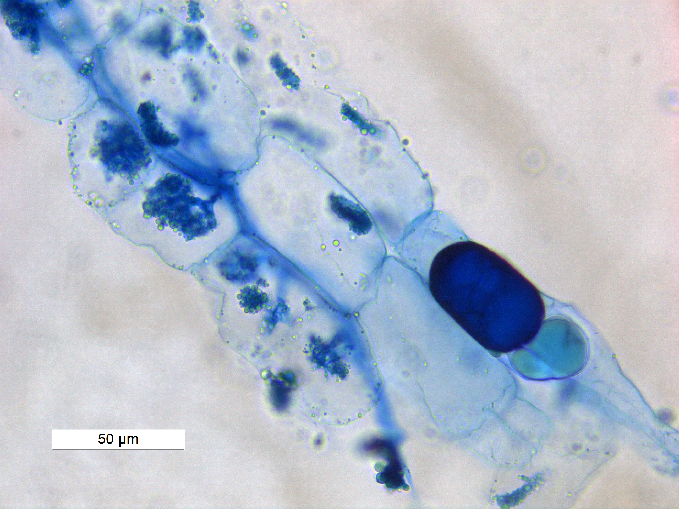
A vital stain in a casual usage may mean a stain that can be applied on living cells without killing them. Vital stains have been useful for diagnostic and surgical techniques in a variety of medical specialties. In supravital staining, living cells have been removed from an organism, whereas intravital staining is done by injecting or otherwise introducing the stain into the body. The term vital stain is used by some authors to refer to an intravital stain, and by others interchangeably with a supravital stain, the core concept being that the cell being examined is still alive. In a more strict sense, the term vital staining has a meaning contrasting with supravital staining. While in supravital staining the living cells take up the stain, in "vital staining" – the most accepted but apparently paradoxical meaning of this term, the living cells exclude the stain i.e. stain negatively and only the dead cells stain positively and thus viability can be assessed by counting the percen ... [...More Info...] [...Related Items...] OR: [Wikipedia] [Google] [Baidu] |
Stain (biology)
Staining is a technique used to enhance contrast in samples, generally at the microscopic level. Stains and dyes are frequently used in histology (microscopic study of biological tissues), in cytology (microscopic study of cells), and in the medical fields of histopathology, hematology, and cytopathology that focus on the study and diagnoses of diseases at the microscopic level. Stains may be used to define biological tissues (highlighting, for example, muscle fibers or connective tissue), cell populations (classifying different blood cells), or organelles within individual cells. In biochemistry, it involves adding a class-specific ( DNA, proteins, lipids, carbohydrates) dye to a substrate to qualify or quantify the presence of a specific compound. Staining and fluorescent tagging can serve similar purposes. Biological staining is also used to mark cells in flow cytometry, and to flag proteins or nucleic acids in gel electrophoresis. Light microscopes are used for viewing ... [...More Info...] [...Related Items...] OR: [Wikipedia] [Google] [Baidu] |
Food Coloring
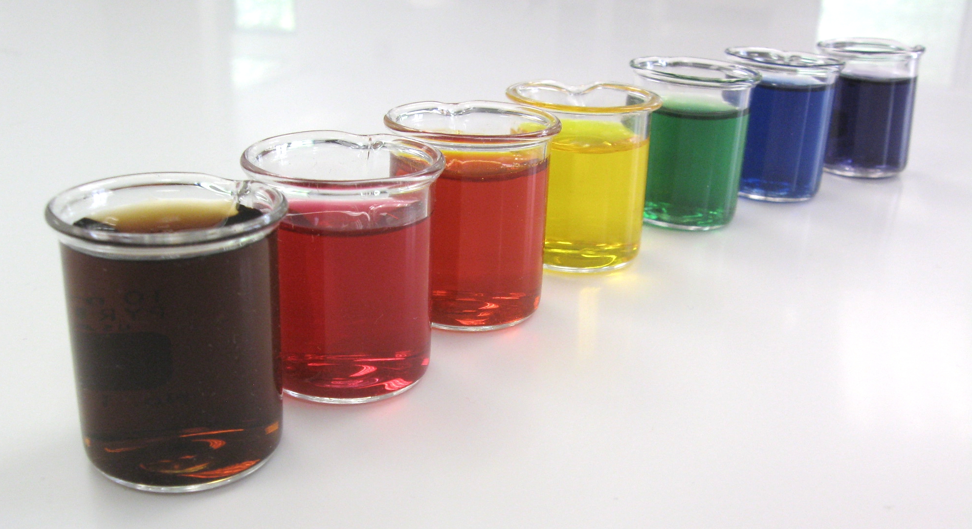
Food coloring, or color additive, is any dye, pigment, or substance that imparts color when it is added to food or drink. They come in many forms consisting of liquids, powders, gels, and pastes. Food coloring is used in both commercial food production and domestic cooking. Food colorants are also used in a variety of non-food applications, including cosmetics, pharmaceuticals, home craft projects, and medical devices. Purpose of food coloring People associate certain colors with certain flavors, and the color of food can influence the perceived flavor in anything from candy to wine. Sometimes, the aim is to simulate a color that is perceived by the consumer as natural, such as adding red coloring to glacé cherries (which would otherwise be beige), but sometimes it is for effect, like the green ketchup that Heinz launched in 2000. Color additives are used in foods for many reasons including: * To make food more attractive, appealing, appetizing, and informative * Offset c ... [...More Info...] [...Related Items...] OR: [Wikipedia] [Google] [Baidu] |
Histology
Histology, also known as microscopic anatomy or microanatomy, is the branch of biology which studies the microscopic anatomy of biological tissues. Histology is the microscopic counterpart to gross anatomy, which looks at larger structures visible without a microscope. Although one may divide microscopic anatomy into ''organology'', the study of organs, ''histology'', the study of tissues, and ''cytology'', the study of cells, modern usage places all of these topics under the field of histology. In medicine, histopathology is the branch of histology that includes the microscopic identification and study of diseased tissue. In the field of paleontology, the term paleohistology refers to the histology of fossil organisms. Biological tissues Animal tissue classification There are four basic types of animal tissues: muscle tissue, nervous tissue, connective tissue, and epithelial tissue. All animal tissues are considered to be subtypes of these four principal tissue types ... [...More Info...] [...Related Items...] OR: [Wikipedia] [Google] [Baidu] |
Cytopathology
Cytopathology (from Greek , ''kytos'', "a hollow"; , ''pathos'', "fate, harm"; and , '' -logia'') is a branch of pathology that studies and diagnoses diseases on the cellular level. The discipline was founded by George Nicolas Papanicolaou in 1928. Cytopathology is generally used on samples of free cells or tissue fragments, in contrast to histopathology, which studies whole tissues. Cytopathology is frequently, less precisely, called "cytology", which means "the study of cells". Cytopathology is commonly used to investigate diseases involving a wide range of body sites, often to aid in the diagnosis of cancer but also in the diagnosis of some infectious diseases and other inflammatory conditions. For example, a common application of cytopathology is the Pap smear, a screening tool used to detect precancerous cervical lesions that may lead to cervical cancer. Cytopathologic tests are sometimes called smear tests because the samples may be smeared across a glass microscope slid ... [...More Info...] [...Related Items...] OR: [Wikipedia] [Google] [Baidu] |
Janus Green B
Janus Green B is a basic dye and vital stain used in histology. It is also used to stain mitochondria supravitally, as was introduced by Leonor Michaelis Leonor Michaelis (16 January 1875 – 8 October 1949) was a German biochemist, physical chemist, and physician, known for his work with Maud Menten on enzyme kinetics in 1913, as well as for work on enzyme inhibition, pH and quinones. Ear ... in 1900. The indicator Janus Green B changes colour according to the amount of oxygen present. When oxygen is present, the indicator oxidizes to a blue colour. In the absence of oxygen, the indicator is reduced and changes to a pink colour. References Staining dyes Azo dyes Phenazines Chlorides Aromatic amines Anilines Diethylamino compounds {{organic-compound-stub ... [...More Info...] [...Related Items...] OR: [Wikipedia] [Google] [Baidu] |
Hematopoietic Stem Cell
Hematopoietic stem cells (HSCs) are the stem cells that give rise to other blood cells. This process is called haematopoiesis. In vertebrates, the very first definitive HSCs arise from the ventral endothelial wall of the embryonic aorta within the (midgestational) aorta-gonad-mesonephros region, through a process known as endothelial-to-hematopoietic transition. In adults, haematopoiesis occurs in the red bone marrow, in the core of most bones. The red bone marrow is derived from the layer of the embryo called the mesoderm. Haematopoiesis is the process by which all mature blood cells are produced. It must balance enormous production needs (the average person produces more than 500 billion blood cells every day) with the need to regulate the number of each blood cell type in the circulation. In vertebrates, the vast majority of hematopoiesis occurs in the bone marrow and is derived from a limited number of hematopoietic stem cells that are multipotent and capable of extensive se ... [...More Info...] [...Related Items...] OR: [Wikipedia] [Google] [Baidu] |
Flow Cytometry
Flow cytometry (FC) is a technique used to detect and measure physical and chemical characteristics of a population of cells or particles. In this process, a sample containing cells or particles is suspended in a fluid and injected into the flow cytometer instrument. The sample is focused to ideally flow one cell at a time through a laser beam, where the light scattered is characteristic to the cells and their components. Cells are often labeled with fluorescent markers so light is absorbed and then emitted in a band of wavelengths. Tens of thousands of cells can be quickly examined and the data gathered are processed by a computer. Flow cytometry is routinely used in basic research, clinical practice, and clinical trials. Uses for flow cytometry include: * Cell counting * Cell sorting * Determining cell characteristics and function * Detecting microorganisms * Biomarker detection * Protein engineering detection * Diagnosis of health disorders such as blood cancers * Measuring ... [...More Info...] [...Related Items...] OR: [Wikipedia] [Google] [Baidu] |
7-Aminoactinomycin D
7-Aminoactinomycin D (7-AAD) is a fluorescent chemical compound with a strong affinity for DNA. It is used as a fluorescent marker for DNA in fluorescence microscopy and flow cytometry. It intercalates in double-stranded DNA, with a high affinity for GC-rich regions, making it useful for chromosome banding studies. Applications With an absorption maximum at 546 nm, 7-AAD is efficiently excited using a 543 nm helium–neon laser; it can also be excited with somewhat lower efficiency using a 488 nm or 514 nm argon laser lines. Its emission has a very large Stokes shift with a maximum in the deep red: 647 nm. 7-AAD is therefore compatible with most blue and green fluorophores – and even many red fluorophores – in multicolour applications. 7-AAD does not readily pass through intact cell membranes; if it is to be used as a stain for imaging DNA fluorescence, the cell membrane must be permeabilized or disrupted. This method can be used in co ... [...More Info...] [...Related Items...] OR: [Wikipedia] [Google] [Baidu] |
Erythrosine
Erythrosine, also known as Red No. 3, is an organoiodine compound, specifically a derivative of fluorone. It is a pink dye which is primarily used for food coloring. It is the disodium salt of 2,4,5,7-tetraiodofluorescein. Its maximum absorbance is at 530 nm in an aqueous solution, and it is subject to photodegradation. Uses It is used as a: * food coloring * printing ink * biological stain * dental plaque disclosing agent * radiopaque medium * sensitizer for orthochromatic photographic films *Visible light photoredox catalyst Erythrosine is commonly used in sweets such as some candies and popsicles, and even more widely used in cake-decorating gels. It is also used to color pistachio shells. As a food additive, it has the E number E127. Health effects As a result of efforts begun in the 1970s, in 1990 the U.S. FDA had instituted a partial ban on erythrosine, citing research that high doses have been found to cause cancer in rats. A 1990 study concluded that "chr ... [...More Info...] [...Related Items...] OR: [Wikipedia] [Google] [Baidu] |
Supravital Stain
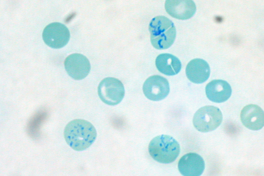
Supravital staining is a method of staining used in microscopy to examine living cells that have been removed from an organism. It differs from intravital staining, which is done by injecting or otherwise introducing the stain into the body. Thus a supravital stain may have a greater toxicity, as only a few cells need to survive it a short while. The term "vital stain" is used by some authors to refer specifically to an intravital stain, and by others interchangeably with a supravital stain, the core concept being that the cell being examined is still alive. As the cells are alive and unfixed, outside the body, supravital stains are temporary in nature. The most common supravital stain is performed on reticulocytes using new methylene blue or brilliant cresyl blue, which makes it possible to see the reticulofilamentous pattern of ribosomes characteristically precipitated in these live immature red blood cells by the supravital stains. By counting the number of such cells the r ... [...More Info...] [...Related Items...] OR: [Wikipedia] [Google] [Baidu] |
Trypan Blue
Trypan blue is an azo dye. It is a direct dye for cotton textiles. In biosciences, it is used as a vital stain to selectively colour dead tissues or cells blue. Live cells or tissues with intact cell membranes are not coloured. Since cells are very selective in the compounds that pass through the membrane, in a viable cell trypan blue is not absorbed; however, it traverses the membrane in a dead cell. Hence, dead cells appear as a distinctive blue colour under a microscope. Since live cells are excluded from staining, this staining method is also described as a dye exclusion method. Background and chemistry Trypan blue is derived from toluidine, that is, any of several isomeric bases, C14H16N2, derived from toluene. Trypan blue is so-called because it can kill trypanosomes, the parasites that cause sleeping sickness. An analog of trypan blue, suramin, is used pharmacologically against trypanosomiasis. Trypan blue is also known as diamine blue and Niagara blue. The extinctio ... [...More Info...] [...Related Items...] OR: [Wikipedia] [Google] [Baidu] |
Cell (biology)
The cell is the basic structural and functional unit of life forms. Every cell consists of a cytoplasm enclosed within a membrane, and contains many biomolecules such as proteins, DNA and RNA, as well as many small molecules of nutrients and metabolites.Cell Movements and the Shaping of the Vertebrate Body in Chapter 21 of Molecular Biology of the Cell '' fourth edition, edited by Bruce Alberts (2002) published by Garland Science. The Alberts text discusses how the "cellular building blocks" move to shape developing embryos. It is also common to describe small molecules such as ... [...More Info...] [...Related Items...] OR: [Wikipedia] [Google] [Baidu] |


