|
Vaginal Squamous-cell Carcinoma
Squamous-cell carcinoma of the vagina is a potentially invasive type of cancer that forms in the tissues of the vagina. Though uncommon, squamous-cell cancer of the vagina (SCCV) is the most common type of vaginal cancer. It is further subdivided into the following subtypes: keratinizing, nonkeratinizing, basaloid, and warty. It forms in squamous cells, the thin, flat cells lining the vagina. Squamous cell vaginal cancer spreads slowly and usually stays near the vagina, but may spread to the lungs, liver, or bone. This is the most common type of vaginal cancer. SCCV accounts for approximately 85% of vaginal cancer cases and initially spreads superficially within the vaginal wall. It can later invade other vaginal tissues. The carcinoma can metastasize to the lungs, and less frequently in liver, bone, or other sites. SCC of the vagina is associated with a high rate of infection with oncogenic strains of human papillomavirus (HPV) and has many risk factors in common with cervical ... [...More Info...] [...Related Items...] OR: [Wikipedia] [Google] [Baidu] |
Oncology
Oncology is a branch of medicine that deals with the study, treatment, diagnosis and prevention of cancer. A medical professional who practices oncology is an ''oncologist''. The name's etymological origin is the Greek word ὄγκος (''ónkos''), meaning "tumor", "volume" or "mass". Oncology is concerned with: * The diagnosis of any cancer in a person (pathology) * Therapy (e.g. surgery, chemotherapy, radiotherapy and other modalities) * Follow-up of cancer patients after successful treatment * Palliative care of patients with terminal malignancies * Ethical questions surrounding cancer care * Screening efforts: ** of populations, or ** of the relatives of patients (in types of cancer that are thought to have a hereditary basis, such as breast cancer) Diagnosis Medical histories remain an important screening tool: the character of the complaints and nonspecific symptoms (such as fatigue, weight loss, unexplained anemia, fever of unknown origin, paraneoplasti ... [...More Info...] [...Related Items...] OR: [Wikipedia] [Google] [Baidu] |
Multiple Sex Partners
Multiple sex partners is the measure and incidence of engaging in sexual activities with two or more people within a specific time period. Sexual activity with MSP can happen simultaneously or serially. MSP includes sexual activity between people of a different gender or the same gender. A person can be said to have multiple sex partners, when the person have sex with more than one person at the same time. Another term, '' polyamorous'', is a behavior and not a measure describing multiple romantically sexually or romantically committed relationships at the same time. Young people having MSP in the last year is an indicator used by the Centers for Disease Control and Prevention (CDC) to evaluate risky sexual behavior in adolescents and monitoring changes in the worldwide HIV/AIDS infection rates and deaths. Definitions and quantification Epidemiologists and clinicians who quantify risks associated with MSP do so to identify those who have had sexual intercourse with more than one ... [...More Info...] [...Related Items...] OR: [Wikipedia] [Google] [Baidu] |
Pelvic Organ Prolapse
Pelvic organ prolapse (POP) is characterized by descent of pelvic organs from their normal positions. In women, the condition usually occurs when the pelvic floor collapses after gynecological cancer treatment, childbirth or heavy lifting. In men, it may occur after the prostate gland is removed. The injury occurs to fascia membranes and other connective structures that can result in cystocele, rectocele or both. Treatment can involve dietary and lifestyle changes, physical therapy, or surgery. Types * Anterior vaginal wall prolapse ** Cystocele (bladder into vagina) ** Urethrocele (urethra into vagina) ** Cystourethrocele (both bladder and urethra) * Posterior vaginal wall prolapse ** Enterocele (small intestine into vagina) ** Rectocele (rectum into vagina) ** Sigmoidocele * Apical vaginal prolapse ** Uterine prolapse (uterus into vagina) ** Vaginal vault prolapse (roof of vagina) – after hysterectomy Grading Pelvic organ prolapses are graded either via the Baden–W ... [...More Info...] [...Related Items...] OR: [Wikipedia] [Google] [Baidu] |
Hysterectomy
Hysterectomy is the surgical removal of the uterus. It may also involve removal of the cervix, ovaries ( oophorectomy), Fallopian tubes ( salpingectomy), and other surrounding structures. Usually performed by a gynecologist, a hysterectomy may be total (removing the body, fundus, and cervix of the uterus; often called "complete") or partial (removal of the uterine body while leaving the cervix intact; also called "supracervical"). Removal of the uterus renders the patient unable to bear children (as does removal of ovaries and fallopian tubes) and has surgical risks as well as long-term effects, so the surgery is normally recommended only when other treatment options are not available or have failed. It is the second most commonly performed gynecological surgical procedure, after cesarean section, in the United States. Nearly 68 percent were performed for conditions such as endometriosis, irregular bleeding, and uterine fibroids. It is expected that the frequency of hysterectom ... [...More Info...] [...Related Items...] OR: [Wikipedia] [Google] [Baidu] |
Radiation Therapy
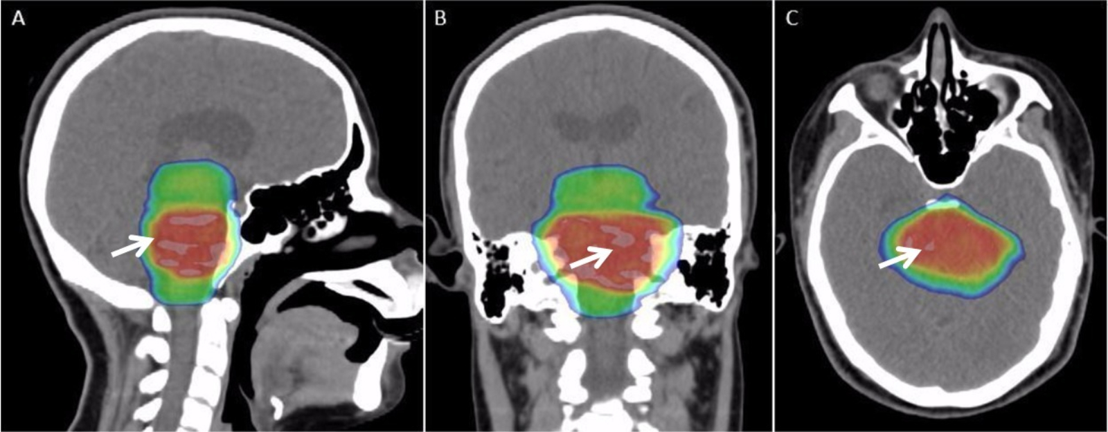
Radiation therapy or radiotherapy, often abbreviated RT, RTx, or XRT, is a therapy using ionizing radiation, generally provided as part of cancer treatment to control or kill malignant cells and normally delivered by a linear accelerator. Radiation therapy may be curative in a number of types of cancer if they are localized to one area of the body. It may also be used as part of adjuvant therapy, to prevent tumor recurrence after surgery to remove a primary malignant tumor (for example, early stages of breast cancer). Radiation therapy is synergistic with chemotherapy, and has been used before, during, and after chemotherapy in susceptible cancers. The subspecialty of oncology concerned with radiotherapy is called radiation oncology. A physician who practices in this subspecialty is a radiation oncologist. Radiation therapy is commonly applied to the cancerous tumor because of its ability to control cell growth. Ionizing radiation works by damaging the DNA of cancerous tissue ... [...More Info...] [...Related Items...] OR: [Wikipedia] [Google] [Baidu] |
Uterus
The uterus (from Latin ''uterus'', plural ''uteri'') or womb () is the organ in the reproductive system of most female mammals, including humans that accommodates the embryonic and fetal development of one or more embryos until birth. The uterus is a hormone-responsive sex organ that contains glands in its lining that secrete uterine milk for embryonic nourishment. In the human, the lower end of the uterus, is a narrow part known as the isthmus that connects to the cervix, leading to the vagina. The upper end, the body of the uterus, is connected to the fallopian tubes, at the uterine horns, and the rounded part above the openings to the fallopian tubes is the fundus. The connection of the uterine cavity with a fallopian tube is called the uterotubal junction. The fertilized egg is carried to the uterus along the fallopian tube. It will have divided on its journey to form a blastocyst that will implant itself into the lining of the uterus – the endometrium, w ... [...More Info...] [...Related Items...] OR: [Wikipedia] [Google] [Baidu] |
Cystoscopy
Cystoscopy is endoscopy of the urinary bladder via the urethra. It is carried out with a cystoscope. The urethra is the tube that carries urine from the bladder to the outside of the body. The cystoscope has lenses like a telescope or microscope. These lenses let the physician focus on the inner surfaces of the urinary tract. Some cystoscopes use optical fibres (flexible glass fibres) that carry an image from the tip of the instrument to a viewing piece at the other end. Cystoscopes range from pediatric to adult and from the thickness of a pencil up to approximately 9 mm and have a light at the tip. Many cystoscopes have extra tubes to guide other instruments for surgical procedures to treat urinary problems. There are two main types of cystoscopy—flexible and rigid—differing in the flexibility of the cystoscope. Flexible cystoscopy is carried out with local anaesthesia on both sexes. Typically, a topical anesthetic, most often xylocaine gel (common brand names are Ane ... [...More Info...] [...Related Items...] OR: [Wikipedia] [Google] [Baidu] |
Positron Emission Tomography
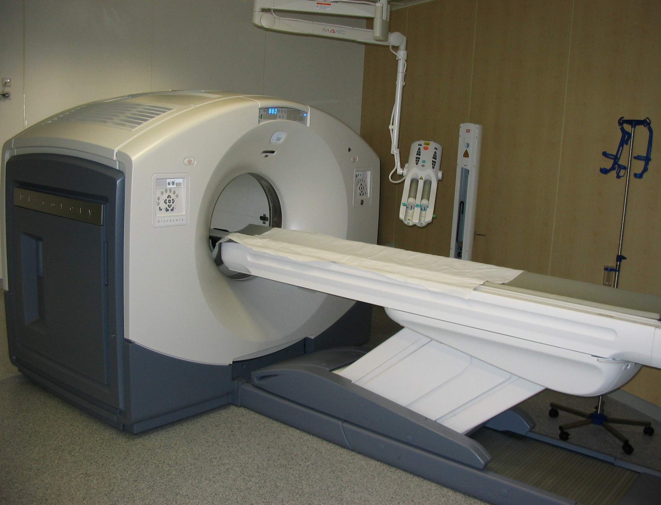
Positron emission tomography (PET) is a functional imaging technique that uses radioactive substances known as radiotracers to visualize and measure changes in metabolic processes, and in other physiological activities including blood flow, regional chemical composition, and absorption. Different tracers are used for various imaging purposes, depending on the target process within the body. For example, -FDG is commonly used to detect cancer, NaF is widely used for detecting bone formation, and oxygen-15 is sometimes used to measure blood flow. PET is a common imaging technique, a medical scintillography technique used in nuclear medicine. A radiopharmaceutical — a radioisotope attached to a drug — is injected into the body as a tracer. When the radiopharmaceutical undergoes beta plus decay, a positron is emitted, and when the positron collides with an ordinary electron, the two particles annihilate and gamma rays are emitted. These gamma rays are detecte ... [...More Info...] [...Related Items...] OR: [Wikipedia] [Google] [Baidu] |
Magnetic Resonance Imaging
Magnetic resonance imaging (MRI) is a medical imaging technique used in radiology to form pictures of the anatomy and the physiological processes of the body. MRI scanners use strong magnetic fields, magnetic field gradients, and radio waves to generate images of the organs in the body. MRI does not involve X-rays or the use of ionizing radiation, which distinguishes it from CT and PET scans. MRI is a medical application of nuclear magnetic resonance (NMR) which can also be used for imaging in other NMR applications, such as NMR spectroscopy. MRI is widely used in hospitals and clinics for medical diagnosis, staging and follow-up of disease. Compared to CT, MRI provides better contrast in images of soft-tissues, e.g. in the brain or abdomen. However, it may be perceived as less comfortable by patients, due to the usually longer and louder measurements with the subject in a long, confining tube, though "Open" MRI designs mostly relieve this. Additionally, implants and ... [...More Info...] [...Related Items...] OR: [Wikipedia] [Google] [Baidu] |
Chest Radiograph
A chest radiograph, called a chest X-ray (CXR), or chest film, is a projection radiograph of the chest used to diagnose conditions affecting the chest, its contents, and nearby structures. Chest radiographs are the most common film taken in medicine. Like all methods of radiography, chest radiography employs ionizing radiation in the form of X-rays to generate images of the chest. The mean radiation dose to an adult from a chest radiograph is around 0.02 mSv (2 mrem) for a front view (PA, or posteroanterior) and 0.08 mSv (8 mrem) for a side view (LL, or latero-lateral). Together, this corresponds to a background radiation equivalent time of about 10 days. Medical uses Conditions commonly identified by chest radiography * Pneumonia * Pneumothorax * Interstitial lung disease * Heart failure * Bone fracture * Hiatal hernia Chest radiographs are used to diagnose many conditions involving the chest wall, including its bones, and also structures contained within the thoracic ... [...More Info...] [...Related Items...] OR: [Wikipedia] [Google] [Baidu] |
Biopsy
A biopsy is a medical test commonly performed by a surgeon, interventional radiologist, or an interventional cardiologist. The process involves extraction of sample cells or tissues for examination to determine the presence or extent of a disease. The tissue is then fixed, dehydrated, embedded, sectioned, stained and mounted before it is generally examined under a microscope by a pathologist; it may also be analyzed chemically. When an entire lump or suspicious area is removed, the procedure is called an excisional biopsy. An incisional biopsy or core biopsy samples a portion of the abnormal tissue without attempting to remove the entire lesion or tumor. When a sample of tissue or fluid is removed with a needle in such a way that cells are removed without preserving the histological architecture of the tissue cells, the procedure is called a needle aspiration biopsy. Biopsies are most commonly performed for insight into possible cancerous or inflammatory conditions. Histor ... [...More Info...] [...Related Items...] OR: [Wikipedia] [Google] [Baidu] |


