|
Visual Pathway Lesions
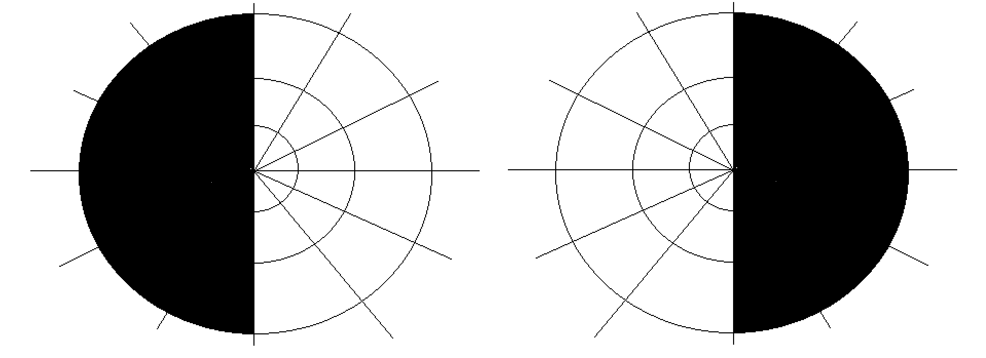
The visual pathway consists of structures that carry visual information from the retina to the brain. Lesions in that pathway cause a variety of visual field defects. In the visual system of human eye, the visual information processed by retinal photoreceptor cells travel in the following way: Retina→Optic nerve→Optic chiasma (here the nasal visual field of both eyes cross over to the opposite side)→Optic tract→Lateral geniculate nucleus, Lateral geniculate body→Optic radiation→Primary visual cortex The type of field defect can help localize where the lesion is located (see picture given in infobox). Optic nerve lesions The optic nerve, also known as cranial nerve II, extends from the optic disc to the optic chiasma. Lesions in optic nerve causes visual field defects and blindness. Causes Causes of optic nerve lesions include optic atrophy, optic neuropathy, head injury, traumatic avulsion, acute optic neuritis etc. Signs and symptoms * Lesions involving the who ... [...More Info...] [...Related Items...] OR: [Wikipedia] [Google] [Baidu] |
Macular Sparing
Macular sparing is visual field loss that preserves vision in the center of the visual field, otherwise known as the macula. It appears in people with damage to one hemisphere of their visual cortex, and occurs simultaneously with bilateral homonymous hemianopia or homonymous quadrantanopia. The exact mechanism behind this phenomenon is still uncertain.Whishaw, I. Q., & Kolb, B. (2015). Fundamentals of Human Neuropsychology (7th ed.). New York, NY: Worth Custom Publishing. The opposing effect, where vision in half of the center of the visual field is lost, is known as macular splitting.Windsor, R. L. (n.d.). Visual Fields in Brain Injury - Hemianopsia.net Everything you need to know about Hemianopsia. Retrieved from http://www.hemianopsia.net/visual-fields-in-brain-injury/ Causes The favored explanation for why the center visual field is preserved after large hemispheric lesions is that the macular regions of the cortex have a double vascular supply from the middle cerebral artery ... [...More Info...] [...Related Items...] OR: [Wikipedia] [Google] [Baidu] |
Lateral Geniculate Nucleus
In neuroanatomy, the lateral geniculate nucleus (LGN; also called the lateral geniculate body or lateral geniculate complex) is a structure in the thalamus and a key component of the mammalian visual pathway. It is a small, ovoid, ventral projection of the thalamus where the thalamus connects with the optic nerve. There are two LGNs, one on the left and another on the right side of the thalamus. In humans, both LGNs have six layers of neurons ( grey matter) alternating with optic fibers (white matter). The LGN receives information directly from the ascending retinal ganglion cells via the optic tract and from the reticular activating system. Neurons of the LGN send their axons through the optic radiation, a direct pathway to the primary visual cortex. In addition, the LGN receives many strong feedback connections from the primary visual cortex. In humans as well as other mammals, the two strongest pathways linking the eye to the brain are those projecting to the dorsal part ... [...More Info...] [...Related Items...] OR: [Wikipedia] [Google] [Baidu] |
Optic Chiasm
In neuroanatomy, the optic chiasm, or optic chiasma (; , ), is the part of the brain where the optic nerves cross. It is located at the bottom of the brain immediately inferior to the hypothalamus. The optic chiasm is found in all vertebrates, although in cyclostomes (lampreys and hagfishes), it is located within the brain. This article is about the optic chiasm of vertebrates, which is the best known nerve chiasm, but not every chiasm denotes a crossing of the body midline (e.g., in some invertebrates, see Chiasm (anatomy)). A midline crossing of nerves inside the brain is called a decussation (see Definition of types of crossings). Structure For the different types of optic chiasm, see In all vertebrates, the optic nerves of the left and the right eye meet in the body midline, ventral to the brain. In many vertebrates the left optic nerve crosses over the right one without fusing with it. In vertebrates with a large overlap of the visual fields of the two eyes, i ... [...More Info...] [...Related Items...] OR: [Wikipedia] [Google] [Baidu] |
Contrast Sensitivity
Contrast is the contradiction in luminance or colour that makes an object (or its representation in an image or display) distinguishable. In visual perception of the real world, contrast is determined by the difference in the colour and brightness of the object and other objects within the same field of view. The human visual system is more sensitive to contrast than absolute luminance; we can perceive the world similarly regardless of the huge changes in illumination over the day or from place to place. The maximum ''contrast'' of an image is the contrast ratio or dynamic range. Images with a contrast ratio close to their medium's maximum possible contrast ratio experience a ''conservation of contrast'', wherein any increase in contrast in some parts of the image must necessarily result in a decrease in contrast elsewhere. Brightening an image will increase contrast in dark areas but decrease contrast in bright areas, while darkening the image will have the opposite effect. Ble ... [...More Info...] [...Related Items...] OR: [Wikipedia] [Google] [Baidu] |
Color Blindness
Color blindness or color vision deficiency (CVD) is the decreased ability to see color or differences in color. It can impair tasks such as selecting ripe fruit, choosing clothing, and reading traffic lights. Color blindness may make some academic activities more difficult. However, issues are generally minor, and the colorblind automatically develop adaptations and coping mechanisms. People with total color blindness (achromatopsia) may also be uncomfortable in bright environments and have decreased visual acuity. The most common cause of color blindness is an inherited problem or variation in the functionality of one or more of the three classes of cone cells in the retina, which mediate color vision. The most common form is caused by a genetic disorder called congenital red–green color blindness. Males are more likely to be color blind than females, because the genes responsible for the most common forms of color blindness are on the X chromosome. Non-color-blind fe ... [...More Info...] [...Related Items...] OR: [Wikipedia] [Google] [Baidu] |
Afferent Pupillary Defect
A relative afferent pupillary defect (RAPD), also known as a Marcus Gunn pupil, is a medical sign observed during the swinging-flashlight test whereupon the patient's pupils dilate when a bright light is swung from the unaffected eye to the affected eye. The affected eye still senses the light and produces pupillary sphincter constriction to some degree, albeit reduced. Depending on severity, different symptoms may appear during the swinging flash light test: Mild RAPD will presents as a weak pupil constriction initially, after which dilation continues to happen. When RAPD is moderate, pupil size will remain, after which it dilates When RAPD is severe, the pupil will dilate quickly Cause The most common cause of Marcus Gunn pupil is a lesion of the optic nerve (between the retina and the optic chiasm) due to glaucoma, or severe retinal disease, or due to multiple sclerosis. It is named after Scottish ophthalmologist Robert Marcus Gunn. A second common cause of Marcus Gunn pup ... [...More Info...] [...Related Items...] OR: [Wikipedia] [Google] [Baidu] |
Scotoma
A scotoma is an area of partial alteration in the field of vision consisting of a partially diminished or entirely degenerated visual acuity that is surrounded by a field of normal – or relatively well-preserved – vision. Every normal mammalian eye has a scotoma in its field of vision, usually termed its blind spot. This is a location with no photoreceptor cells, where the retinal ganglion cell axons that compose the optic nerve exit the retina. This location is called the optic disc. There is no direct conscious awareness of visual scotomas. They are simply regions of reduced information within the visual field. Rather than recognizing an incomplete image, patients with scotomas report that things "disappear" on them. The presence of the blind spot scotoma can be demonstrated subjectively by covering one eye, carefully holding fixation with the open eye, and placing an object (such as one's thumb) in the lateral and horizontal visual field, about 15 degrees from f ... [...More Info...] [...Related Items...] OR: [Wikipedia] [Google] [Baidu] |
Tunnel Vision
Tunnel vision is the loss of peripheral vision with retention of central vision, resulting in a constricted circular tunnel-like field of vision. Causes Tunnel vision can be caused by: Eyeglass users Eyeglass users experience tunnel vision to varying degrees due to the corrective lens only providing a small area of proper focus, with the rest of the field of view beyond the lenses being unfocused and blurry. Where a naturally sighted person only needs to move their eyes to see an object far to the side or far down, the eyeglass wearer may need to move their whole head to point the eyeglasses towards the target object. The eyeglass frame also blocks the view of the world with a thin opaque boundary separating the lens area from the rest of the field of view. The eyeglass frame is capable of obscuring small objects and details in the peripheral field. Mask, goggle, and helmet users Activities which require a protective mask, safety goggles, or fully enclosing protective ... [...More Info...] [...Related Items...] OR: [Wikipedia] [Google] [Baidu] |
Visual Field Central Scotoma
The visual system comprises the sensory organ (the eye) and parts of the central nervous system (the retina containing photoreceptor cells, the optic nerve, the optic tract and the visual cortex) which gives organisms the sense of sight (the ability to detect and process visible light) as well as enabling the formation of several non-image photo response functions. It detects and interprets information from the optical spectrum perceptible to that species to "build a representation" of the surrounding environment. The visual system carries out a number of complex tasks, including the reception of light and the formation of monocular neural representations, colour vision, the neural mechanisms underlying stereopsis and assessment of distances to and between objects, the identification of a particular object of interest, motion perception, the analysis and integration of visual information, pattern recognition, accurate motor coordination under visual guidance, and m ... [...More Info...] [...Related Items...] OR: [Wikipedia] [Google] [Baidu] |
Retinitis Pigmentosa Visual Field
Retinitis is inflammation of the retina in the eye, which can permanently damage the retina and lead to blindness. The retina is the eye's "sensing" tissue. Retinitis may be caused by a number of different infectious agents. Its most common form, called retinitis pigmentosa, has a prevalence of one in every 2,500–7,000 people. This condition is one of the leading causes that leads to blindness in patients in the age range of 20–60 years old. Retinitis may be caused by several infectious agents, including toxoplasmosis, cytomegalovirus and candida. Cytomegalovirus retinitis is an important cause of blindness in AIDS Human immunodeficiency virus infection and acquired immunodeficiency syndrome (HIV/AIDS) is a spectrum of conditions caused by infection with the human immunodeficiency virus (HIV), a retrovirus. Following initial infection an individual m ... patients. Candida spreading to the retina from the bloodstream usually results in the production of sev ... [...More Info...] [...Related Items...] OR: [Wikipedia] [Google] [Baidu] |
Head Injury
A head injury is any injury that results in trauma to the skull or brain. The terms ''traumatic brain injury'' and ''head injury'' are often used interchangeably in the medical literature. Because head injuries cover such a broad scope of injuries, there are many causes—including accidents, falls, physical assault, or traffic accidents—that can cause head injuries. The number of new cases is 1.7 million in the United States each year, with about 3% of these incidents leading to death. Adults have head injuries more frequently than any age group resulting from falls, motor vehicle crashes, colliding or being struck by an object, or assaults. Children, however, may experience head injuries from accidental falls or intentional causes (such as being struck or shaken) leading to hospitalization. Acquired brain injury (ABI) is a term used to differentiate brain injuries occurring after birth from injury, from a genetic disorder, or from a congenital disorder. Unlike a broke ... [...More Info...] [...Related Items...] OR: [Wikipedia] [Google] [Baidu] |
Optic Neuropathy
Optic neuropathy is damage to the optic nerve from any cause. The optic nerve is a bundle of millions of fibers in the retina that sends visual signals to the brain. Damage and death of these nerve cells, or neurons, leads to characteristic features of optic neuropathy. The main symptom is loss of vision, with colors appearing subtly washed out in the affected eye. A pale disc is characteristic of long-standing optic neuropathy. In many cases, only one eye is affected and patients may not be aware of the loss of color vision until the doctor asks them to cover the healthy eye. Optic neuropathy is often called optic atrophy, to describe the loss of some or most of the fibers of the optic nerve. Ischemic optic neuropathy In ischemic optic neuropathies, there is insufficient blood flow (ischemia) to the optic nerve. The anterior optic nerve is supplied by the short posterior ciliary artery and choroidal circulation, while the retrobulbar optic nerve is supplied intraorbitally by ... [...More Info...] [...Related Items...] OR: [Wikipedia] [Google] [Baidu] |


