|
Vesical Arteries
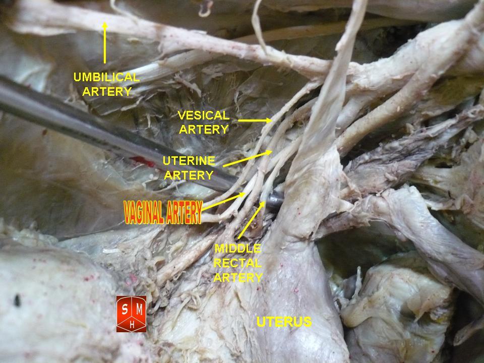
Vesical arteries are variable in number. They supply the bladder and terminal ureter. The two most prominent are the superior vesical artery and the inferior vesical artery. The superior vesical artery comes off of the internal iliac artery and sometimes the umbilical artery. The inferior vesical artery comes off of the internal iliac artery. The inferior vesical artery is a pelvic branch of the internal iliac artery in men; and in women it branches from the vaginal artery The vaginal artery is an artery in females that supplies blood to the vagina and the base of the bladder. Structure The vaginal artery is usually a branch of the internal iliac artery. Some sources say that the vaginal artery can arise from the .... This literature has been reviewed recently with observations of variation in pelvic vascularization and the close relationship between vaginal and bladder vascularization in women. References Arteries of the abdomen {{circulatory-stub ... [...More Info...] [...Related Items...] OR: [Wikipedia] [Google] [Baidu] |
Bladder
The urinary bladder, or simply bladder, is a hollow organ in humans and other vertebrates that stores urine from the kidneys before disposal by urination. In humans the bladder is a distensible organ that sits on the pelvic floor. Urine enters the bladder via the ureters and exits via the urethra. The typical adult human bladder will hold between 300 and (10.14 and ) before the urge to empty occurs, but can hold considerably more. The Latin phrase for "urinary bladder" is ''vesica urinaria'', and the term ''vesical'' or prefix ''vesico -'' appear in connection with associated structures such as vesical veins. The modern Latin word for "bladder" – ''cystis'' – appears in associated terms such as cystitis (inflammation of the bladder). Structure In humans, the bladder is a hollow muscular organ situated at the base of the pelvis. In gross anatomy, the bladder can be divided into a broad , a body, an apex, and a neck. The apex (also called the vertex) is directed forw ... [...More Info...] [...Related Items...] OR: [Wikipedia] [Google] [Baidu] |
Ureter
The ureters are tubes made of smooth muscle that propel urine from the kidneys to the urinary bladder. In a human adult, the ureters are usually long and around in diameter. The ureter is lined by urothelial cells, a type of transitional epithelium, and has an additional smooth muscle layer that assists with peristalsis in its lowest third. The ureters can be affected by a number of diseases, including urinary tract infections and kidney stone. is when a ureter is narrowed, due to for example chronic inflammation. Congenital abnormalities that affect the ureters can include the development of two ureters on the same side or abnormally placed ureters. Additionally, reflux of urine from the bladder back up the ureters is a condition commonly seen in children. The ureters have been identified for at least two thousand years, with the word "ureter" stemming from the stem relating to urinating and seen in written records since at least the time of Hippocrates. It is, ho ... [...More Info...] [...Related Items...] OR: [Wikipedia] [Google] [Baidu] |
Superior Vesical Artery
The superior vesical artery supplies numerous branches to the upper part of the bladder. This artery often also gives branches to the vas deferens and can provide minor collateral circulation for the testicles. Anatomy The superior vesical artery is a branch of the umbilical artery. The vesiculo-prostatic artery usually arises from the superior vesical artery in men. Distribution Other branches supply the ureter. Variation The middle vesical artery, usually a branch of the superior vesical artery, is distributed to the fundus ''Fundus'' (Latin for "bottom") is an anatomical term referring to that part of a concavity in any organ, which is at the far end from its opening. It may refer to: Anatomy * Fundus (brain), the deepest part of any sulcus of the cerebral cortex * ... of the bladder and the seminal vesicles. This artery is not usually described in modern anatomy textbooks. Instead, it is described that the superior vesical artery may exist as multiple vessels th ... [...More Info...] [...Related Items...] OR: [Wikipedia] [Google] [Baidu] |
Inferior Vesical Artery
The inferior vesical artery (or inferior vesicle artery) is an artery of the pelvis which arises from the internal iliac artery and supplies parts of the urinary bladder as well as other structures of the urinary system and structures of the male reproductive system. Some sources consider this vessel to be present only in males, and cite the vaginal artery as the homologous structure in females; others consider it to be present in both sexes, with the vessel taking the form of a small branch of a vaginal artery in females. Structure Origin The inferior vesical artery is a branch of the anterior division of the internal iliac artery. It frequently has a common origin with the middle rectal artery. Course The inferior vesical artery passes medially across the pelvic floor. Distribution The inferior vesical artery is distributed to the trigone and inferior portion of the urinary bladder, the ureter, prostate, vas deferens, and seminal vesicles.vas deferens The vas ... [...More Info...] [...Related Items...] OR: [Wikipedia] [Google] [Baidu] |
Internal Iliac Artery
The internal iliac artery (formerly known as the hypogastric artery) is the main artery of the pelvis. Structure The internal iliac artery supplies the walls and viscera of the pelvis, the buttock, the reproductive organs, and the medial compartment of the thigh. The vesicular branches of the internal iliac arteries supply the bladder. It is a short, thick vessel, smaller than the external iliac artery, and about 3 to 4 cm in length. Course The internal iliac artery arises at the bifurcation of the common iliac artery, opposite the lumbosacral articulation, and, passing downward to the upper margin of the greater sciatic foramen, divides into two large trunks, an anterior and a posterior. It is posterior to the ureter, anterior to the internal iliac vein, anterior to the lumbosacral trunk, and anterior to the piriformis muscle. Near its origin, it is medial to the external iliac vein, which lies between it and the psoas major muscle. It is above the obturator nerve ... [...More Info...] [...Related Items...] OR: [Wikipedia] [Google] [Baidu] |
Umbilical Artery
The umbilical artery is a paired artery (with one for each half of the body) that is found in the abdominal and pelvic regions. In the fetus, it extends into the umbilical cord. Structure Development The umbilical arteries supply deoxygenated blood from the fetus to the placenta. Although this blood is typically referred to as deoxygenated, this blood is fetal systemic arterial blood and will have the same amount of oxygen and nutrients as blood distributed to the other fetal tissues. There are usually two umbilical arteries present together with one umbilical vein in the umbilical cord. The umbilical arteries surround the urinary bladder and then carry all the deoxygenated blood out of the fetus through the umbilical cord. Inside the placenta, the umbilical arteries connect with each other at a distance of approximately 5 mm from the cord insertion in what is called the ''Hyrtl anastomosis''. Subsequently, they branch into chorionic arteries or ''intraplacental fetal arte ... [...More Info...] [...Related Items...] OR: [Wikipedia] [Google] [Baidu] |
Pelvis
The pelvis (plural pelves or pelvises) is the lower part of the trunk, between the abdomen and the thighs (sometimes also called pelvic region), together with its embedded skeleton (sometimes also called bony pelvis, or pelvic skeleton). The pelvic region of the trunk includes the bony pelvis, the pelvic cavity (the space enclosed by the bony pelvis), the pelvic floor, below the pelvic cavity, and the perineum, below the pelvic floor. The pelvic skeleton is formed in the area of the back, by the sacrum and the coccyx and anteriorly and to the left and right sides, by a pair of hip bones. The two hip bones connect the spine with the lower limbs. They are attached to the sacrum posteriorly, connected to each other anteriorly, and joined with the two femurs at the hip joints. The gap enclosed by the bony pelvis, called the pelvic cavity, is the section of the body underneath the abdomen and mainly consists of the reproductive organs (sex organs) and the rectum, while the p ... [...More Info...] [...Related Items...] OR: [Wikipedia] [Google] [Baidu] |
Vaginal Artery
The vaginal artery is an artery in females that supplies blood to the vagina and the base of the bladder. Structure The vaginal artery is usually a branch of the internal iliac artery. Some sources say that the vaginal artery can arise from the uterine artery, but the phrase vaginal branches of uterine artery is the term for blood supply to the vagina coming from the uterine artery. The vaginal artery is frequently represented by two or three branches. These descend to the vagina, supplying its mucous membrane. They anastomose with branches from the uterine artery. It can send branches to the bulb of the vestibule, the fundus of the bladder, and the contiguous part of the rectum. Function The vaginal artery supplies oxygenated blood to the muscular wall of the vagina, along with the uterine artery and the internal pudendal artery. It also supplies the cervix, along with the uterine artery. Other animals In horses, the vaginal artery may haemorrhage after birth, which can ... [...More Info...] [...Related Items...] OR: [Wikipedia] [Google] [Baidu] |
Vascularization
Angiogenesis is the physiological process through which new blood vessels form from pre-existing vessels, formed in the earlier stage of vasculogenesis. Angiogenesis continues the growth of the vasculature by processes of sprouting and splitting. Vasculogenesis is the embryonic formation of endothelial cells from mesoderm cell precursors, and from neovascularization, although discussions are not always precise (especially in older texts). The first vessels in the developing embryo form through vasculogenesis, after which angiogenesis is responsible for most, if not all, blood vessel growth during development and in disease. Angiogenesis is a normal and vital process in growth and development, as well as in wound healing and in the formation of granulation tissue. However, it is also a fundamental step in the transition of tumors from a benign state to a malignant one, leading to the use of angiogenesis inhibitors in the treatment of cancer. The essential role of angiogenesis in ... [...More Info...] [...Related Items...] OR: [Wikipedia] [Google] [Baidu] |
Vagina
In mammals, the vagina is the elastic, muscular part of the female genital tract. In humans, it extends from the vestibule to the cervix. The outer vaginal opening is normally partly covered by a thin layer of mucosal tissue called the hymen. At the deep end, the cervix (neck of the uterus) bulges into the vagina. The vagina allows for sexual intercourse and birth. It also channels menstrual flow, which occurs in humans and closely related primates as part of the menstrual cycle. Although research on the vagina is especially lacking for different animals, its location, structure and size are documented as varying among species. Female mammals usually have two external openings in the vulva; these are the urethral opening for the urinary tract and the vaginal opening for the genital tract. This is different from male mammals, who usually have a single urethral opening for both urination and reproduction. The vaginal opening is much larger than the nearby urethral ... [...More Info...] [...Related Items...] OR: [Wikipedia] [Google] [Baidu] |


