|
Verocay Body
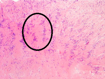
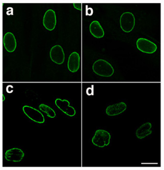
Verocay bodies were first described by Uruguayan neuro-pathologist José Juan Verocay (born: 16 June 1876, Nuevo Paysandú, Uruguay; died: 1927) in 1910. It is a required histopathological finding for diagnosing schwannomas. Verocay bodies are a component of "Antoni A" which are the dense areas of schwannomas located between palisading spindle cells found in neoplasms. Two nuclear palisading regions and an anuclear zone make up one Verocay body. Originally Verocay bodies were called 'neuromas', a term coined by Louis Odier in 1803. The name changed to ‘neuro-fibroma’ under Von Recklinghausen and later in 1935 to ‘neurilemmomas’ under Arthur Purdy Stout. When Harkin and Reed coined the term 'schwannoma' in 1968, Verocay bodies received their present-day name. Features on histopathological examination include: 1. Eosinophilic acellular area due to overexpression of lamin Lamins, also known as nuclear lamins are fibrous proteins in type V intermediate filaments, provid ... [...More Info...] [...Related Items...] OR: [Wikipedia] [Google] [Baidu] |
Antoni A Area Of Schwannoma With Verocay Bodies - Annotated
Antoni is a Catalan, Polish, and Slovene given name and a surname used in the eastern part of Spain, Poland and Slovenia. As a Catalan given name it is a variant of the male names Anton and Antonio. As a Polish given name it is a variant of the female names Antonia and Antonina. As a Slovene name it is a variant of the male names Anton, Antonij and Antonijo and the female name Antonija. As a surname it is derived from the Antonius root name. It may refer to: Given name * Antoni Brzeżańczyk, Polish football player and manager * Antoni Derezinski, Northern Irish Strongman * Antoni Gaudi, Catalan architect * Antoni Kenar, Polish sculptor * Antoni Lima, Catalan footballer * Antoni Lomnicki, Polish mathematician * Antoni Melchior Fijałkowski, Polish bishop * Antoni Niemczak, Polish long-distance runner * Józef Antoni Poniatowski, Polish prince and Marshal of France * Antoni Porowski, Polish-Canadian chef, actor, and television personality * Antoni Radziwiłł, Polish politician ... [...More Info...] [...Related Items...] OR: [Wikipedia] [Google] [Baidu] |
José Juan Verocay
José is a predominantly Spanish and Portuguese form of the given name Joseph. While spelled alike, this name is pronounced differently in each language: Spanish ; Portuguese (or ). In French, the name ''José'', pronounced , is an old vernacular form of Joseph, which is also in current usage as a given name. José is also commonly used as part of masculine name composites, such as José Manuel, José Maria or Antonio José, and also in female name composites like Maria José or Marie-José. The feminine written form is ''Josée'' as in French. In Netherlandic Dutch, however, ''José'' is a feminine given name and is pronounced ; it may occur as part of name composites like Marie-José or as a feminine first name in its own right; it can also be short for the name ''Josina'' and even a Dutch hypocorism of the name ''Johanna''. In England, Jose is originally a Romano-Celtic surname, and people with this family name can usually be found in, or traced to, the English county of C ... [...More Info...] [...Related Items...] OR: [Wikipedia] [Google] [Baidu] |
Histopathology
Histopathology (compound of three Greek words: ''histos'' "tissue", πάθος ''pathos'' "suffering", and -λογία '' -logia'' "study of") refers to the microscopic examination of tissue in order to study the manifestations of disease. Specifically, in clinical medicine, histopathology refers to the examination of a biopsy or surgical specimen by a pathologist, after the specimen has been processed and histological sections have been placed onto glass slides. In contrast, cytopathology examines free cells or tissue micro-fragments (as "cell blocks"). Collection of tissues Histopathological examination of tissues starts with surgery, biopsy, or autopsy. The tissue is removed from the body or plant, and then, often following expert dissection in the fresh state, placed in a fixative which stabilizes the tissues to prevent decay. The most common fixative is 10% neutral buffered formalin (corresponding to 3.7% w/v formaldehyde in neutral buffered water, such as phosphate buf ... [...More Info...] [...Related Items...] OR: [Wikipedia] [Google] [Baidu] |
Schwannoma
A schwannoma (or neurilemmoma) is a usually benign nerve sheath tumor composed of Schwann cells, which normally produce the insulating myelin sheath covering peripheral nerves. Schwannomas are homogeneous tumors, consisting only of Schwann cells. The tumor cells always stay on the outside of the nerve, but the tumor itself may either push the nerve aside and/or up against a bony structure (thereby possibly causing damage). Schwannomas are relatively slow-growing. For reasons not yet understood, schwannomas are mostly benign and less than 1% become malignant, degenerating into a form of cancer known as neurofibrosarcoma. These masses are generally contained within a capsule, so surgical removal is often successful. Schwannomas can be associated with neurofibromatosis type II, which may be due to a loss-of-function mutation in the protein merlin. They are universally S-100 positive, which is a marker for cells of neural crest cell origin. Schwannomas of the head and neck are a fa ... [...More Info...] [...Related Items...] OR: [Wikipedia] [Google] [Baidu] |
Neoplasm
A neoplasm () is a type of abnormal and excessive growth of tissue. The process that occurs to form or produce a neoplasm is called neoplasia. The growth of a neoplasm is uncoordinated with that of the normal surrounding tissue, and persists in growing abnormally, even if the original trigger is removed. This abnormal growth usually forms a mass, when it may be called a tumor. ICD-10 classifies neoplasms into four main groups: benign neoplasms, in situ neoplasms, malignant neoplasms, and neoplasms of uncertain or unknown behavior. Malignant neoplasms are also simply known as cancers and are the focus of oncology. Prior to the abnormal growth of tissue, as neoplasia, cells often undergo an abnormal pattern of growth, such as metaplasia or dysplasia. However, metaplasia or dysplasia does not always progress to neoplasia and can occur in other conditions as well. The word is from Ancient Greek 'new' and 'formation, creation'. Types A neoplasm can be benign, potentially ma ... [...More Info...] [...Related Items...] OR: [Wikipedia] [Google] [Baidu] |
Lamin
Lamins, also known as nuclear lamins are fibrous proteins in type V intermediate filaments, providing structural function and transcriptional regulation in the cell nucleus. Nuclear lamins interact with inner nuclear membrane proteins to form the nuclear lamina on the interior of the nuclear envelope. Lamins have elastic and mechanosensitive properties, and can alter gene regulation in a feedback response to mechanical cues. Lamins are present in all animals but are not found in microorganisms, plants or fungi. Lamin proteins are involved in the disassembling and reforming of the nuclear envelope during mitosis, the positioning of nuclear pores, and programmed cell death. Mutations in lamin genes can result in several genetic laminopathies, which may be life-threatening. History Lamins were first identified in the cell nucleus, using electron-microscopy. However, they were not recognized as vital components of nuclear structural support until 1975. During this time period, inv ... [...More Info...] [...Related Items...] OR: [Wikipedia] [Google] [Baidu] |
Basement Membrane
The basement membrane is a thin, pliable sheet-like type of extracellular matrix that provides cell and tissue support and acts as a platform for complex signalling. The basement membrane sits between Epithelium, epithelial tissues including mesothelium and endothelium, and the underlying connective tissue. Structure As seen with the electron microscope, the basement membrane is composed of two layers, the basal lamina and the reticular lamina. The underlying connective tissue attaches to the basal lamina with collagen VII anchoring fibrils and fibrillin microfibrils. The basal lamina layer can further be subdivided into two layers based on their visual appearance in electron microscopy. The lighter-colored layer closer to the epithelium is called the lamina lucida, while the denser-colored layer closer to the connective tissue is called the lamina densa. The Electron microscope, electron-dense lamina densa layer is about 30–70 nanometers thick and consists of an underlying ... [...More Info...] [...Related Items...] OR: [Wikipedia] [Google] [Baidu] |


