|
Vascular Space
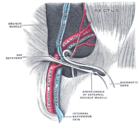
The vascular lacuna (Latin: ''lacuna vasorum'') is the compartment beneath the inguinal ligament which allows for passage of the femoral vessels, lymph vessels and lymph nodes. Its boundaries are the iliopectineal arch, the inguinal ligament, the lacunar ligament, and the superior border of the pubis. The structures found in the vascular lacuna, from medial to lateral, are: * Cloquet's node; * Femoral vein; * Femoral artery; and * Femoral branch of the genitofemoral nerve The vascular lacuna is separated from the muscular lacuna The muscular lacuna (Latin: ''lacuna musculorum'') is the lateral compartment of the thigh inferior to the inguinal ligament, for the passage of the iliopsoas muscle, the femoral nerve and the lateral cutaneous nerve of the thigh The lateral ... by the iliopectineal arch. The lacunar ligament can be a site of entrapment for femoral hernias.Ross, L.M., Lamperti, E.D. (2006). Thieme: Atlas of Anatomy: 489 References Muscular system ... [...More Info...] [...Related Items...] OR: [Wikipedia] [Google] [Baidu] |
Inguinal Ligament
The inguinal ligament (), also known as Poupart's ligament or groin ligament, is a band running from the pubic tubercle to the anterior superior iliac spine. It forms the base of the inguinal canal through which an indirect inguinal hernia may develop. Structure The inguinal ligament runs from the anterior superior iliac crest of the ilium to the pubic tubercle of the pubic bone. It is formed by the external abdominal oblique aponeurosis and is continuous with the fascia lata of the thigh. There is some dispute over the attachments. Structures that pass deep to the inguinal ligament include: *Psoas major, iliacus, pectineus *Femoral nerve, artery, and vein *Lateral cutaneous nerve of thigh *Lymphatics Function The ligament serves to contain soft tissues as they course anteriorly from the trunk to the lower extremity. This structure demarcates the superior border of the femoral triangle. It demarcates the inferior border of the inguinal triangle. The midpoint of the ingui ... [...More Info...] [...Related Items...] OR: [Wikipedia] [Google] [Baidu] |
Femoral Vessels
The femoral vessels are those blood vessels passing through the femoral ring into the femoral canal thereby passing down the length of the thigh until behind the knee. These large vessel are the: * Femoral artery (also known in this location as the common femoral artery) and * Femoral vein Lymphatic vessels found in the thigh aren’t usually included in this collective noun. As the blood vessels pass along the thigh, they branch, with their main branches remaining closely associated, where they are still referred to collectively as femoral vessels. The adjective femoral, in this case, relates to the thigh, which contains the femur. The relative position of these two large vessels is very important in medicine and surgery, because several medical interventions involve puncturing one or the other of them. Reliably distinguishing between them is therefore important. The location of the vessel is also used as an anatomical landmark for the femoral nerve The femoral nerve is a n ... [...More Info...] [...Related Items...] OR: [Wikipedia] [Google] [Baidu] |
Lymph Node
A lymph node, or lymph gland, is a kidney-shaped organ of the lymphatic system and the adaptive immune system. A large number of lymph nodes are linked throughout the body by the lymphatic vessels. They are major sites of lymphocytes that include B and T cells. Lymph nodes are important for the proper functioning of the immune system, acting as filters for foreign particles including cancer cells, but have no detoxification function. In the lymphatic system a lymph node is a secondary lymphoid organ. A lymph node is enclosed in a fibrous capsule and is made up of an outer cortex and an inner medulla. Lymph nodes become inflamed or enlarged in various diseases, which may range from trivial throat infections to life-threatening cancers. The condition of lymph nodes is very important in cancer staging, which decides the treatment to be used and determines the prognosis. Lymphadenopathy refers to glands that are enlarged or swollen. When inflamed or enlarged, lymph nodes can be ... [...More Info...] [...Related Items...] OR: [Wikipedia] [Google] [Baidu] |
Iliopectineal Arch
The Iliopectineal arch is a thickened band of fused iliac fascia and psoas fascia passing from the posterior aspect of the inguinal ligament anteriorly across the front of the femoral nerve to attach to the iliopubic eminence of the hip bone posteriorly. The iliopectineal arch thus forms a septum which subdivides the space deep to the inguinal ligament into a lateral muscular lacuna and a medial vascular lacuna. When a psoas minor muscle is present, its tendon of insertion blends with the iliopectineal arch It is sometimes transected in treatment of femoral nerve The femoral nerve is a nerve in the thigh that supplies skin on the upper thigh and inner leg, and the muscles that extend the knee. Structure The femoral nerve is the major nerve supplying the anterior compartment of the thigh. It is the largest ... entrapment. Additional Images File:Slide2gala.JPG, ILIOPECTINEAL ARCH.Deep dissection.Anterior view. References {{Authority control Pelvis Fascia ... [...More Info...] [...Related Items...] OR: [Wikipedia] [Google] [Baidu] |
Lacunar Ligament
The lacunar ligament, also named Gimbernat’s ligament, is a ligament in the inguinal region. It connects the inguinal ligament to the pectineal ligament, near the point where they both insert on the pubic tubercle. Structure The lacunar ligament is the part of the aponeurosis of the external oblique muscle that is reflected backward and laterally and is attached to the pectineal line of the pubis. It is about 1.25 cm. long, larger in the male than in the female, almost horizontal in direction in the erect posture, and of a triangular form with the base directed laterally. Its ''base'' is concave, thin, and sharp, and forms the medial boundary of the femoral ring. Its ''apex'' corresponds to the pubic tubercle. Its ''posterior margin'' is attached to the pectineal line, and is continuous with the pectineal ligament. Its ''anterior margin'' is attached to the inguinal ligament. Its surfaces are directed upward and downward. Clinical significance The lacunar ligament ... [...More Info...] [...Related Items...] OR: [Wikipedia] [Google] [Baidu] |
Cloquet's Node
Inguinal lymph nodes are lymph nodes in the human groin. Located in the femoral triangle of the inguinal region, they are grouped into superficial and deep lymph nodes. The superficial have three divisions: the superomedial, superolateral, and inferior superficial. Superficial inguinal lymph nodes * The superficial inguinal lymph nodes are the inguinal lymph nodes that form a chain immediately below the inguinal ligament. They lie deep to the fascia of Camper that overlies the femoral vessels at the medial aspect of the thigh. They are bounded superiorly by the inguinal ligament in the femoral triangle; laterally by the border of the sartorius muscle, and medially by the adductor longus muscle. They are divided into three groups: * inferior – inferior of the saphenous opening of the leg, receive drainage from lower legs * superolateral – on the side of the saphenous opening, receive drainage from the side buttocks and the lower abdominal wall. * superomedial – located a ... [...More Info...] [...Related Items...] OR: [Wikipedia] [Google] [Baidu] |
Femoral Vein
In the human body, the femoral vein is a blood vessel that accompanies the femoral artery in the femoral sheath. It begins at the adductor hiatus (an opening in the adductor magnus muscle) as the continuation of the popliteal vein. It ends at the inferior margin of the inguinal ligament where it becomes the external iliac vein. The femoral vein bears valves which are mostly bicuspid and whose number is variable between individuals and often between left and right leg. Structure Segments *The common femoral vein is the segment of the femoral vein between the branching point of the deep femoral vein and the inferior margin of the inguinal ligament.Page 590 in: *The subsartorial vein or superficial femoral vein are designations ... [...More Info...] [...Related Items...] OR: [Wikipedia] [Google] [Baidu] |
Femoral Artery
The femoral artery is a large artery in the thigh and the main arterial supply to the thigh and leg. The femoral artery gives off the deep femoral artery or profunda femoris artery and descends along the anteromedial part of the thigh in the femoral triangle. It enters and passes through the adductor canal, and becomes the popliteal artery as it passes through the adductor hiatus in the adductor magnus near the junction of the middle and distal thirds of the thigh. Structure The femoral artery enters the thigh from behind the inguinal ligament as the continuation of the external iliac artery. Here, it lies midway between the anterior superior iliac spine and the symphysis pubis (Mid-inguinal point). Segments In clinical parlance, the femoral artery has the following segments: *The common femoral artery (CFA) is the segment of the femoral artery between the inferior margin of the inguinal ligament and the branching point of the deep femoral artery/profunda femoris artery. Its ... [...More Info...] [...Related Items...] OR: [Wikipedia] [Google] [Baidu] |
Genitofemoral Nerve
The genitofemoral nerve refers to a nerve that is found in the abdomen. Its branches, the genital branch and femoral branch supply sensation to the upper anterior thigh, as well as the skin of the anterior scrotum in males and mons pubis in females. The femoral branch is different from the femoral nerve, which also arises from the lumbar plexus. Anatomy The genitofemoral nerve originates from the upper L1-2 segments of the lumbar plexus. It passes downwards, pierces the psoas major and emerges from its anterior surface. The nerve divides into two branches, the genital branch and the lumboinguinal nerve also known as the femoral branch, both of which then continue downwards and medially to the inguinal and femoral canal respectively. Genital Branch The genital branch continues downward on the surface of the psoas major muscle and then enters the inguinal canal through the deep inguinal ring. In men, the genital branch supplies the cremaster and scrotal skin. In women, the genit ... [...More Info...] [...Related Items...] OR: [Wikipedia] [Google] [Baidu] |
Muscular Lacuna
The muscular lacuna (Latin: ''lacuna musculorum'') is the lateral compartment of the thigh inferior to the inguinal ligament, for the passage of the iliopsoas muscle, the femoral nerve and the lateral cutaneous nerve of the thigh; it is separated by the iliopectineal arch from the vascular lacuna The vascular lacuna (Latin: ''lacuna vasorum'') is the compartment beneath the inguinal ligament which allows for passage of the femoral vessels, lymph vessels and lymph nodes. Its boundaries are the iliopectineal arch, the inguinal ligament, .... Muscular system {{musculoskeletal-stub ... [...More Info...] [...Related Items...] OR: [Wikipedia] [Google] [Baidu] |

.jpg)
