|
Vadasaurus Herzogi Skull
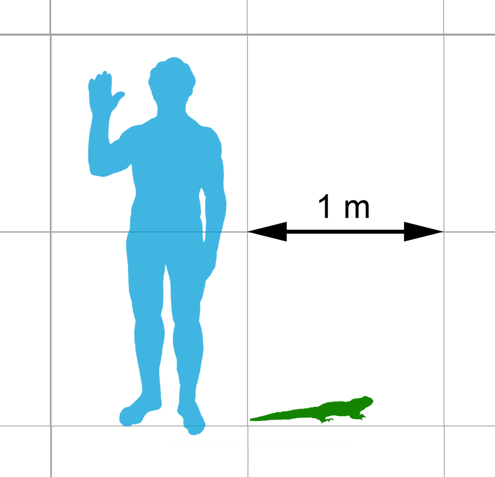
''Vadasaurus'' is an extinct genus of rhynchocephalian closely related to the aquatic pleurosaurids. Although this genus was not as specialized as the eel-like pleurosaurs for aquatic life, various skeletal features support the idea that it had a semiaquatic lifestyle. The type species, ''Vadasaurus herzogi'', was described and named in 2017. It was discovered in the Solnhofen Limestone in Germany, which is dated to the Late Jurassic. The generic name "''Vadasaurus"'' is derived from "''vadare''", which is Latin for "to go" or "to walk forth", and "''saurus''", which means "lizard" (although rhynchocephalians are not lizards). "''Vadare"'' is the root of the English word "wade", which is the reason it was chosen for this genus, in reference to its perceived semiaquatic habits. The specific name, "''herzogi''", refers to Werner Herzog, a Bavarian filmmaker. Description The holotype of ''Vadasaurus herzogi'' is AMNH FARB 32768, a well-preserved but slightly flattened skeleton ... [...More Info...] [...Related Items...] OR: [Wikipedia] [Google] [Baidu] |
Late Jurassic
The Late Jurassic is the third epoch of the Jurassic Period, and it spans the geologic time from 163.5 ± 1.0 to 145.0 ± 0.8 million years ago (Ma), which is preserved in Upper Jurassic strata.Owen 1987. In European lithostratigraphy, the name "Malm" indicates rocks of Late Jurassic age. In the past, ''Malm'' was also used to indicate the unit of geological time, but this usage is now discouraged to make a clear distinction between lithostratigraphic and geochronologic/chronostratigraphic units. Subdivisions The Late Jurassic is divided into three ages, which correspond with the three (faunal) stages of Upper Jurassic rock: Paleogeography During the Late Jurassic Epoch, Pangaea broke up into two supercontinents, Laurasia to the north, and Gondwana to the south. The result of this break-up was the spawning of the Atlantic Ocean. However, at this time, the Atlantic Ocean was relatively narrow. Life forms of the epoch This epoch is well known for many famous types of dinosau ... [...More Info...] [...Related Items...] OR: [Wikipedia] [Google] [Baidu] |
Holotype
A holotype is a single physical example (or illustration) of an organism, known to have been used when the species (or lower-ranked taxon) was formally described. It is either the single such physical example (or illustration) or one of several examples, but explicitly designated as the holotype. Under the International Code of Zoological Nomenclature (ICZN), a holotype is one of several kinds of name-bearing types. In the International Code of Nomenclature for algae, fungi, and plants (ICN) and ICZN, the definitions of types are similar in intent but not identical in terminology or underlying concept. For example, the holotype for the butterfly '' Plebejus idas longinus'' is a preserved specimen of that subspecies, held by the Museum of Comparative Zoology at Harvard University. In botany, an isotype is a duplicate of the holotype, where holotype and isotypes are often pieces from the same individual plant or samples from the same gathering. A holotype is not necessarily "typ ... [...More Info...] [...Related Items...] OR: [Wikipedia] [Google] [Baidu] |
Squamosal Bone
The squamosal is a skull bone found in most reptiles, amphibians, and birds. In fishes, it is also called the pterotic bone. In most tetrapods, the squamosal and quadratojugal bones form the cheek series of the skull. The bone forms an ancestral component of the dermal roof and is typically thin compared to other skull bones. The squamosal bone lies ventral to the temporal series and otic notch, and is bordered anteriorly by the postorbital. Posteriorly, the squamosal articulates with the quadrate and pterygoid bones. The squamosal is bordered anteroventrally by the jugal and ventrally by the quadratojugal. Function in reptiles In reptiles, the quadrate and articular bones of the skull articulate to form the jaw joint. The squamosal bone lies anterior to the quadrate bone. Anatomy in synapsids Non-mammalian synapsids In non-mammalian synapsids, the jaw is composed of four bony elements and referred to as a quadro-articular jaw because the joint is between the articular an ... [...More Info...] [...Related Items...] OR: [Wikipedia] [Google] [Baidu] |
Jugal Bone
The jugal is a skull bone found in most reptiles, amphibians and birds. In mammals, the jugal is often called the malar or zygomatic. It is connected to the quadratojugal and maxilla, as well as other bones, which may vary by species. Anatomy The jugal bone is located on either side of the skull in the circumorbital region. It is the origin of several masticatory muscles in the skull. The jugal and lacrimal bones are the only two remaining from the ancestral circumorbital series: the prefrontal, postfrontal, postorbital, jugal, and lacrimal bones. During development, the jugal bone originates from dermal bone. In dinosaurs This bone is considered key in the determination of general traits in cases in which the entire skull has not been found intact (for instance, as with dinosaurs in paleontology). In some dinosaur genera the jugal also forms part of the lower margin of either the antorbital fenestra or the infratemporal fenestra, or both. Most commonly, this bone articu ... [...More Info...] [...Related Items...] OR: [Wikipedia] [Google] [Baidu] |
Postorbital Bone
The ''postorbital'' is one of the bones in vertebrate skulls which forms a portion of the dermal skull roof and, sometimes, a ring about the orbit. Generally, it is located behind the postfrontal and posteriorly to the orbital fenestra. In some vertebrates, the postorbital is fused with the postfrontal to create a postorbitofrontal. Birds have a separate postorbital as an embryo, but the bone fuses with the frontal Front may refer to: Arts, entertainment, and media Films * ''The Front'' (1943 film), a 1943 Soviet drama film * ''The Front'', 1976 film Music * The Front (band), an American rock band signed to Columbia Records and active in the 1980s and e ... before it hatches. References * Roemer, A. S. 1956. ''Osteology of the Reptiles''. University of Chicago Press. 772 pp. Skull {{Vertebrate anatomy-stub ... [...More Info...] [...Related Items...] OR: [Wikipedia] [Google] [Baidu] |
Tuatara
Tuatara (''Sphenodon punctatus'') are reptiles endemic to New Zealand. Despite their close resemblance to lizards, they are part of a distinct lineage, the order Rhynchocephalia. The name ''tuatara'' is derived from the Māori language and means "peaks on the back". The single extant species of tuatara is the only surviving member of its order. Rhynchocephalians originated during the Triassic (~250 million years ago), reached worldwide distribution and peak diversity during the Jurassic and, with the exception of tuatara, were extinct by 60 million years ago. Their closest living relatives are squamates (lizards and snakes). For this reason, tuatara are of interest in the study of the evolution of lizards and snakes, and for the reconstruction of the appearance and habits of the earliest diapsids, a group of amniote tetrapods that also includes dinosaurs (including birds) and crocodilians. Tuatara are greenish brown and grey, and measure up to from head to tail-tip and wei ... [...More Info...] [...Related Items...] OR: [Wikipedia] [Google] [Baidu] |
Parietal Eye
A parietal eye, also known as a third eye or pineal eye, is a part of the epithalamus present in some vertebrates. The eye is located at the top of the head, is photoreceptive and is associated with the pineal gland, regulating circadian rhythmicity and hormone production for thermoregulation. The hole in the head which contains the eye is known as a pineal foramen or parietal foramen, since it is often enclosed by the parietal bones. Presence in various animals The parietal eye is found in the tuatara, most lizards, frogs, salamanders, certain bony fish, sharks, and lampreys. It is absent in mammals, but was present in their closest extinct relatives, the therapsids, suggesting it was lost during the course of the mammalian evolution due to it being useless in endothermic animals. It is also absent in the ancestrally endothermic ("warm-blooded") archosaurs such as birds. The parietal eye is also lost in ectothermic ("cold-blooded") archosaurs like crocodilians, and in turtles, ... [...More Info...] [...Related Items...] OR: [Wikipedia] [Google] [Baidu] |
Sagittal Crest
A sagittal crest is a ridge of bone running lengthwise along the midline of the top of the skull (at the sagittal suture) of many mammalian and reptilian skulls, among others. The presence of this ridge of bone indicates that there are exceptionally strong jaw muscles. The sagittal crest serves primarily for attachment of the temporalis muscle, which is one of the main chewing muscles. Development of the sagittal crest is thought to be connected to the development of this muscle. A sagittal crest usually develops during the juvenile stage of an animal in conjunction with the growth of the temporalis muscle, as a result of convergence and gradual heightening of the temporal lines. Function A sagittal crest tends to be present on the skulls of adult animals that rely on powerful biting and clenching of their teeth, usually as a part of their hunting strategy. Skulls of some dinosaur species, including tyrannosaurs, possessed well developed sagittal crests. Among mammals, dogs, cats, ... [...More Info...] [...Related Items...] OR: [Wikipedia] [Google] [Baidu] |
Parietal Bone
The parietal bones () are two bones in the Human skull, skull which, when joined at a fibrous joint, form the sides and roof of the Human skull, cranium. In humans, each bone is roughly quadrilateral in form, and has two surfaces, four borders, and four angles. It is named from the Latin ''paries'' (''-ietis''), wall. Surfaces External The external surface [Fig. 1] is convex, smooth, and marked near the center by an eminence, the parietal eminence (''tuber parietale''), which indicates the point where ossification commenced. Crossing the middle of the bone in an arched direction are two curved lines, the superior and inferior temporal lines; the former gives attachment to the temporal fascia, and the latter indicates the upper limit of the muscular origin of the temporal muscle. Above these lines the bone is covered by a tough layer of fibrous tissue – the epicranial aponeurosis; below them it forms part of the temporal fossa, and affords attachment to the temporal muscle. ... [...More Info...] [...Related Items...] OR: [Wikipedia] [Google] [Baidu] |
Frontal Bone
The frontal bone is a bone in the human skull. The bone consists of two portions.''Gray's Anatomy'' (1918) These are the vertically oriented squamous part, and the horizontally oriented orbital part, making up the bony part of the forehead, part of the bony orbital cavity holding the eye, and part of the bony part of the nose respectively. The name comes from the Latin word ''frons'' (meaning " forehead"). Structure of the frontal bone The frontal bone is made up of two main parts. These are the squamous part, and the orbital part. The squamous part marks the vertical, flat, and also the biggest part, and the main region of the forehead. The orbital part is the horizontal and second biggest region of the frontal bone. It enters into the formation of the roofs of the orbital and nasal cavities. Sometimes a third part is included as the nasal part of the frontal bone, and sometimes this is included with the squamous part. The nasal part is between the brow ridges, and ends in ... [...More Info...] [...Related Items...] OR: [Wikipedia] [Google] [Baidu] |
Nasal Bone
The nasal bones are two small oblong bones, varying in size and form in different individuals; they are placed side by side at the middle and upper part of the face and by their junction, form the bridge of the upper one third of the nose. Each has two surfaces and four borders. Structure The two nasal bones are joined at the midline internasal suture and make up the bridge of the nose. Surfaces The ''outer surface'' is concavo-convex from above downward, convex from side to side; it is covered by the procerus and nasalis muscles, and perforated about its center by a foramen, for the transmission of a small vein. The ''inner surface'' is concave from side to side, and is traversed from above downward, by a groove for the passage of a branch of the nasociliary nerve. Articulations The nasal articulates with four bones: two of the cranium, the frontal and ethmoid, and two of the face, the opposite nasal and the maxilla. Other animals In primitive bony fish and tetrapod ... [...More Info...] [...Related Items...] OR: [Wikipedia] [Google] [Baidu] |
Cusp (anatomy)
A cusp is a pointed, projecting, or elevated feature. In animals, it is usually used to refer to raised points on the crowns of teeth. The concept is also used with regard to the leaflets of the four heart valves. The mitral valve, which has two cusps, is also known as the bicuspid valve, and the tricuspid valve has three cusps. In humans A cusp is an occlusal or incisal eminence on a tooth. Canine teeth, otherwise known as cuspids, each possess a single cusp, while premolars, otherwise known as bicuspids, possess two each. Molars normally possess either four or five cusps. In certain populations the maxillary molars, especially first molars, will possess a fifth cusp situated on the mesiolingual cusp known as the Cusp of Carabelli. Buccal Cusp- One other variation of the upper first premolar is the 'Uto-Aztecan' upper premolar. It is a bulge on the buccal cusp that is only found in Native American Indians, with highest frequencies of occurrence in Arizona. The name is no ... [...More Info...] [...Related Items...] OR: [Wikipedia] [Google] [Baidu] |


.jpg)


.jpg)