|
Urotensin-II Receptor
The urotensin-2 receptor (UR-II-R) also known as GPR14 is a class A rhodopsin family G protein coupled-receptor (GPCR) that is 386 amino acids long which binds primarily to the neuropeptide urotensin II. /sup> The receptor quickly rose to prominence when it was found that when activated by urotensin II it induced the most potent vasoconstriction effect ever seen. While the precise function of the urotensin II receptor is not fully known it has been linked to cardiovascular effects, stress, and REM sleep. Ligands There are two known endogenous agonists for the urotensin II receptor. One is urotensin II whose mRNA is found in a variety of tissues including the brain and also blood vessels. It is a potent vasoconstrictor and can increase REM cycles. The other is urotensin II-Related Peptide (URP) which is found in a variety of tissues as well although at less concentrations then urotensin II. The one exception is in human reproductive tissue where the levels of URP are much highe ... [...More Info...] [...Related Items...] OR: [Wikipedia] [Google] [Baidu] |
Neuropeptide
Neuropeptides are chemical messengers made up of small chains of amino acids that are synthesized and released by neurons. Neuropeptides typically bind to G protein-coupled receptors (GPCRs) to modulate neural activity and other tissues like the gut, muscles, and heart. There are over 100 known neuropeptides, representing the largest and most diverse class of signaling molecules in the nervous system. Neuropeptides are synthesized from large precursor proteins which are cleaved and post-translationally processed then packaged into dense core vesicles. Neuropeptides are often co-released with other neuropeptides and neurotransmitters in a single neuron, yielding a multitude of effects. Once released, neuropeptides can diffuse widely to affect a broad range of targets. Synthesis Neuropeptides are synthesized from large, inactive precursor proteins called prepropeptides. Prepropeptides contain sequences for a family of distinct peptides and often contain repeated copies of the sa ... [...More Info...] [...Related Items...] OR: [Wikipedia] [Google] [Baidu] |
Laterodorsal Tegmental Nucleus
The laterodorsal tegmental nucleus (or lateroposterior tegmental nucleus) is a nucleus situated in the brainstem, spanning the midbrain tegmentum and the pontine tegmentum. Its location is one-third of the way from the pedunculopontine nucleus to the thalamus, inferior to the pineal gland. Function The laterodorsal tegmental nucleus (LDT) sends cholinergic (acetylcholine) projections to many subcortical and cortical structures, including the thalamus, hypothalamus, substantia nigra (dopamine neurons), ventral tegmental area (dopamine neurons), cortex (with bidirectional connections with the prefrontal cortex). The laterodorsal tegmental nucleus may be involved in modulating sustained attention or in mediating alerting responses, and also in the generation of REM sleep (along with the pedunculopontine nucleus The pedunculopontine nucleus (PPN) or pedunculopontine tegmental nucleus (PPT or PPTg) is a collection of neurons located in the upper pons in the brainstem. It lies caudal ... [...More Info...] [...Related Items...] OR: [Wikipedia] [Google] [Baidu] |
Intron
An intron is any nucleotide sequence within a gene that is not expressed or operative in the final RNA product. The word ''intron'' is derived from the term ''intragenic region'', i.e. a region inside a gene."The notion of the cistron .e., gene... must be replaced by that of a transcription unit containing regions which will be lost from the mature messenger – which I suggest we call introns (for intragenic regions) – alternating with regions which will be expressed – exons." (Gilbert 1978) The term ''intron'' refers to both the DNA sequence within a gene and the corresponding RNA sequence in RNA transcripts. The non-intron sequences that become joined by this RNA processing to form the mature RNA are called exons. Introns are found in the genes of most organisms and many viruses and they can be located in both protein-coding genes and genes that function as RNA (noncoding genes). There are four main types of introns: tRNA introns, group I introns, group II introns, and ... [...More Info...] [...Related Items...] OR: [Wikipedia] [Google] [Baidu] |
Ventricular Hypertrophy
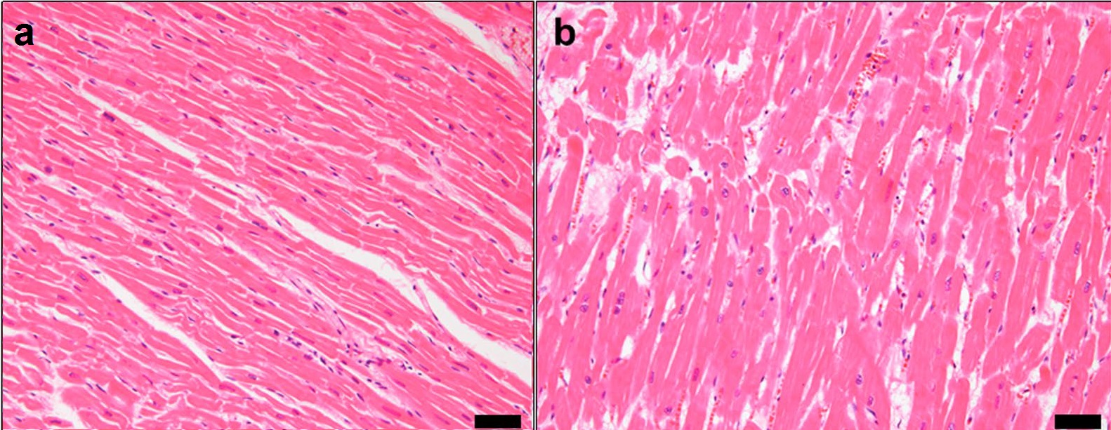
Ventricular hypertrophy (VH) is thickening of the walls of a ventricle (lower chamber) of the heart. Although left ventricular hypertrophy (LVH) is more common, right ventricular hypertrophy (RVH), as well as concurrent hypertrophy of both ventricles can also occur. Ventricular hypertrophy can result from a variety of conditions, both adaptive and maladaptive. For example, it occurs in what is regarded as a physiologic, adaptive process in pregnancy in response to increased blood volume; but can also occur as a consequence of ventricular remodeling following a heart attack. Importantly, pathologic and physiologic remodeling engage different cellular pathways in the heart and result in different gross cardiac phenotypes. Presentation In individuals with eccentric hypertrophy there may be little or no indication that hypertrophy has occurred as it is generally a healthy response to increased demands on the heart. Conversely, concentric hypertrophy can make itself known in a variety ... [...More Info...] [...Related Items...] OR: [Wikipedia] [Google] [Baidu] |
Aorta
The aorta ( ) is the main and largest artery in the human body, originating from the left ventricle of the heart and extending down to the abdomen, where it splits into two smaller arteries (the common iliac arteries). The aorta distributes oxygenated blood to all parts of the body through the systemic circulation. Structure Sections In anatomical sources, the aorta is usually divided into sections. One way of classifying a part of the aorta is by anatomical compartment, where the thoracic aorta (or thoracic portion of the aorta) runs from the heart to the diaphragm. The aorta then continues downward as the abdominal aorta (or abdominal portion of the aorta) from the diaphragm to the aortic bifurcation. Another system divides the aorta with respect to its course and the direction of blood flow. In this system, the aorta starts as the ascending aorta, travels superiorly from the heart, and then makes a hairpin turn known as the aortic arch. Following the aortic arch ... [...More Info...] [...Related Items...] OR: [Wikipedia] [Google] [Baidu] |
Slow Wave Sleep
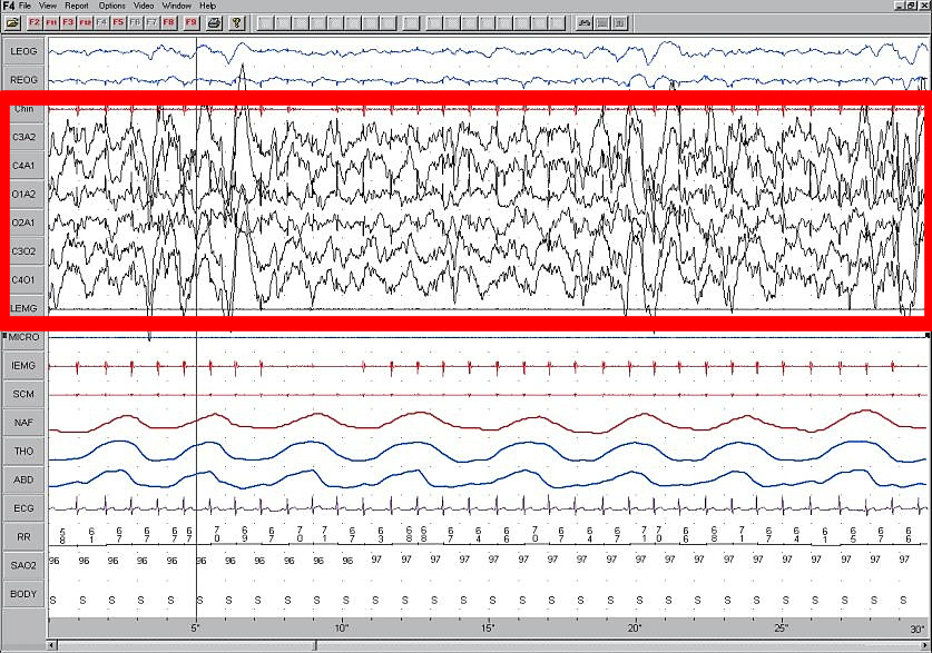
Slow-wave sleep (SWS), often referred to as deep sleep, consists of stage three of non-rapid eye movement sleep. It usually lasts between 70 and 90 minutes and takes place during the first hours of the night. Initially, SWS consisted of both Stage 3, which has 20–50 percent delta wave activity, and Stage 4, which has more than 50 percent delta wave activity. Overview This period of sleep is called slow-wave sleep because the EEG activity is synchronized, characterised by slow waves with a frequency range of 0.5–4.5 Hz, relatively high amplitude power with peak-to-peak amplitude greater than 75µV. The first section of the wave signifies a "down state", an inhibition or hyperpolarizing phase in which the neurons in the neocortex are silent. This is the period when the neocortical neurons are able to rest. The second section of the wave signifies an "up state", an excitation or depolarizing phase in which the neurons fire briefly at a high rate. The principal character ... [...More Info...] [...Related Items...] OR: [Wikipedia] [Google] [Baidu] |
Wakefulness
Wakefulness is a daily recurring Human brain, brain state and state of consciousness in which an individual is conscious and engages in coherent cognition, cognitive and behavioral responses to the external world. Being awake is the opposite of being asleep, in which most external inputs to the brain are excluded from neural processing. Effects upon the brain The longer the brain has been awake, the greater the synchronous firing rates of cerebral cortex neurons. After sustained periods of sleep, both the speed and synchronicity of the neurons firing are shown to decrease. Another effect of wakefulness is the reduction of glycogen held in the astrocytes, which supply energy to the neurons. Studies have shown that one of sleep's underlying functions is to replenish this glycogen energy source. Maintenance by the brain Wakefulness is produced by a complex interaction between multiple neurotransmitter systems arising in the brainstem and ascending through the midbrain, hypothalam ... [...More Info...] [...Related Items...] OR: [Wikipedia] [Google] [Baidu] |
Electrophysiology
Electrophysiology (from Greek , ''ēlektron'', "amber" etymology of "electron"">Electron#Etymology">etymology of "electron" , ''physis'', "nature, origin"; and , '' -logia'') is the branch of physiology that studies the electrical properties of biological cells and tissues. It involves measurements of voltage changes or electric current or manipulations on a wide variety of scales from single ion channel proteins to whole organs like the heart. In neuroscience, it includes measurements of the electrical activity of neurons, and, in particular, action potential activity. Recordings of large-scale electric signals from the nervous system, such as electroencephalography, may also be referred to as electrophysiological recordings. They are useful for electrodiagnosis and monitoring. Definition and scope Classical electrophysiological techniques Principle and mechanisms Electrophysiology is the branch of physiology that pertains broadly to the flow of ions (ion current) in biologi ... [...More Info...] [...Related Items...] OR: [Wikipedia] [Google] [Baidu] |
Hypothalamic–pituitary–adrenal Axis
The hypothalamic–pituitary–adrenal axis (HPA axis or HTPA axis) is a complex set of direct influences and feedback interactions among three components: the hypothalamus (a part of the brain located below the thalamus), the pituitary gland (a pea-shaped structure located below the hypothalamus), and the adrenal (also called "suprarenal") glands (small, conical organs on top of the kidneys). These organs and their interactions constitute the HPA axis. The HPA axis is a major neuroendocrine system that controls reactions to stress and regulates many body processes, including digestion, the immune system, mood and emotions, sexuality, and energy storage and expenditure. It is the common mechanism for interactions among glands, hormones, and parts of the midbrain that mediate the general adaptation syndrome (GAS). While steroid hormones are produced mainly in vertebrates, the physiological role of the HPA axis and corticosteroids in stress response is so fundamental that anal ... [...More Info...] [...Related Items...] OR: [Wikipedia] [Google] [Baidu] |
C-Fos
Protein c-Fos is a proto-oncogene that is the human homolog of the retroviral oncogene v-fos. It is encoded in humans by the ''FOS'' gene. It was first discovered in rat fibroblasts as the transforming gene of the FBJ MSV (Finkel–Biskis–Jinkins murine osteogenic sarcoma virus) (Curran and Tech, 1982). It is a part of a bigger Fos family of transcription factors which includes c-Fos, FosB, Fra-1 and Fra-2. It has been mapped to chromosome region 14q21→q31. c-Fos encodes a 62 kDa protein, which forms heterodimer with c-jun (part of Jun family of transcription factors), resulting in the formation of AP-1 (Activator Protein-1) complex which binds DNA at AP-1 specific sites at the promoter and enhancer regions of target genes and converts extracellular signals into changes of gene expression. It plays an important role in many cellular functions and has been found to be overexpressed in a variety of cancers. Structure and function c-Fos is a 380 amino acid protein with a basic ... [...More Info...] [...Related Items...] OR: [Wikipedia] [Google] [Baidu] |
Corticotropin
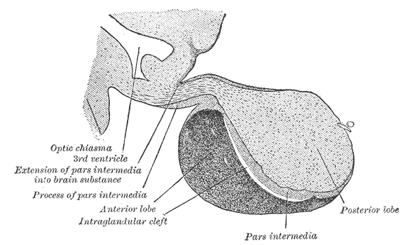
Adrenocorticotropic hormone (ACTH; also adrenocorticotropin, corticotropin) is a Peptide, polypeptide tropic hormone produced by and secreted by the Anterior pituitary, anterior pituitary gland. It is also used as a Adrenocorticotropic hormone (medication), medication and diagnostic agent. ACTH is an important component of the hypothalamic-pituitary-adrenal axis and is often produced in response to biological stress (along with its precursor corticotropin-releasing hormone from the hypothalamus). Its principal effects are increased production and release of cortisol by the Adrenal cortex, cortex of the adrenal gland. ACTH is also related to the circadian rhythm in many organisms. Deficiency of ACTH is an indicator of secondary adrenal insufficiency (suppressed production of ACTH due to an impairment of the pituitary gland or hypothalamus, cf. hypopituitarism) or tertiary adrenal insufficiency (disease of the hypothalamus, with a decrease in the release of corticotropin releasing ... [...More Info...] [...Related Items...] OR: [Wikipedia] [Google] [Baidu] |
Paraventricular Nucleus Of Hypothalamus
The paraventricular nucleus (PVN, PVA, or PVH) is a nucleus in the hypothalamus. Anatomically, it is adjacent to the third ventricle and many of its neurons project to the posterior pituitary. These projecting neurons secrete oxytocin and a smaller amount of vasopressin, otherwise the nucleus also secretes corticotropin-releasing hormone (CRH) and thyrotropin-releasing hormone (TRH). CRH and TRH are secreted into the hypophyseal portal system and act on different targets neurons in the anterior pituitary. PVN is thought to mediate many diverse functions through these different hormones, including osmoregulation, appetite, and the response of the body to stress. Location The paraventricular nucleus lies adjacent to the third ventricle. It lies within the periventricular zone and is not to be confused with the periventricular nucleus, which occupies a more medial position, beneath the third ventricle. The PVN is highly vascularised and is protected by the blood–brain barrier, althou ... [...More Info...] [...Related Items...] OR: [Wikipedia] [Google] [Baidu] |


