|
Ureterovesical Valve
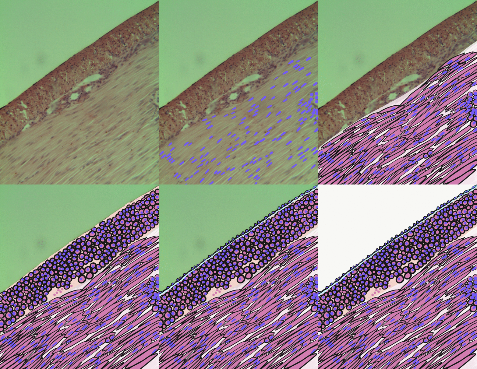
The ureters are tubes made of Smooth muscle tissue, smooth muscle that propel urine from the kidneys to the urinary bladder. In a human adult, the ureters are usually long and around in diameter. The ureter is lined by urothelium, urothelial cells, a type of transitional epithelium, and has an additional smooth muscle layer that assists with peristalsis in its lowest third. The ureters can be affected by a number of diseases, including urinary tract infections and kidney stone. is when a ureter is narrowed, due to for example chronic inflammation. Congenital abnormalities that affect the ureters can include the development of two ureters on the same side or abnormally placed ureters. Additionally, reflux of urine from the bladder back up the ureters is a condition commonly seen in children. The ureters have been identified for at least two thousand years, with the word "ureter" stemming from the stem relating to urinating and seen in written records since at least the time o ... [...More Info...] [...Related Items...] OR: [Wikipedia] [Google] [Baidu] |
Urine
Urine is a liquid by-product of metabolism in humans and in many other animals. Urine flows from the kidneys through the ureters to the urinary bladder. Urination results in urine being excretion, excreted from the body through the urethra. Cell (biology), Cellular metabolism generates many by-products that are rich in nitrogen and must be clearance (medicine), cleared from the Circulatory system, bloodstream, such as urea, uric acid, and creatinine. These by-products are expelled from the body during urination, which is the primary method for excreting water-soluble chemicals from the body. A urinalysis can detect nitrogenous wastes of the mammalian body. Urine plays an important role in the earth's nitrogen cycle. In balanced ecosystems, urine fertilizes the soil and thus helps plants to grow. Therefore, Reuse of excreta, urine can be used as a fertilizer. Some animals use it to territory (animal)#Scent marking, mark their territories. Historically, aged or fermented urine (kn ... [...More Info...] [...Related Items...] OR: [Wikipedia] [Google] [Baidu] |
Hippocrates
Hippocrates of Kos (; grc-gre, Ἱπποκράτης ὁ Κῷος, Hippokrátēs ho Kôios; ), also known as Hippocrates II, was a Greek physician of the classical period who is considered one of the most outstanding figures in the history of medicine. He is traditionally referred to as the "Father of Medicine" in recognition of his lasting contributions to the field, such as the use of prognosis and clinical observation, the systematic categorization of diseases, or the formulation of humoral theory. The Hippocratic school of medicine revolutionized ancient Greek medicine, establishing it as a discipline distinct from other fields with which it had traditionally been associated (theurgy and philosophy), thus establishing medicine as a profession. However, the achievements of the writers of the Hippocratic Corpus, the practitioners of Hippocratic medicine, and the actions of Hippocrates himself were often conflated; thus very little is known about what Hippocrates actually t ... [...More Info...] [...Related Items...] OR: [Wikipedia] [Google] [Baidu] |
Peritoneum
The peritoneum is the serous membrane forming the lining of the abdominal cavity or coelom in amniotes and some invertebrates, such as annelids. It covers most of the intra-abdominal (or coelomic) organs, and is composed of a layer of mesothelium supported by a thin layer of connective tissue. This peritoneal lining of the cavity supports many of the abdominal organs and serves as a conduit for their blood vessels, lymphatic vessels, and nerves. The abdominal cavity (the space bounded by the vertebrae, abdominal muscles, diaphragm, and pelvic floor) is different from the intraperitoneal space (located within the abdominal cavity but wrapped in peritoneum). The structures within the intraperitoneal space are called "intraperitoneal" (e.g., the stomach and intestines), the structures in the abdominal cavity that are located behind the intraperitoneal space are called "retroperitoneal" (e.g., the kidneys), and those structures below the intraperitoneal space are called "subp ... [...More Info...] [...Related Items...] OR: [Wikipedia] [Google] [Baidu] |
Psoas Major Muscle
The psoas major ( or ; from grc, ψόᾱ, psóā, muscles of the loins) is a long fusiform muscle located in the lateral lumbar region between the vertebral column and the brim of the lesser pelvis. It joins the iliacus muscle to form the iliopsoas. In animals, this muscle is equivalent to the tenderloin. Structure The psoas major is divided into a superficial and a deep part. The deep part originates from the transverse processes of lumbar vertebrae L1–L5. The superficial part originates from the lateral surfaces of the last thoracic vertebra, lumbar vertebrae L1–L4, and the neighboring intervertebral discs. The lumbar plexus lies between the two layers. Together, the iliacus muscle and the psoas major form the iliopsoas, which is surrounded by the iliac fascia. The iliopsoas runs across the iliopubic eminence through the muscular lacuna to its insertion on the lesser trochanter of the femur. The iliopectineal bursa separates the tendon of the iliopsoas muscle from ... [...More Info...] [...Related Items...] OR: [Wikipedia] [Google] [Baidu] |
Internal Urethral Orifice
The internal urethral orifice is the opening of the urinary bladder into the urethra. It is placed at the apex of the trigonum vesicae, in the most dependent part of the bladder, and is usually somewhat crescent-shaped; the mucous membrane immediately behind it presents a slight elevation in males, the uvula vesicae, caused by the middle lobe of the prostate. See also * Internal sphincter muscle of urethra The internal urethral sphincter is a urethral sphincter muscle which constricts the internal urethral orifice. It is located at the junction of the urethra with the urinary bladder and is continuous with the detrusor muscle, but anatomically and ... References External links * - "The Male Pelvis: The Urethra" Urinary system Urethra {{genitourinary-stub ... [...More Info...] [...Related Items...] OR: [Wikipedia] [Google] [Baidu] |
Smooth Muscle
Smooth muscle is an involuntary non-striated muscle, so-called because it has no sarcomeres and therefore no striations (''bands'' or ''stripes''). It is divided into two subgroups, single-unit and multiunit smooth muscle. Within single-unit muscle, the whole bundle or sheet of smooth muscle cells contracts as a syncytium. Smooth muscle is found in the walls of hollow organs, including the stomach, intestines, bladder and uterus; in the walls of passageways, such as blood, and lymph vessels, and in the tracts of the respiratory, urinary, and reproductive systems. In the eyes, the ciliary muscles, a type of smooth muscle, dilate and contract the iris and alter the shape of the lens. In the skin, smooth muscle cells such as those of the arrector pili cause hair to stand erect in response to cold temperature or fear. Structure Gross anatomy Smooth muscle is grouped into two types: single-unit smooth muscle, also known as visceral smooth muscle, and multiunit smooth muscle. ... [...More Info...] [...Related Items...] OR: [Wikipedia] [Google] [Baidu] |
Common Iliac Artery
The common iliac artery is a large artery of the abdomen paired on each side. It originates from the aortic bifurcation at the level of the 4th lumbar vertebra. It ends in front of the sacroiliac joint, one on either side, and each bifurcates into the external and internal iliac arteries. Structure The common iliac artery are about 4 cm long in adults and more than a centimeter in diameter. It begins as a branch of the aorta. This is at the level of the 4th lumbar vertebra. It runs inferolaterally, along the medial border of the psoas muscles. It bifurcates into the external iliac artery and the internal iliac artery at the pelvic brim, in front of the sacroiliac joints. The common iliac artery, and all of its branches, exist as paired structures (that is to say, there is one on the left side and one on the right). The distribution of the common iliac artery is basically the pelvis and lower limb (as the femoral artery) on the corresponding side. Relations Both common il ... [...More Info...] [...Related Items...] OR: [Wikipedia] [Google] [Baidu] |
Pelvic Brim
The pelvic brim is the edge of the pelvic inlet. It is an approximately Mickey Mouse head-shaped line passing through the prominence of the sacrum, the arcuate and pectineal lines, and the upper margin of the pubic symphysis. Structure The pelvic brim is an approximately Mickey Mouse head-shaped line passing through the prominence of the sacrum, the arcuate and pectineal lines, and the upper margin of the pubic symphysis. The pelvic brim is obtusely pointed in front, diverging on either side, and encroached upon behind by the projection forward of the promontory of the sacrum. The oblique plane passing approximately through the pelvic brim divides the internal part of the pelvis (pelvic cavity) into the false or greater pelvis and the true or lesser pelvis. The false pelvis, which is above that plane, is sometimes considered to be a part of the abdominal cavity, rather than a part of the pelvic cavity. In this case, the pelvic cavity coincides with the true pelvis, which is b ... [...More Info...] [...Related Items...] OR: [Wikipedia] [Google] [Baidu] |
Psoas Major
The psoas major ( or ; from grc, ψόᾱ, psóā, muscles of the loins) is a long fusiform muscle located in the lateral lumbar region between the vertebral column and the brim of the lesser pelvis. It joins the iliacus muscle to form the iliopsoas. In animals, this muscle is equivalent to the tenderloin. Structure The psoas major is divided into a superficial and a deep part. The deep part originates from the transverse processes of lumbar vertebrae L1–L5. The superficial part originates from the lateral surfaces of the last thoracic vertebra, lumbar vertebrae L1–L4, and the neighboring intervertebral discs. The lumbar plexus lies between the two layers. Together, the iliacus muscle and the psoas major form the iliopsoas, which is surrounded by the iliac fascia. The iliopsoas runs across the iliopubic eminence through the muscular lacuna to its insertion on the lesser trochanter of the femur. The iliopectineal bursa separates the tendon of the iliopsoas muscle from th ... [...More Info...] [...Related Items...] OR: [Wikipedia] [Google] [Baidu] |
Renal Pelvis
The renal pelvis or pelvis of the kidney is the funnel-like dilated part of the ureter in the kidney. It is formed by the covnvergence of the major calyces, acting as a funnel for urine flowing from the major calyces to the ureter. It has a mucous membrane and is covered with transitional epithelium and an underlying lamina propria of loose-to-dense connective tissue. The renal pelvis is situated within the renal sinus alongside the other structures of the renal sinus. The renal pelvis is the location of several kinds of kidney cancer and is affected by infection in pyelonephritis. Clinical significance The renal pelvis is the location of several kinds of kidney cancer and is affected by infection in pyelonephritis. A large "staghorn" kidney stone may block all or part of the renal pelvis. The size of the renal pelvis plays a major role in the grading of hydronephrosis. Normally, the anteroposterior diameter of the renal pelvis is less than 4 mm in fetuses up to 32 weeks ... [...More Info...] [...Related Items...] OR: [Wikipedia] [Google] [Baidu] |
Ureteroscopy
Ureteroscopy is an examination of the upper urinary tract, usually performed with a ureteroscope that is passed through the urethra and the bladder, and then directly into the ureter. The procedure is useful in the diagnosis and treatment of disorders such as kidney stones and urothelial carcinoma of the upper urinary tract. Smaller stones in the bladder or lower ureter can be removed in one piece, while bigger ones are usually broken before removal during ureteroscopy. The examination may be performed with either a flexible, semi-rigid or rigid device while the patient is under anesthesia. In specific cases, the patient is free to go home after the examination.Ureteropyeloscopy. Baylor College of Medicine. 2018 ccessed 2018 Mar 5br>/ref> In pyeloscopy, the endoscope is designed to reach all the way to the renal pelvis The renal pelvis or pelvis of the kidney is the funnel-like dilated part of the ureter in the kidney. It is formed by the covnvergence of the major calyces, acti ... [...More Info...] [...Related Items...] OR: [Wikipedia] [Google] [Baidu] |
Urinary Tract Ultrasound
Ultrasound of the urinary tract involves the use of an ultrasound probe to image parts of the urinary tract. The urinary tract is the path that urine follows after it is formed within the kidneys, and involves a left and right ureter, the bladder, and the urethra. Procedure Ultrasound involves a probe that is placed near a structure and uses the transmission of ultrasound waves through a structure to produce images, after computer processing. Ultrasound of the urinary tract is performed by a probe placed on the abdominal wall, called a transducer. This occurs while a person is lying down. It is then moved around to better visualise different parts of the urinary tract. Gel is used on the abdominal wall, allowing smooth movement and improve sound conduction. The images are usually taken by a trained ultrasonographer, and then reported by a specialist radiologist. Prior to the test, a medical practitioner will have conducted a medical history to evaluate for symptoms that may rel ... [...More Info...] [...Related Items...] OR: [Wikipedia] [Google] [Baidu] |
