|
Urachal Cyst
A urachal cyst is a sinus remaining from the allantois during embryogenesis. It is a cyst which occurs in the remnants between the umbilicus and bladder. This is a type of cyst occurring in a persistent portion of the urachus, presenting as an extraperitoneal mass in the umbilical region. It is characterized by abdominal pain, and fever if infected. It may rupture, leading to peritonitis, or it may drain through the umbilicus. Urachal cysts are usually silent clinically until infection, calculi or adenocarcinoma develop. Symptoms and signs * Lower abdominal pain * Pain on urination * Persistent umbilical discharge * Fever * Urinary tract infection * Lump * Hematuria Diagnosis Urachal cysts are rare defects found mostly in young children and hence medical ultrasound of the abdomen, bladder and pelvis is the most used diagnostic tool combined with MRI scan and CT scan in older patients who can remain still during a scan. See also * urachus * cyst A cyst is a closed sac, havi ... [...More Info...] [...Related Items...] OR: [Wikipedia] [Google] [Baidu] |
Sinus (anatomy)
A sinus is a sac or cavity in any organ or tissue, or an abnormal cavity or passage caused by the destruction of tissue. In common usage, "sinus" usually refers to the paranasal sinuses, which are air cavities in the cranial bones, especially those near the nose and connecting to it. Most individuals have four paired cavities located in the cranial bone or skull. Etymology ''Sinus'' is Latin for "bay", "pocket", "curve", or "bosom". In anatomy, the term is used in various contexts. The word "sinusitis" is used to indicate that one or more of the membrane linings found in the sinus cavities has become inflamed or infected. It is however distinct from a fistula, which is a tract connecting two epithelial surfaces. If left untreated, infections occurring in the sinus cavities can affect the chest and lungs. Sinuses in the body * Paranasal sinuses ** Maxillary ** Ethmoid ** Sphenoid ** Frontal * Dural venous sinuses ** Anterior midline *** Cavernous *** Superior petrosal *** I ... [...More Info...] [...Related Items...] OR: [Wikipedia] [Google] [Baidu] |
Calculus (medicine)
A calculus (plural calculi), often called a stone, is a concretion of material, usually mineral salts, that forms in an organ or duct of the body. Formation of calculi is known as lithiasis (). Stones can cause a number of medical conditions. Some common principles (below) apply to stones at any location, but for specifics see the particular stone type in question. Calculi are not to be confused with gastroliths. Types * Calculi in the urinary system are called urinary calculi and include kidney stones (also called renal calculi or nephroliths) and bladder stones (also called vesical calculi or cystoliths). They can have any of several compositions, including mixed. Principal compositions include oxalate and urate. * Calculi of the gallbladder and bile ducts are called gallstones and are primarily developed from bile salts and cholesterol derivatives. * Calculi in the nasal passages (rhinoliths) are rare. * Calculi in the gastrointestinal tract ( enteroliths) can be enormous. In ... [...More Info...] [...Related Items...] OR: [Wikipedia] [Google] [Baidu] |
CT Scan
A computed tomography scan (CT scan; formerly called computed axial tomography scan or CAT scan) is a medical imaging technique used to obtain detailed internal images of the body. The personnel that perform CT scans are called radiographers or radiology technologists. CT scanners use a rotating X-ray tube and a row of detectors placed in a gantry (medical), gantry to measure X-ray Attenuation#Radiography, attenuations by different tissues inside the body. The multiple X-ray measurements taken from different angles are then processed on a computer using tomographic reconstruction algorithms to produce Tomography, tomographic (cross-sectional) images (virtual "slices") of a body. CT scans can be used in patients with metallic implants or pacemakers, for whom magnetic resonance imaging (MRI) is Contraindication, contraindicated. Since its development in the 1970s, CT scanning has proven to be a versatile imaging technique. While CT is most prominently used in medical diagnosis, ... [...More Info...] [...Related Items...] OR: [Wikipedia] [Google] [Baidu] |
Magnetic Resonance Imaging
Magnetic resonance imaging (MRI) is a medical imaging technique used in radiology to form pictures of the anatomy and the physiological processes of the body. MRI scanners use strong magnetic fields, magnetic field gradients, and radio waves to generate images of the organs in the body. MRI does not involve X-rays or the use of ionizing radiation, which distinguishes it from CT and PET scans. MRI is a medical application of nuclear magnetic resonance (NMR) which can also be used for imaging in other NMR applications, such as NMR spectroscopy. MRI is widely used in hospitals and clinics for medical diagnosis, staging and follow-up of disease. Compared to CT, MRI provides better contrast in images of soft-tissues, e.g. in the brain or abdomen. However, it may be perceived as less comfortable by patients, due to the usually longer and louder measurements with the subject in a long, confining tube, though "Open" MRI designs mostly relieve this. Additionally, implants and oth ... [...More Info...] [...Related Items...] OR: [Wikipedia] [Google] [Baidu] |
Medical Ultrasound
Medical ultrasound includes diagnostic techniques (mainly imaging techniques) using ultrasound, as well as therapeutic applications of ultrasound. In diagnosis, it is used to create an image of internal body structures such as tendons, muscles, joints, blood vessels, and internal organs, to measure some characteristics (e.g. distances and velocities) or to generate an informative audible sound. Its aim is usually to find a source of disease or to exclude pathology. The usage of ultrasound to produce visual images for medicine is called medical ultrasonography or simply sonography. The practice of examining pregnant women using ultrasound is called obstetric ultrasonography, and was an early development of clinical ultrasonography. Ultrasound is composed of sound waves with frequencies which are significantly higher than the range of human hearing (>20,000 Hz). Ultrasonic images, also known as sonograms, are created by sending pulses of ultrasound into tissue using a pr ... [...More Info...] [...Related Items...] OR: [Wikipedia] [Google] [Baidu] |
Hematuria
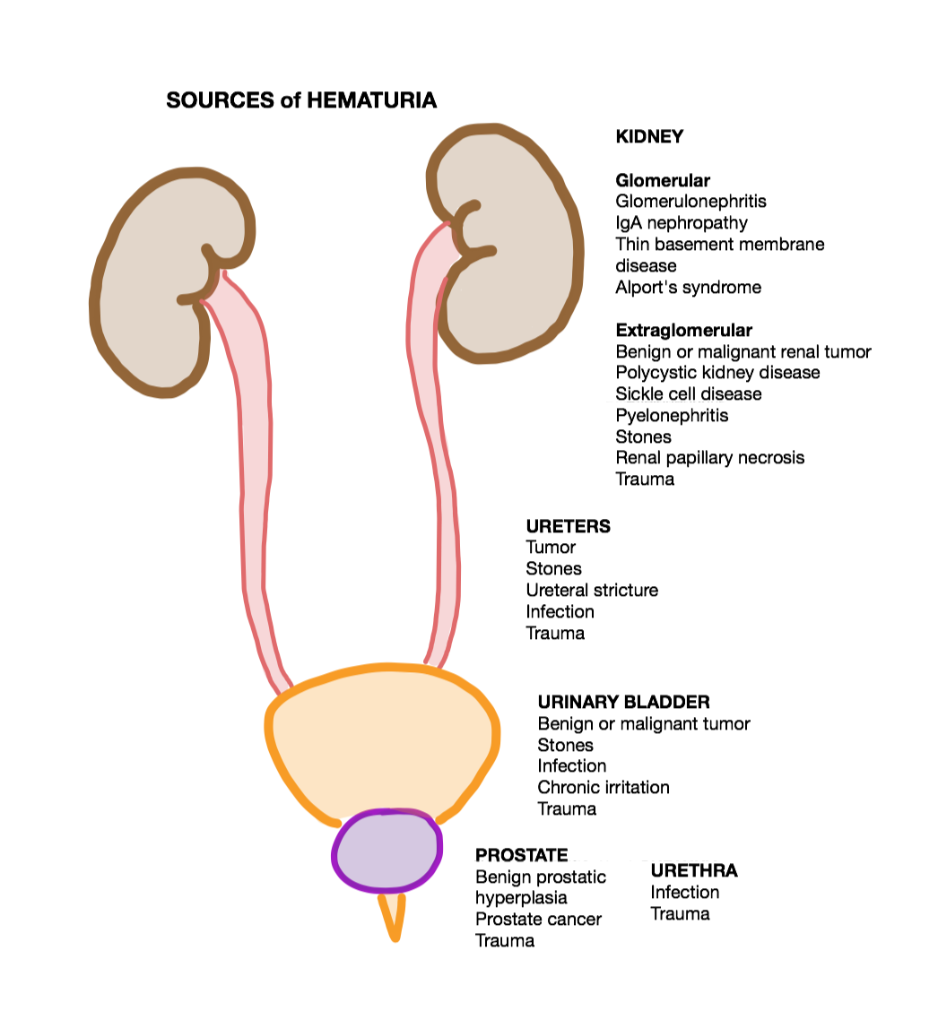
Hematuria or haematuria is defined as the presence of blood or red blood cells in the urine. “Gross hematuria” occurs when urine appears red, brown, or tea-colored due to the presence of blood. Hematuria may also be subtle and only detectable with a microscope or laboratory test. Blood that enters and mixes with the urine can come from any location within the urinary system, including the kidney, ureter, urinary bladder, urethra, and in men, the prostate. Common causes of hematuria include urinary tract infection (UTI), kidney stones, viral illness, trauma, bladder cancer, and exercise. These causes are grouped into glomerular and non-glomerular causes, depending on the involvement of the glomerulus of the kidney. But not all red urine is hematuria. Other substances such as certain medications and foods (e.g. blackberries, beets, food dyes) can cause urine to appear red. Menstruation in women may also cause the appearance of hematuria and may result in a positive urine dipstick ... [...More Info...] [...Related Items...] OR: [Wikipedia] [Google] [Baidu] |
Urinary Tract Infection
A urinary tract infection (UTI) is an infection that affects part of the urinary tract. When it affects the lower urinary tract it is known as a bladder infection (cystitis) and when it affects the upper urinary tract it is known as a kidney infection (pyelonephritis). Symptoms from a lower urinary tract infection include pain with urination, frequent urination, and feeling the need to urinate despite having an empty bladder. Symptoms of a kidney infection include fever and flank pain usually in addition to the symptoms of a lower UTI. Rarely the urine may appear bloody. In the very old and the very young, symptoms may be vague or non-specific. The most common cause of infection is ''Escherichia coli'', though other bacteria or fungi may sometimes be the cause. Risk factors include female anatomy, sexual intercourse, diabetes, obesity, and family history. Although sexual intercourse is a risk factor, UTIs are not classified as sexually transmitted infections (STIs). Kidney ... [...More Info...] [...Related Items...] OR: [Wikipedia] [Google] [Baidu] |
General Electric
General Electric Company (GE) is an American multinational conglomerate founded in 1892, and incorporated in New York state and headquartered in Boston. The company operated in sectors including healthcare, aviation, power, renewable energy, digital industry, additive manufacturing and venture capital and finance, but has since divested from several areas, now primarily consisting of the first four segments. In 2020, GE ranked among the Fortune 500 as the 33rd largest firm in the United States by gross revenue. In 2011, GE ranked among the Fortune 20 as the 14th most profitable company, but later very severely underperformed the market (by about 75%) as its profitability collapsed. Two employees of GE – Irving Langmuir (1932) and Ivar Giaever (1973) – have been awarded the Nobel Prize. On November 9, 2021, the company announced it would divide itself into three investment-grade public companies. On July 18, 2022, GE unveiled the brand names of the companies it will ... [...More Info...] [...Related Items...] OR: [Wikipedia] [Google] [Baidu] |
Adenocarcinoma
Adenocarcinoma (; plural adenocarcinomas or adenocarcinomata ) (AC) is a type of cancerous tumor that can occur in several parts of the body. It is defined as neoplasia of epithelial tissue that has glandular origin, glandular characteristics, or both. Adenocarcinomas are part of the larger grouping of carcinomas, but are also sometimes called by more precise terms omitting the word, where these exist. Thus invasive ductal carcinoma, the most common form of breast cancer, is adenocarcinoma but does not use the term in its name—however, esophageal adenocarcinoma does to distinguish it from the other common type of esophageal cancer, esophageal squamous cell carcinoma. Several of the most common forms of cancer are adenocarcinomas, and the various sorts of adenocarcinoma vary greatly in all their aspects, so that few useful generalizations can be made about them. In the most specific usage (narrowest sense), the glandular origin or traits are exocrine; endocrine gland tumors, suc ... [...More Info...] [...Related Items...] OR: [Wikipedia] [Google] [Baidu] |
Peritonitis
Peritonitis is inflammation of the localized or generalized peritoneum, the lining of the inner wall of the abdomen and cover of the abdominal organs. Symptoms may include severe pain, swelling of the abdomen, fever, or weight loss. One part or the entire abdomen may be tender. Complications may include shock and acute respiratory distress syndrome. Causes include perforation of the intestinal tract, pancreatitis, pelvic inflammatory disease, stomach ulcer, cirrhosis, or a ruptured appendix. Risk factors include ascites (the abnormal build-up of fluid in the abdomen) and peritoneal dialysis. Diagnosis is generally based on examination, blood tests, and medical imaging. Treatment often includes antibiotics, intravenous fluids, pain medication, and surgery. Other measures may include a nasogastric tube or blood transfusion. Without treatment death may occur within a few days. About 20% of people with cirrhosis who are hospitalized have peritonitis. Signs and symptoms Abd ... [...More Info...] [...Related Items...] OR: [Wikipedia] [Google] [Baidu] |
Allantois
The allantois (plural ''allantoides'' or ''allantoises'') is a hollow sac-like structure filled with clear fluid that forms part of a developing amniote's conceptus (which consists of all embryonic and extraembryonic tissues). It helps the embryo exchange gases and handle liquid waste. The allantois, along with the amnion, chorion, and yolk sac (other extraembryonic membranes), identify humans and other mammals, birds, and other reptiles as amniotes. Fish and amphibians are anamniotes, and lack the allantois. In mammals the extraembryonic membranes are known as the fetal membranes. Function This sac-like structure, whose name is the New Latin equivalent of "sausage" (in reference to its shape when first formed) is primarily involved in nutrition and excretion, and is webbed with blood vessels. The function of the allantois is to collect liquid waste from the embryo, as well as to exchange gases used by the embryo. In mammals In mammals excluding egg-laying monotremes, the all ... [...More Info...] [...Related Items...] OR: [Wikipedia] [Google] [Baidu] |
Navel
The navel (clinically known as the umbilicus, commonly known as the belly button or tummy button) is a protruding, flat, or hollowed area on the abdomen at the attachment site of the umbilical cord. All placental mammals have a navel, although it is generally more conspicuous in humans. Structure The umbilicus is used to visually separate the abdomen into quadrants. The umbilicus is a prominent scar on the abdomen, with its position being relatively consistent among humans. The skin around the waist at the level of the umbilicus is supplied by the tenth thoracic spinal nerve (T10 dermatome). The umbilicus itself typically lies at a vertical level corresponding to the junction between the L3 and L4 vertebrae, with a normal variation among people between the L3 and L5 vertebrae. Parts of the adult navel include the "umbilical cord remnant" or "umbilical tip", which is the often protruding scar left by the detachment of the umbilical cord. This is located in the center of the ... [...More Info...] [...Related Items...] OR: [Wikipedia] [Google] [Baidu] |


