|
Tubuli Seminiferi Recti
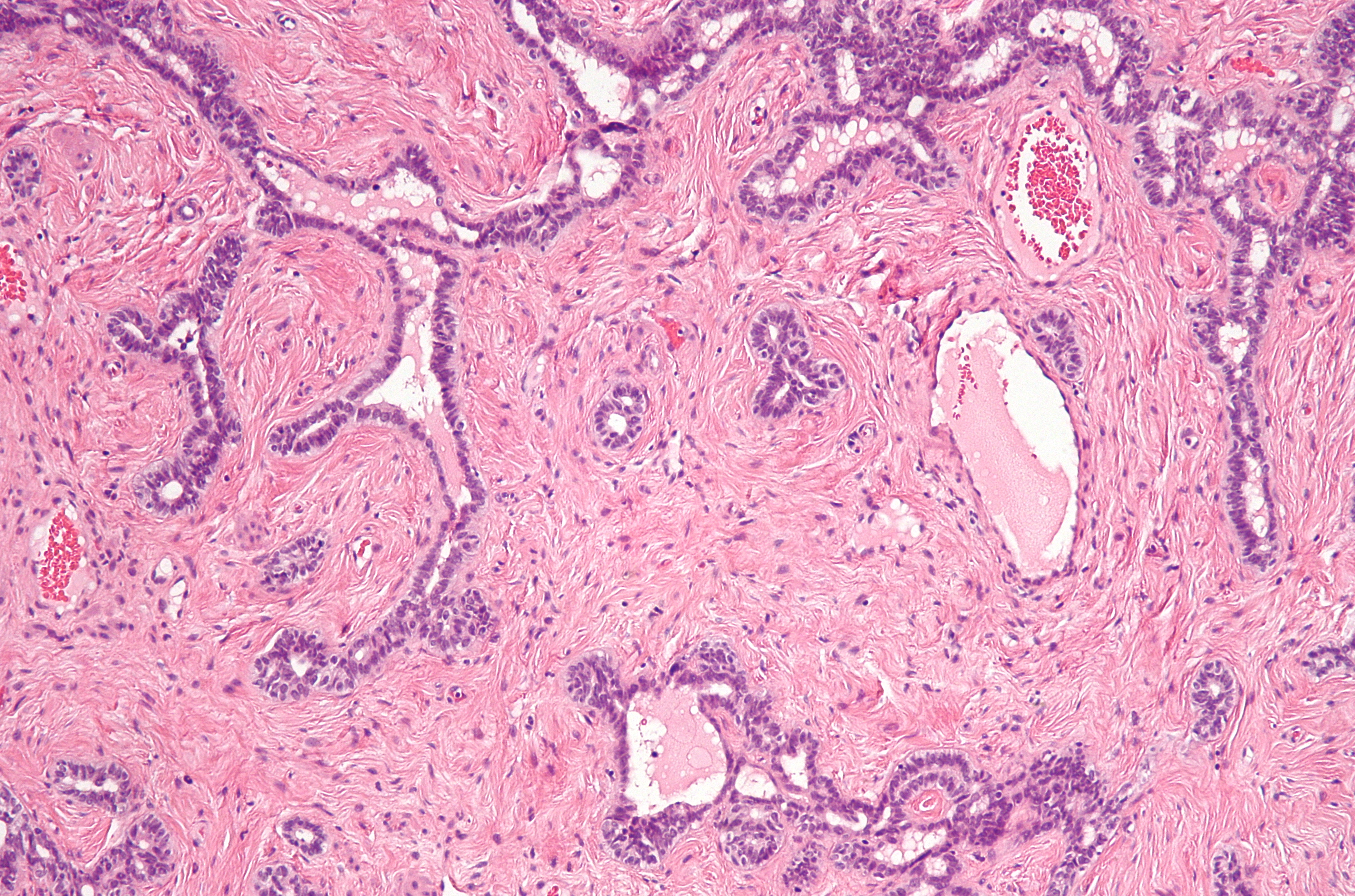
The tubuli seminiferi recti (also known as the tubuli recti, tubulus rectus, or straight seminiferous tubules) are structures in the testicle connecting the convoluted region of the seminiferous tubules to the rete testis, although the tubuli recti have a different appearance distinguishing them from these two structures. They enter the fibrous tissue of the mediastinum, and pass upward and backward, forming, in their ascent, a close network of anastomosing tubes which are merely channels in the fibrous stroma, lined by flattened epithelium, and having no proper walls; this constitutes the rete testis The rete testis ( ) is an anastomosing network of delicate tubules located in the hilum of the testicle (mediastinum testis) that carries sperm from the seminiferous tubules to the efferent ducts. It is the counterpart of the rete ovarii in females .... Only Sertoli cells line the terminal ends of the seminiferous tubules (tubuli recti). References Mammal male reproductive ... [...More Info...] [...Related Items...] OR: [Wikipedia] [Google] [Baidu] |
Tunica Albuginea Of Testis
The tunica albuginea is the fibrous tissue covering of the testis. It is a dense blue-grey membrane, composed of bundles of white fibrous connective tissue, from which it derives its name '' albuginea'', which interlace in every direction. Structure The tunica albuginea is a layer of fibrous tissue capsule covering the testis. It is covered by the tunica vaginalis, except at the points of attachment of the epididymis to the testis, and along its posterior border, where the spermatic vessels enter the gland. It is thicker than the tunica albuginea of the ovary. The tunica albuginea is applied to the tunica vasculosa over the glandular substance of the testis, and, at its posterior border, is reflected into the interior of the gland, forming an incomplete vertical septum, called the mediastinum testis (corpus Highmori). Additional images File:Gray1145.png, Transverse section through the left side of the scrotum and the left testis. File:Gray1114.png, Section of a genital cor ... [...More Info...] [...Related Items...] OR: [Wikipedia] [Google] [Baidu] |
Convoluted Seminiferous Tubules
Seminiferous tubules are located within the testes, and are the specific location of meiosis, and the subsequent creation of male gametes, namely spermatozoa. Structure The epithelium of the tubule consists of a type of sustentacular cells known as Sertoli cells, which are tall, columnar type cells that line the tubule. In between the Sertoli cells are spermatogenic cells, which differentiate through meiosis to sperm cells. Sertoli cells function to nourish the developing sperm cells. They secrete androgen-binding protein, a binding protein which increases the concentration of testosterone. There are two types: convoluted and straight, convoluted toward the lateral side, and straight as the tubule comes medially to form ducts that will exit the testis. The seminiferous tubules are formed from the testis cords that develop from the primitive gonadal cords, formed from the gonadal ridge. Function Spermatogenesis, the process for producing spermatozoa, takes place ... [...More Info...] [...Related Items...] OR: [Wikipedia] [Google] [Baidu] |
Stroma (animal Tissue)
Stroma () is the part of a tissue or organ with a structural or connective role. It is made up of all the parts without specific functions of the organ - for example, connective tissue, blood vessels, ducts, etc. The other part, the parenchyma, consists of the cells that perform the function of the tissue or organ. There are multiple ways of classifying tissues: one classification scheme is based on tissue functions and another analyzes their cellular components. Stromal tissue falls into the "functional" class that contributes to the body's support and movement. The cells which make up stroma tissues serve as a matrix in which the other cells are embedded. Stroma is made of various types of stromal cells. Examples of stroma include: * stroma of iris * stroma of cornea * stroma of ovary * stroma of thyroid gland * stroma of thymus * stroma of bone marrow * lymph node stromal cell * multipotent stromal cell (mesenchymal stem cell) Structure Stromal connective tissues are foun ... [...More Info...] [...Related Items...] OR: [Wikipedia] [Google] [Baidu] |
Anastomosing
An anastomosis (, plural anastomoses) is a connection or opening between two things (especially cavities or passages) that are normally diverging or branching, such as between blood vessels, leaf veins, or streams. Such a connection may be normal (such as the foramen ovale in a fetus's heart) or abnormal (such as the patent foramen ovale in an adult's heart); it may be acquired (such as an arteriovenous fistula) or innate (such as the arteriovenous shunt of a metarteriole); and it may be natural (such as the aforementioned examples) or artificial (such as a surgical anastomosis). The reestablishment of an anastomosis that had become blocked is called a reanastomosis. Anastomoses that are abnormal, whether congenital or acquired, are often called fistulas. The term is used in medicine, biology, mycology, geology, and geography. Etymology Anastomosis: medical or Modern Latin, from Greek ἀναστόμωσις, anastomosis, "outlet, opening", Gr ana- "up, on, upon", stoma "mouth", ... [...More Info...] [...Related Items...] OR: [Wikipedia] [Google] [Baidu] |
Mediastinum
The mediastinum (from ) is the central compartment of the thoracic cavity. Surrounded by loose connective tissue, it is an undelineated region that contains a group of structures within the thorax, namely the heart and its vessels, the esophagus, the trachea, the phrenic nerve, phrenic and cardiac nerves, the thoracic duct, the thymus and the lymph nodes of the central chest. Anatomy The mediastinum lies within the thorax and is enclosed on the right and left by pulmonary pleurae, pleurae. It is surrounded by the chest wall in front, the lungs to the sides and the Spine (anatomy), spine at the back. It extends from the sternum in front to the vertebral column behind. It contains all the organs of the thorax except the lungs. It is continuous with the loose connective tissue of the neck. The mediastinum can be divided into an upper (or superior) and lower (or inferior) part: * The superior mediastinum starts at the superior thoracic aperture and ends at the #Thoracic plane, t ... [...More Info...] [...Related Items...] OR: [Wikipedia] [Google] [Baidu] |
Rete Testis
The rete testis ( ) is an anastomosing network of delicate tubules located in the hilum of the testicle (mediastinum testis) that carries sperm from the seminiferous tubules to the efferent ducts. It is the counterpart of the rete ovarii in females. Its function is to provide a site for fluid reabsorption. Structure The rete testis is the network of interconnecting tubules where the straight seminiferous tubules (the terminal part of the seminiferous tubules) empty. It is located within a highly vascular connective tissue in the mediastinum testis. The epithelial cells form a single layer that lines the inner surface of the tubules. These cells are cuboidal, with microvilli and a single cilium on their surface. Development In the development of the urinary and reproductive organs, the testis is developed in much the same way as the ovary, originating from mesothelium as well as mesonephros. Like the ovary, in its earliest stages it consists of a central mass covered by a surfac ... [...More Info...] [...Related Items...] OR: [Wikipedia] [Google] [Baidu] |
Seminiferous Tubules
Seminiferous tubules are located within the testes, and are the specific location of meiosis, and the subsequent creation of male gametes, namely spermatozoa. Structure The epithelium of the tubule consists of a type of sustentacular cells known as Sertoli cells, which are tall, columnar type cells that line the tubule. In between the Sertoli cells are spermatogenic cells, which differentiate through meiosis to sperm cells. Sertoli cells function to nourish the developing sperm cells. They secrete androgen-binding protein, a binding protein which increases the concentration of testosterone. There are two types: convoluted and straight, convoluted toward the lateral side, and straight as the tubule comes medially to form ducts that will exit the testis. The seminiferous tubules are formed from the testis cords that develop from the primitive gonadal cords, formed from the gonadal ridge. Function Spermatogenesis, the process for producing spermatozoa, takes place in the semin ... [...More Info...] [...Related Items...] OR: [Wikipedia] [Google] [Baidu] |
Testicle
A testicle or testis (plural testes) is the male reproductive gland or gonad in all bilaterians, including humans. It is homologous to the female ovary. The functions of the testes are to produce both sperm and androgens, primarily testosterone. Testosterone release is controlled by the anterior pituitary luteinizing hormone, whereas sperm production is controlled both by the anterior pituitary follicle-stimulating hormone and gonadal testosterone. Structure Appearance Males have two testicles of similar size contained within the scrotum, which is an extension of the abdominal wall. Scrotal asymmetry, in which one testicle extends farther down into the scrotum than the other, is common. This is because of the differences in the vasculature's anatomy. For 85% of men, the right testis hangs lower than the left one. Measurement and volume The volume of the testicle can be estimated by palpating it and comparing it to ellipsoids of known sizes. Another method is to use caliper ... [...More Info...] [...Related Items...] OR: [Wikipedia] [Google] [Baidu] |
Efferent Ductules
The efferent ducts (or efferent ductules or ductuli efferentes or ductus efferentes or vasa efferentia) connect the rete testis with the initial section of the epididymis.Hess 2018 There are two basic designs for efferent ductule structure: * a) multiple entries into the epididymis, as seen in most large mammals. In humans and other large mammals, there are approximately 15 to 20 efferent ducts, which also occupy nearly one third of the head of the epididymis. * b) single entry, as seen in most small animals such as rodent Rodents (from Latin , 'to gnaw') are mammals of the order Rodentia (), which are characterized by a single pair of continuously growing incisors in each of the upper and lower jaws. About 40% of all mammal species are rodents. They are n ...s, where by the 3–6 ductules merge into a single small ductule prior to entering the epididymis. The ductuli are unilaminar and composed of columnar ciliated and non-ciliated (absorptive) cells. The ciliate ... [...More Info...] [...Related Items...] OR: [Wikipedia] [Google] [Baidu] |
Testicular Lobules
The glandular structure of the testis consists of numerous lobules. Their number, in a single testis, is estimated by Berres at 250, and by Krause at 400. Anatomic studies have demonstrated figures of 250–290 for the same. They differ in size according to their position, those in the middle of the gland being larger and longer. The lobules are conical in shape, the base being directed toward the circumference of the organ, the apex toward the mediastinum testis. Each lobule is contained in one of the intervals between the fibrous septa which extend between the mediastinum testis and the tunica albuginea, and consists of from one to three, or more, minute convoluted tubes, the tubuli seminiferi Seminiferous tubules are located within the testes, and are the specific location of meiosis, and the subsequent creation of male gametes, namely spermatozoa. Structure The epithelium of the tubule consists of a type of sustentacular cells known .... Additional images File:Gray11 ... [...More Info...] [...Related Items...] OR: [Wikipedia] [Google] [Baidu] |
Testicular Septa
Each Lobules of testis, lobule of the testis is contained in one of the intervals between the fibrous septa which extend between the mediastinum testis and the Tunica albuginea of testis, tunica albuginea, and consists of from one to three, or more, minute convoluted tubes, the tubuli seminiferi. Additional images File:Gray1145.png, Transverse section through the left side of the scrotum and the left testis. File:Gray1149.png, Vertical section of the testis, to show the arrangement of the ducts. References External links * - "Inguinal Region, Scrotum and Testes: The Cross-Section of the Testis" Mammal male reproductive system {{genitourinary-stub ... [...More Info...] [...Related Items...] OR: [Wikipedia] [Google] [Baidu] |
Septula Testis
Each lobule of the testis is contained in one of the intervals between the fibrous septa which extend between the mediastinum testis and the tunica albuginea, and consists of from one to three, or more, minute convoluted tubes, the tubuli seminiferi Seminiferous tubules are located within the testes, and are the specific location of meiosis, and the subsequent creation of male gametes, namely spermatozoa. Structure The epithelium of the tubule consists of a type of sustentacular cells known .... Additional images File:Gray1145.png, Transverse section through the left side of the scrotum and the left testis. File:Gray1149.png, Vertical section of the testis, to show the arrangement of the ducts. References External links * - "Inguinal Region, Scrotum and Testes: The Cross-Section of the Testis" Mammal male reproductive system {{genitourinary-stub ... [...More Info...] [...Related Items...] OR: [Wikipedia] [Google] [Baidu] |


