|
Transversalis Fascia
The transversalis fascia (or transverse fascia) is a thin aponeurotic membrane of the abdomen. It lies between the inner surface of the transverse abdominal muscle and the parietal peritoneum. It forms part of the general layer of fascia lining the abdominal parietes. It is directly continuous with the iliac fascia, the internal spermatic fascia, and pelvic fasciae. Structure In the inguinal region, the transversalis fascia is thick and dense. It is joined by fibers from the aponeurosis of the transverse abdominal muscle. It becomes thin as it ascends to the diaphragm and blends with the fascia covering the under surface of this muscle. It is directly continuous with the iliac fascia, the internal spermatic fascia, and pelvic fasciae. Borders Behind, it is lost in the fat which covers the posterior surfaces of the kidneys. Below, it has the following attachments: posteriorly, to the whole length of the iliac crest, between the attachments of the transverse abdominal an ... [...More Info...] [...Related Items...] OR: [Wikipedia] [Google] [Baidu] |
Abdominal Inguinal Ring
The inguinal canals are the two passages in the Anatomical terms of location#Anterior and posterior, anterior abdominal wall of humans and animals which in males convey the spermatic cords and in females the round ligament of the uterus. The inguinal canals are larger and more prominent in males. There is one inguinal canal on each side of the Anatomical terms of location#Planes, midline. Structure The inguinal canals are situated just above the medial half of the inguinal ligament. In both sexes the canals transmit the ilioinguinal nerves. The canals are approximately 3.75 to 4 cm long. , angled anteroinferiorly and medially. In males, its diameter is normally 2 cm (±1 cm in standard deviation) at the deep inguinal ring.The diameter has been estimated to be ±2.2cm ±1.08cm in Africans, and 2.1 cm ±0.41cm in Europeans. A first-order approximation is to visualize each canal as a cylinder. Walls To help define the boundaries, these canals are often further approximated as b ... [...More Info...] [...Related Items...] OR: [Wikipedia] [Google] [Baidu] |
Femoral Vessel
The femoral vessels are those blood vessels passing through the femoral ring into the femoral canal thereby passing down the length of the thigh until behind the knee. These large vessel are the: * Femoral artery (also known in this location as the common femoral artery) and * Femoral vein Lymphatic vessels found in the thigh aren’t usually included in this collective noun. As the blood vessels pass along the thigh, they branch, with their main branches remaining closely associated, where they are still referred to collectively as femoral vessels. The adjective femoral, in this case, relates to the thigh, which contains the femur. The relative position of these two large vessels is very important in medicine and surgery, because several medical interventions involve puncturing one or the other of them. Reliably distinguishing between them is therefore important. The location of the vessel is also used as an anatomical landmark for the femoral nerve The femoral nerve is a n ... [...More Info...] [...Related Items...] OR: [Wikipedia] [Google] [Baidu] |
Inguinal Canal
The inguinal canals are the two passages in the anterior abdominal wall of humans and animals which in males convey the spermatic cords and in females the round ligament of the uterus. The inguinal canals are larger and more prominent in males. There is one inguinal canal on each side of the midline. Structure The inguinal canals are situated just above the medial half of the inguinal ligament. In both sexes the canals transmit the ilioinguinal nerves. The canals are approximately 3.75 to 4 cm long. , angled anteroinferiorly and medially. In males, its diameter is normally 2 cm (±1 cm in standard deviation) at the deep inguinal ring.The diameter has been estimated to be ±2.2cm ±1.08cm in Africans, and 2.1 cm ±0.41cm in Europeans. A first-order approximation is to visualize each canal as a cylinder. Walls To help define the boundaries, these canals are often further approximated as boxes with six sides. Not including the two rings, the remaining four sides are usually ca ... [...More Info...] [...Related Items...] OR: [Wikipedia] [Google] [Baidu] |
Churchill Livingstone
Churchill Livingstone is an academic publisher. It was formed in 1971 from the merger of Longman's medical list, E & S Livingstone (Edinburgh, Scotland) and J & A Churchill (London, England) and was owned by Pearson. Harcourt acquired Churchill Livingstone in 1997. It is now integrated as an imprint in Elsevier's health science division after Elsevier acquired Harcourt in 2001. In the past it published a number of classic medical texts, including Sir William Osler's textbook '' The Principles and Practice of Medicine, Gray's Anatomy,'' and ''Myles In Greek mythology, Myles (; Ancient Greek: Μύλης means 'mill-man') was an ancient king of Laconia. He was the son of the King Lelex and possibly the naiad Queen Cleocharia, and brother of Polycaon. Myles was the father of Eurotas who begott ...' Textbook for Midwives.'' In the 1980s, in addition to new texts in all areas of clinical medicine, it published an extensive list of medical and nursing textbooks in low-cost editions ... [...More Info...] [...Related Items...] OR: [Wikipedia] [Google] [Baidu] |
Inguinal Canal
The inguinal canals are the two passages in the anterior abdominal wall of humans and animals which in males convey the spermatic cords and in females the round ligament of the uterus. The inguinal canals are larger and more prominent in males. There is one inguinal canal on each side of the midline. Structure The inguinal canals are situated just above the medial half of the inguinal ligament. In both sexes the canals transmit the ilioinguinal nerves. The canals are approximately 3.75 to 4 cm long. , angled anteroinferiorly and medially. In males, its diameter is normally 2 cm (±1 cm in standard deviation) at the deep inguinal ring.The diameter has been estimated to be ±2.2cm ±1.08cm in Africans, and 2.1 cm ±0.41cm in Europeans. A first-order approximation is to visualize each canal as a cylinder. Walls To help define the boundaries, these canals are often further approximated as boxes with six sides. Not including the two rings, the remaining four sides are usually ca ... [...More Info...] [...Related Items...] OR: [Wikipedia] [Google] [Baidu] |
Deep Inguinal Ring
The inguinal canals are the two passages in the anterior abdominal wall of humans and animals which in males convey the spermatic cords and in females the round ligament of the uterus. The inguinal canals are larger and more prominent in males. There is one inguinal canal on each side of the midline. Structure The inguinal canals are situated just above the medial half of the inguinal ligament. In both sexes the canals transmit the ilioinguinal nerves. The canals are approximately 3.75 to 4 cm long. , angled anteroinferiorly and medially. In males, its diameter is normally 2 cm (±1 cm in standard deviation) at the deep inguinal ring.The diameter has been estimated to be ±2.2cm ±1.08cm in Africans, and 2.1 cm ±0.41cm in Europeans. A first-order approximation is to visualize each canal as a cylinder. Walls To help define the boundaries, these canals are often further approximated as boxes with six sides. Not including the two rings, the remaining four sides are usually cal ... [...More Info...] [...Related Items...] OR: [Wikipedia] [Google] [Baidu] |
Round Ligament Of The Uterus
The round ligament of the uterus is a ligament that connects the uterus to the labia majora. Structure The round ligament of the uterus originates at the uterine horns, in the parametrium. The round ligament exits the pelvis via the deep inguinal ring. It passes through the inguinal canal, and continues on to the labia majora. At the labia majora, its fibers spread and mix with the tissue of the mons pubis. Development The round ligament develops from the gubernaculum which attaches the gonad to the labioscrotal swellings in the embryo. Blood supply The round ligament is supplied by the artery of the round ligament of uterus, also known as ''Sampson's artery''. Function The function of the round ligament is maintenance of the anteversion of the uterus (a position where the fundus of the uterus is turned forward at the junction of cervix and vagina) during pregnancy. Normally, the cardinal ligament is what supports the uterine angle (angle of anteversion). When the uterus gro ... [...More Info...] [...Related Items...] OR: [Wikipedia] [Google] [Baidu] |
Spermatic Cord
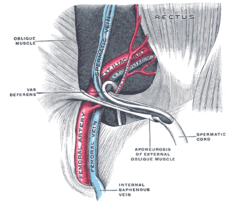
The spermatic cord is the cord-like structure in males formed by the vas deferens (''ductus deferens'') and surrounding tissue that runs from the deep inguinal ring down to each testicle. Its serosal covering, the tunica vaginalis, is an extension of the peritoneum that passes through the transversalis fascia. Each testicle develops in the lower thoracic and upper lumbar region and migrates into the scrotum. During its descent it carries along with it the vas deferens, its vessels, nerves etc. There is one on each side. Structure The spermatic cord is ensheathed in three layers of tissue: * ''external spermatic fascia'', an extension of the innominate fascia that overlies the aponeurosis of the external oblique muscle. * ''cremasteric muscle and fascia'', formed from a continuation of the internal oblique muscle and its fascia. * ''internal spermatic fascia'', continuous with the transversalis fascia. The normal diameter of the spermatic cord is about 16 mm (range 11 to 22 mm). It ... [...More Info...] [...Related Items...] OR: [Wikipedia] [Google] [Baidu] |
Iliopubic Tract
The iliopubic tract is a thickened band of fibers curving over the external iliac vessels, at the spot where they become femoral, on the abdominal side of the inguinal ligaments and loosely connected with it. It is apparently a thickening of the transversalis fascia joined laterally to the iliac crest, and arching across the front of the femoral sheath to be inserted by a broad attachment into the pubic tubercle and pectineal line, behind the conjoint tendon. In some subjects this structure is not very prominently marked, and not infrequently it is altogether wanting. It can be of clinical significance in hernia repair Hernia repair refers to a surgical operation for the correction of a hernia—a bulging of internal organs or tissues through the wall that contains it. It can be of two different types: herniorrhaphy; or hernioplasty. This operation may be perfo .... References External links Iliopubic Tract Hernia Repair Images at vesalius.com Abdomen {{muscle-stub ... [...More Info...] [...Related Items...] OR: [Wikipedia] [Google] [Baidu] |
Femoral Sheath
The femoral sheath (also called the crural sheath) is a funnel-shaped downward extension of abdominal fascia within which the femoral artery and femoral vein pass between the abdomen and the thigh. The femoral sheath is subdivided by two vertical partitions to form three compartments (medial, intermediate, and lateral); the medial compartment is known as the femoral canal and contains lymphatic vessels and a lymph node, whereas the intermediate canal and the lateral canal accommodate the femoral vein and the femoral artery (respectively). Some neurovascular structures perforate the femoral sheath. Topographically, the femoral sheath is contained within in the femoral triangle. Structure The femoral sheath is funnel-shaped fascial structure, with the wide end directed superior-ward. The femoral sheath is formed by an inferior-ward prolongation - posterior to the inguinal ligament - of abdominal fascia, with transverse fascia being continued down anterior to the femoral vessels, ... [...More Info...] [...Related Items...] OR: [Wikipedia] [Google] [Baidu] |
Inguinal Falx
The conjoint tendon (previously known as the inguinal aponeurotic falx) is a sheath of connective tissue formed from the lower part of the common aponeurosis of the abdominal internal oblique muscle and the transversus abdominis muscle, joining the muscle to the pelvis. It forms the medial part of the posterior wall of the inguinal canal. Structure The conjoint tendon is formed from the lower part of the common aponeurosis of the abdominal internal oblique muscle and the transversus abdominis muscle. It inserts into the pubic crest and the pectineal line immediately behind the superficial inguinal ring. It is usually conjoint with the tendon of the internal oblique muscle, but they may be separate as well. It forms the medial part of the posterior wall of the inguinal canal. Clinical significance The conjoint tendon serves to protect what would otherwise be a weak point in the abdominal wall. A weakening of the conjoint tendon can precipitate a direct inguinal hernia. A direct ... [...More Info...] [...Related Items...] OR: [Wikipedia] [Google] [Baidu] |
Pectineal Line (pubis)
The pectineal line of the pubis (also pecten pubis) is a ridge on the superior ramus of the pubic bone. It forms part of the pelvic brim. Lying across from the pectineal line are fibers of the pectineal ligament, and the proximal origin of the pectineus muscle. In combination with the arcuate line, it makes the iliopectineal line The iliopectineal line is the border of the iliopubic eminence. It can be defined as a compound structure of the arcuate line (from the ilium) and pectineal line (from the pubis). With the sacral promontory, it makes up the linea terminalis The .... References External links * () {{Authority control Bones of the pelvis Pubis (bone) ... [...More Info...] [...Related Items...] OR: [Wikipedia] [Google] [Baidu] |

