|
Tooth Germ
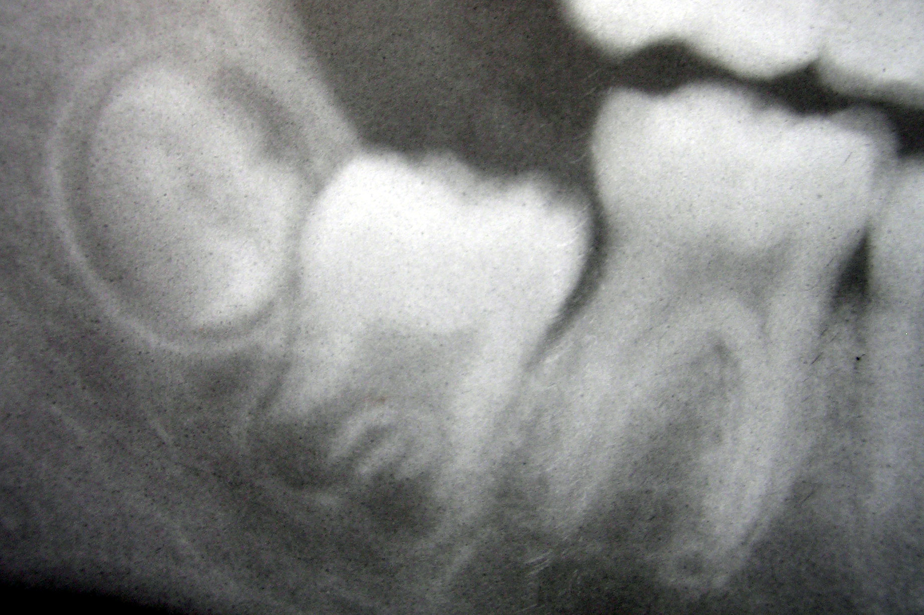
Tooth development or odontogenesis is the complex process by which teeth form from embryonic cells, grow, and erupt into the mouth. For human teeth to have a healthy oral environment, all parts of the tooth must develop during appropriate stages of fetal development. Primary (baby) teeth start to form between the sixth and eighth week of prenatal development, and permanent teeth begin to form in the twentieth week.Ten Cate's Oral Histology, Nanci, Elsevier, 2013, pages 70-94 If teeth do not start to develop at or near these times, they will not develop at all, resulting in hypodontia or anodontia. A significant amount of research has focused on determining the processes that initiate tooth development. It is widely accepted that there is a factor within the tissues of the first pharyngeal arch that is necessary for the development of teeth. Overview The tooth germ is an aggregation of cells that eventually forms a tooth.University of Texas Medical Branch. These cells are der ... [...More Info...] [...Related Items...] OR: [Wikipedia] [Google] [Baidu] |
Enamel Organ
The enamel organ, also known as the dental organ, is a cellular aggregation seen in a developing tooth and it lies above the dental papilla. The enamel organ which is differentiated from the primitive oral epithelium lining the stomodeum.The enamel organ is responsible for the formation of enamel, initiation of dentine formation, establishment of the shape of a tooth's crown, and establishment of the dentoenamel junction. The enamel organ has four layers; the inner enamel epithelium, outer enamel epithelium, stratum intermedium, and the stellate reticulum. The dental papilla, the differentiated ectomesenchyme deep to the enamel organ, will produce dentin and the dental pulp. The surrounding ectomesenchyme tissue, the dental follicle, is the primitive cementum, periodontal ligament and alveolar bone beneath the tooth root. The site where the internal enamel epithelium and external enamel epithelium coalesce is the cervical root, important in proliferation of the dental root. Toot ... [...More Info...] [...Related Items...] OR: [Wikipedia] [Google] [Baidu] |
Primary Enamel Cuticle
Primary enamel cuticle, also called Nasmyth's membrane, is thin membrane of tissue also known as reduced enamel epithelium (REE) produced by the ameloblast, that covers the tooth once it has erupted. This tissue is primarily basal lamina. It is usually worn away by mastication and cleaning. The primary enamel cuticle protects enamel from resorption by cells of the dental sac and also secretes desmolytic enzymes for elimination of the dental sac, allowing fusion between reduced enamel epithelium and oral epithelium. This process allows eruption of the tooth without bleeding. See also * Tooth development Tooth development or odontogenesis is the complex process by which teeth form from embryonic cells, grow, and erupt into the mouth. For human teeth to have a healthy oral environment, all parts of the tooth must develop during appropriate stag ... References * * Oral Pathology Handbook 2008-2009- L.C. Schneider Epithelium Dental enamel {{dentistry-stub ... [...More Info...] [...Related Items...] OR: [Wikipedia] [Google] [Baidu] |
Teeth Enamel
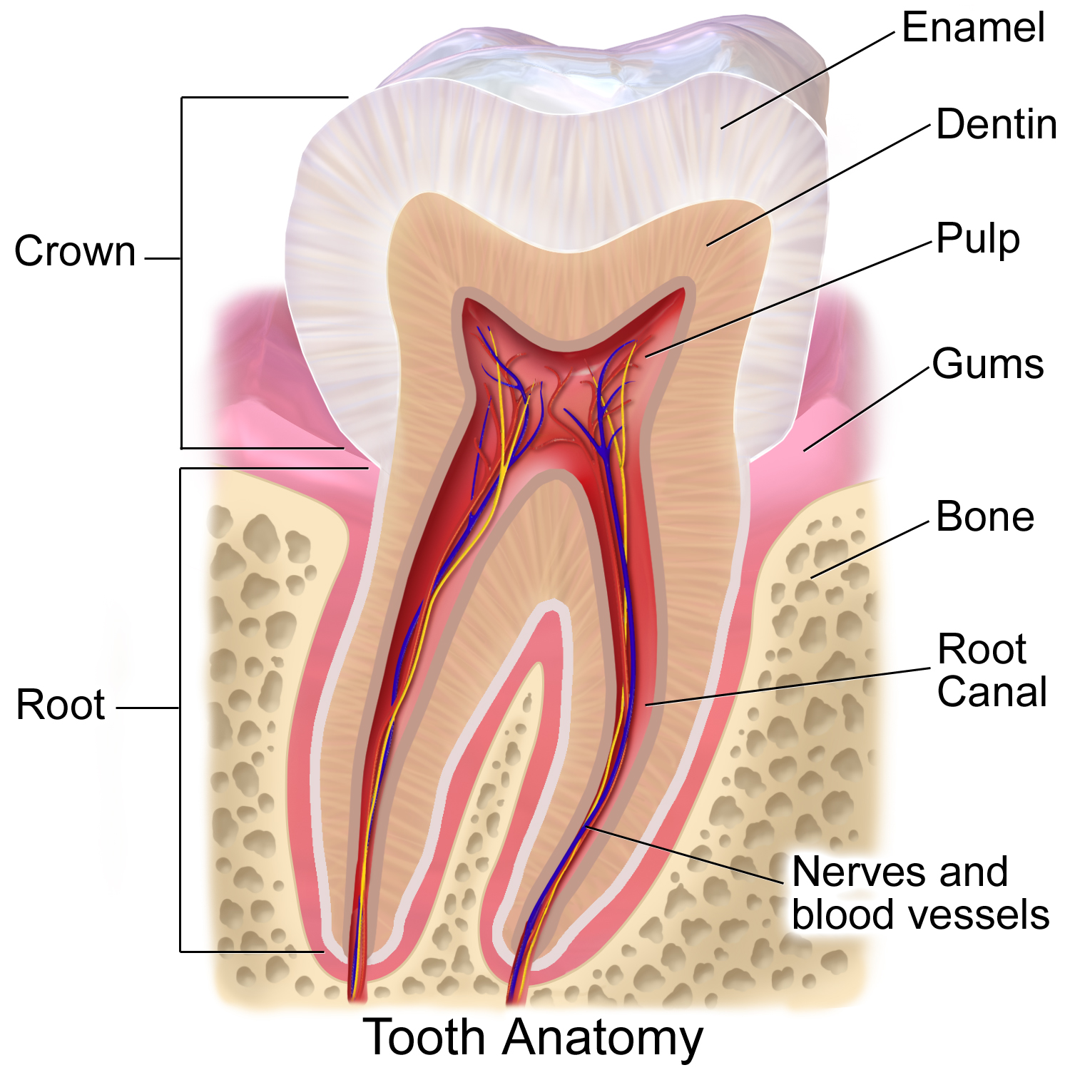
Tooth enamel is one of the four major tissues that make up the tooth in humans and many other animals, including some species of fish. It makes up the normally visible part of the tooth, covering the crown. The other major tissues are dentin, cementum, and dental pulp. It is a very hard, white to off-white, highly mineralised substance that acts as a barrier to protect the tooth but can become susceptible to degradation, especially by acids from food and drink. Calcium hardens the tooth enamel. In rare circumstances enamel fails to form, leaving the underlying dentin exposed on the surface. Features Enamel is the hardest substance in the human body and contains the highest percentage of minerals (at 96%),Ross ''et al.'', p. 485 with water and organic material composing the rest.Ten Cate's Oral Histology, Nancy, Elsevier, pp. 70–94 The primary mineral is hydroxyapatite, which is a crystalline calcium phosphate. Enamel is formed on the tooth while the tooth develops within th ... [...More Info...] [...Related Items...] OR: [Wikipedia] [Google] [Baidu] |
Keratin
Keratin () is one of a family of structural fibrous proteins also known as ''scleroproteins''. Alpha-keratin (α-keratin) is a type of keratin found in vertebrates. It is the key structural material making up scales, hair, nails, feathers, horns, claws, hooves, and the outer layer of skin among vertebrates. Keratin also protects epithelial cells from damage or stress. Keratin is extremely insoluble in water and organic solvents. Keratin monomers assemble into bundles to form intermediate filaments, which are tough and form strong unmineralized epidermal appendages found in reptiles, birds, amphibians, and mammals. Excessive keratinization participate in fortification of certain tissues such as in horns of cattle and rhinos, and armadillos' osteoderm. The only other biological matter known to approximate the toughness of keratinized tissue is chitin. Keratin comes in two types, the primitive, softer forms found in all vertebrates and harder, derived forms found only amon ... [...More Info...] [...Related Items...] OR: [Wikipedia] [Google] [Baidu] |
Hertwig's Epithelial Root Sheath
The Hertwig epithelial root sheath (HERS) or epithelial root sheath is a proliferation of epithelial cells located at the cervical loop of the enamel organ in a developing tooth. Hertwig epithelial root sheath initiates the formation of dentin in the root of a tooth by causing the differentiation of odontoblasts from the dental papilla. The root sheath eventually disintegrates with the periodontal ligament, but residual pieces that do not completely disappear are seen as epithelial cell rests of Malassez (ERM). These rests can become cystic, presenting future periodontal infections. Structure Hertwig epithelial root sheath is derived from the inner and outer enamel epithelium of the enamel organ.Illustrated Dental Embryology, Histology, and Anatomy, Bath-Balogh and Fehrenbach, Elsevier, 2011, p. 66 Function The sheath is also responsible for multiple or accessory roots (medial growth) and lateral or accessory canals in the root (break in epithelium).Ten Cate's Oral Histology ... [...More Info...] [...Related Items...] OR: [Wikipedia] [Google] [Baidu] |
Cervical Loop
The cervical loop is the location on an enamel organ in a developing tooth where the outer enamel epithelium and the inner enamel epithelium join. The cervical loop is a histologic term indicating a specific epithelial structure at the apical side of the tooth germ, consisting of loosely aggregated stellate reticulum in the center surrounded by stratum intermedium. These tissues are enveloped by a basal layer of epithelium known on the outside of the tooth as outer enamel epithelium and on the inside as inner enamel epithelium. During root formation the inner layers of epithelium disappear and only the basal layers are left creating Hertwig's epithelial root sheath (HERS). At this point it is usually referred to as HERS instead of the cervical loop to indicate the structural difference. Cervical loop as epithelial stem cell niche It is thought that the central epithelial tissue of the cervical loop, the stellate reticulum, acts as a stem cell In multicellular organisms, stem ... [...More Info...] [...Related Items...] OR: [Wikipedia] [Google] [Baidu] |
Reduced Enamel Epithelium
The reduced enamel epithelium, sometimes called reduced dental epithelium, overlies a developing tooth and is formed by two layers: a layer of ameloblast cells and the adjacent layer of cuboidal cells (outer enamel epithelium) from the dental lamina. As the cells of the reduced enamel epithelium degenerate, the tooth is revealed progressively with its eruption into the mouth. The degeneration of reduced enamel epithelium also mediates the initial epithelial attachment to the tooth, which is called the junctional epithelium. The reduced enamel epithelium consist of: * Inner enamel epithelium * Outer enamel epithelium The outer enamel epithelium, also known as the external enamel epithelium, is a layer of cuboidal cells located on the periphery of the enamel organ in a developing tooth. This layer is first seen during the bell stage. The rim of the enamel org ... References *Cate, A.R. Ten. Oral Histology: development, structure, and function. 5th ed. 1998. . *Brand, Richar ... [...More Info...] [...Related Items...] OR: [Wikipedia] [Google] [Baidu] |
Ameloblast
Ameloblasts are cells present only during tooth development that deposit tooth enamel, which is the hard outermost layer of the tooth forming the surface of the crown. Structure Each ameloblast is a columnar cell approximately 4 micrometers in diameter, 40 micrometers in length and is hexagonal in cross section. The secretory end of the ameloblast ends in a six-sided pyramid-like projection known as the Tomes' process. The angulation of the Tomes' process is significant in the orientation of enamel rods, the basic unit of tooth enamel. Distal terminal bars are junctional complexes that separate the Tomes' processes from ameloblast proper. Development Ameloblasts are derived from oral epithelium tissue of ectodermal origin. Their differentiation from preameloblasts (whose origin is from inner enamel epithelium) is a result of signaling from the ectomesenchymal cells of the dental papilla. Initially the preameloblasts will differentiate into presecretory ameloblasts and then into s ... [...More Info...] [...Related Items...] OR: [Wikipedia] [Google] [Baidu] |
Stratum Intermedium
The stratum intermedium in a developing tooth is a layer of two or three cells between the inner enamel epithelium and the newly forming cells of the stellate reticulum. It first appears during the early bell stage of tooth development, at around the 14th week of intrauterine life. These cells are closely attached by desmosomes and gap junctions .The stratum intermedium has a notably high alkaline phosphatase activity. This layer, along with the inner enamel epithelium, is responsible for the tooth enamel formation Formation may refer to: Linguistics * Back-formation, the process of creating a new lexeme by removing or affixes * Word formation, the creation of a new word by adding affixes Mathematics and science * Cave formation or speleothem, a secondar .... It is a part of the dental (enamel) organ. Stratum intermedium stores glycogen. It is absent in the part of the tooth germ that outlines the root portions of the tooth which does not form enamel. References *Cate, A.R ... [...More Info...] [...Related Items...] OR: [Wikipedia] [Google] [Baidu] |
Stellate Reticulum
The stellate reticulum is a group of cells located in the center of the enamel organ of a developing tooth. These cells are star-shaped and synthesize glycosaminoglycans. As glycosaminoglycans are produced, water is drawn in between the cells, stretching them apart. As they are moved further away from one another, the stellate reticular cells maintain contact with one another through desmosome A desmosome (; "binding body"), also known as a macula adherens (plural: maculae adherentes) (Latin for ''adhering spot''), is a cell structure specialized for cell-to-cell adhesion. A type of junctional complex, they are localized spot-like adh ...s, resulting in their unique appearance. The stellate reticulum is lost after the first layer of enamel is laid down. This brings cells in the inner enamel epithelium closer to blood vessels at the periphery. References * Orbans Oral histology and embryology – 10th ed. * Cate, A.R. Ten. Oral Histology: development, structure, and functio ... [...More Info...] [...Related Items...] OR: [Wikipedia] [Google] [Baidu] |
Inner Enamel Epithelium
The inner enamel epithelium, also known as the internal enamel epithelium, is a layer of columnar cells located on the rim nearest the dental papilla of the enamel organ in a developing tooth. This layer is first seen during the cap stage, in which these inner enamel epithelium cells are pre-ameloblast cells. These will differentiate into Ameloblasts which are responsible for secretion of enamel during tooth development. The location of the enamel organ where the outer and inner enamel epithelium join is called the cervical loop The cervical loop is the location on an enamel organ in a developing tooth where the outer enamel epithelium and the inner enamel epithelium join. The cervical loop is a histologic term indicating a specific epithelial structure at the apical s .... References *Cate, A.R. Ten. ''Oral Histology: development, structure, and function.'' 5th ed. 1998. . *Ross, Michael H., Gordon I. Kaye, and Wojciech Pawlina. ''Histology: a text and atlas.'' 4th edition ... [...More Info...] [...Related Items...] OR: [Wikipedia] [Google] [Baidu] |


