|
Thenar Eminence
The thenar eminence is the mound formed at the base of the thumb on the palm of the hand by the intrinsic group of muscles of the thumb. The skin overlying this region is the area stimulated when trying to elicit a palmomental reflex. The word thenar comes . Structure The following three muscles are considered part of the thenar eminence: * Abductor pollicis brevis abducts the thumb. This muscle is the most superficial of the thenar group. * Flexor pollicis brevis, which lies next to the abductor, will flex the thumb, curling it up in the palm. (The Flexor pollicis longus, which is inserted into the distal phalanx of the thumb, is not considered part of the thenar eminence.) * Opponens pollicis lies deep to abductor pollicis brevis. As its name suggests it opposes the thumb, bringing it against the fingers. This is a very important movement, as most of human hand dexterity comes from this action. Another muscle that controls movement of the thumb is adductor pollicis. It lies ... [...More Info...] [...Related Items...] OR: [Wikipedia] [Google] [Baidu] |
Wrist
In human anatomy, the wrist is variously defined as (1) the Carpal bones, carpus or carpal bones, the complex of eight bones forming the proximal skeletal segment of the hand; "The wrist contains eight bones, roughly aligned in two rows, known as the carpal bones." (2) the wrist joint or radiocarpal joint, the joint between the radius (bone), radius and the Carpal bones, carpus and; (3) the anatomical region surrounding the carpus including the distal parts of the bones of the forearm and the proximal parts of the metacarpus or five metacarpal bones and the series of joints between these bones, thus referred to as ''wrist joints''. "With the large number of bones composing the wrist (ulna, radius, eight carpas, and five metacarpals), it makes sense that there are many, many joints that make up the structure known as the wrist." This region also includes the carpal tunnel, the anatomical snuff box, bracelet lines, the Flexor retinaculum of the hand, flexor retinaculum, and the ex ... [...More Info...] [...Related Items...] OR: [Wikipedia] [Google] [Baidu] |
Anatomical Terms Of Motion
Motion, the process of movement, is described using specific anatomical terms. Motion includes movement of organs, joints, limbs, and specific sections of the body. The terminology used describes this motion according to its direction relative to the anatomical position of the body parts involved. Anatomists and others use a unified set of terms to describe most of the movements, although other, more specialized terms are necessary for describing unique movements such as those of the hands, feet, and eyes. In general, motion is classified according to the anatomical plane it occurs in. ''Flexion'' and ''extension'' are examples of ''angular'' motions, in which two axes of a joint are brought closer together or moved further apart. ''Rotational'' motion may occur at other joints, for example the shoulder, and are described as ''internal'' or ''external''. Other terms, such as ''elevation'' and ''depression'', describe movement above or below the horizontal plane. Many anatomica ... [...More Info...] [...Related Items...] OR: [Wikipedia] [Google] [Baidu] |
Lumbricals Of The Hand
The lumbricals are intrinsic muscles of the hand that flex the metacarpophalangeal joints, and extend the interphalangeal joints. p. 97 The lumbrical muscles of the foot also have a similar action, though they are of less clinical concern. Structure The lumbricals are four, small, worm-like muscles on each hand. These muscles are unusual in that they do not attach to bone. Instead, they attach proximally to the tendons of flexor digitorum profundus, and distally to the extensor expansions. The first and second lumbricals are unipennate, while the third and fourth lumbricals are bipennate. Nerve supply The first and second lumbricals (the most radial two) are innervated by the median nerve. The third and fourth lumbricals (most ulnar two) are innervated by the deep branch of ulnar nerve. This is the usual innervation of the lumbricals (occurring in 60% of individuals). However 1:3 (median:ulnar - 20% of individuals) and 3:1 (median:ulnar - 20% of individuals) also exist ... [...More Info...] [...Related Items...] OR: [Wikipedia] [Google] [Baidu] |
Recurrent Branch Of The Median Nerve
The recurrent branch of the median nerve is the branch of the median nerve which supplies the thenar muscles. It is also occasionally referred to as the thenar branch of the median nerve, or the thenar muscular branch of the median nerve. Structure An earlier branch of the median nerve also supplies the lumbricals 1 & 2. All other intrinsic muscles of the hand receive their motor innervation from branches of the ulnar nerve. It usually passes distal to the transverse carpal ligament. It ends in the opponens pollicis. Function In the thenar eminence, the recurrent branch of the median nerve provides motor innervation to: * opponens pollicis muscle. * abductor pollicis brevis muscle. * the superficial part of flexor pollicis brevis muscle. Clinical significance The recurrent branch of the median nerve may be affected in carpal tunnel syndrome, or from its own separate peripheral neuropathies. Surgery The recurrent branch of the median nerve is also colloquially called the ... [...More Info...] [...Related Items...] OR: [Wikipedia] [Google] [Baidu] |
Hypothenar Eminence
The hypothenar muscles are a group of three muscles of the hand, palm that control the motion of the little finger. The three muscles are: * Abductor minimi digiti muscle (hand), Abductor digiti minimi * Flexor digiti minimi brevis (hand), Flexor digiti minimi brevis * Opponens digiti minimi Structure The muscles of hypothenar eminence are from medial to lateral: * Opponens digiti minimi muscle, Opponens digiti minimi * Flexor digiti minimi brevis muscle (hand), Flexor digiti minimi brevis * Abductor digiti minimi muscle of hand, Abductor digiti minimi The intrinsic muscles of hand can be remembered using the mnemonic, "A OF A OF A" for, Abductor pollicis brevis, Opponens pollicis, Flexor pollicis brevis (the three thenar muscles), Adductor pollicis, and the three hypothenar muscles, Opponens digiti minimi, Flexor digiti minimi brevis, Abductor digiti minimi. Clinical significance "Hypothenar atrophy" is associated with the lesion of the ulnar nerve, which supplies the three hyp ... [...More Info...] [...Related Items...] OR: [Wikipedia] [Google] [Baidu] |
Thoracic Spinal Nerve 1
The thoracic spinal nerve 1 (T1) is a spinal nerve of the thoracic segment. Nervous System -- Groups of Nerves It originates from the spinal column from below the thoracic vertebra 1
In vertebrates, thoracic vertebrae compose the middle segment of the vertebral column, between the cervical vertebrae and the lumbar vertebrae. In humans, there are twelve thoracic vertebrae and they are intermediate in size between the cervical ... (T1).
Additional Images References [...More Info...] [...Related Items...] OR: [Wikipedia] [Google] [Baidu] |
Cervical Spinal Nerve 8
The cervical spinal nerve 8 (C8) is a spinal nerve of the cervical segment. Nervous System -- Groups of Nerves It originates from the spinal column from below the (C7). Innervation The C8 nerve forms part of the and s via the , and ther ...[...More Info...] [...Related Items...] OR: [Wikipedia] [Google] [Baidu] |
Deep Branch Of Ulnar Nerve
Deep or The Deep may refer to: Places United States * Deep Creek (Appomattox River tributary), Virginia * Deep Creek (Great Salt Lake), Idaho and Utah * Deep Creek (Mahantango Creek tributary), Pennsylvania * Deep Creek (Mojave River tributary), California * Deep Creek (Pine Creek tributary), Pennsylvania * Deep Creek (Soque River tributary), Georgia * Deep Creek (Texas), a tributary of the Colorado River * Deep Creek (Washington), a tributary of the Spokane River * Deep River (Indiana), a tributary of the Little Calumet River * Deep River (Iowa), a minor tributary of the English River * Deep River (North Carolina) * Deep River (Washington), a minor tributary of the Columbia River * Deep Voll Brook, New Jersey, also known as Deep Brook Elsewhere * Deep Creek (Bahamas) * Deep Creek (Melbourne, Victoria), Australia, a tributary of the Maribyrnong River * Deep River (Western Australia) People * Deep (given name) * Deep (rapper), Punjabi rapper from Houston, Texas * Ravi Deep (b ... [...More Info...] [...Related Items...] OR: [Wikipedia] [Google] [Baidu] |
Median Nerve
The median nerve is a nerve in humans and other animals in the upper limb. It is one of the five main nerves originating from the brachial plexus. The median nerve originates from the lateral and medial cords of the brachial plexus, and has contributions from ventral roots of C5-C7 (lateral cord) and C8 and T1 (medial cord). The median nerve is the only nerve that passes through the carpal tunnel. Carpal tunnel syndrome is the disability that results from the median nerve being pressed in the carpal tunnel. Structure The median nerve arises from the branches from lateral and medial cords of the brachial plexus, courses through the anterior part of arm, forearm, and hand, and terminates by supplying the muscles of the hand. Arm After receiving inputs from both the lateral and medial cords of the brachial plexus, the median nerve enters the arm from the axilla at the inferior margin of the teres major muscle. It then passes vertically down and courses lateral to the brachial ar ... [...More Info...] [...Related Items...] OR: [Wikipedia] [Google] [Baidu] |
Ulnar Nerve
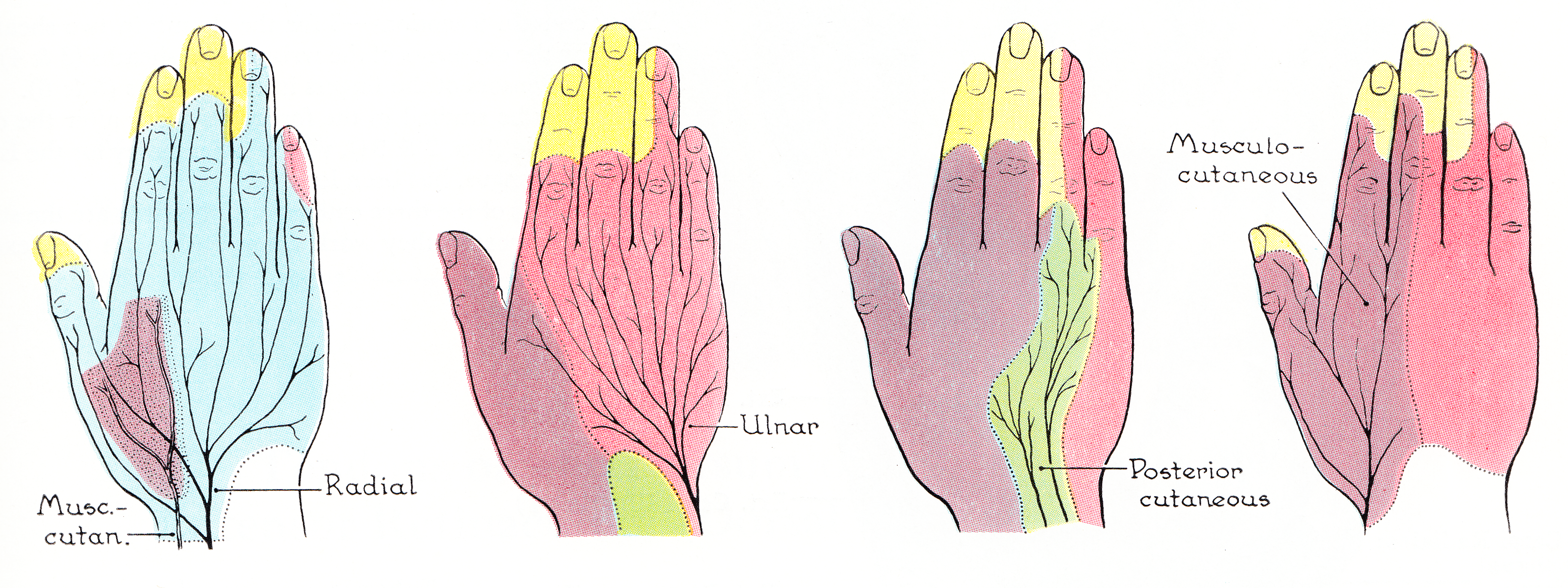
In human anatomy, the ulnar nerve is a nerve that runs near the ulna bone. The ulnar collateral ligament of elbow joint is in relation with the ulnar nerve. The nerve is the largest in the human body unprotected by muscle or bone, so injury is common. This nerve is directly connected to the little finger, and the adjacent half of the ring finger, innervating the palmar aspect of these fingers, including both front and back of the tips, perhaps as far back as the fingernail beds. This nerve can cause an electric shock-like sensation by striking the medial epicondyle of the humerus posteriorly, or inferiorly with the elbow flexed. The ulnar nerve is trapped between the bone and the overlying skin at this point. This is commonly referred to as bumping one's "funny bone". This name is thought to be a pun, based on the sound resemblance between the name of the bone of the upper arm, the humerus, and the word "humorous". Alternatively, according to the Oxford English Dictionary, i ... [...More Info...] [...Related Items...] OR: [Wikipedia] [Google] [Baidu] |


