|
Tenascin
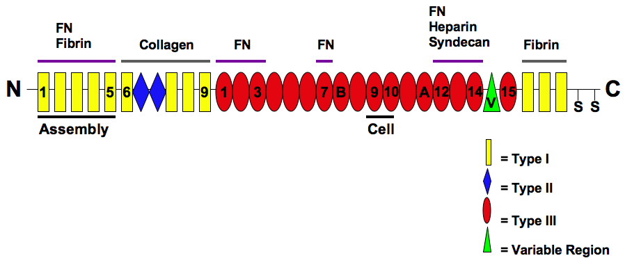
Tenascins are extracellular matrix glycoproteins. They are abundant in the extracellular matrix of developing vertebrate embryos and they reappear around healing wounds and in the stroma of some tumors. Types There are four members of the tenascin gene family: tenascin-C, tenascin-R, tenascin-X and tenascin-W. * Tenascin-C is the founding member of the gene family. In the embryo it is made by migrating cells like the neural crest; it is also abundant in developing tendons, bone and cartilage. * Tenascin-R is found in the developing and adult nervous system. * Tenascin-X is found primarily in loose connective tissue; mutations in the human tenascin-X gene can lead to a form of Ehlers-Danlos syndrome. * Tenascin-W is found in the kidney and in developing bone. The basic structure is 14 EGF-like repeats towards the N-terminal end, and 8 or more fibronectin-III domains which vary upon species and variant. Tenascin-C is the most intensely studied member of the family. It has ... [...More Info...] [...Related Items...] OR: [Wikipedia] [Google] [Baidu] |
Tenascin C
Tenascin C (TN-C) is a glycoprotein that in humans is encoded by the ''TNC'' gene. It is expressed in the extracellular matrix of various tissues during development, disease or injury, and in restricted neurogenic areas of the central nervous system. Tenascin-C is the founding member of the tenascin protein family. In the embryo it is made by migrating cells like the neural crest; it is also abundant in developing tendons, bone and cartilage. Gene and expression The human tenascin C gene, ''TN-C'', is located on chromosome 9 with location of the cytogenic band at the 9q33. The entire Tenascin family coding region spans approximately 80 kilobases translating into 2203 amino acids. Expression of TN-C changes from development to adulthood. TN-C is highly expressed during embryogenesis and is briefly expressed during organogenesis, while in developed organs, expression is absent or in trace amounts. TN-C has been shown to be upregulated under pathological conditions caused by in ... [...More Info...] [...Related Items...] OR: [Wikipedia] [Google] [Baidu] |
Tenascin-X
A member of the tenascin family, tenascin X (TN-X) also known as flexillin or hexabrachion-like protein is a 450kDa glycoprotein that is expressed in connective tissues. TN-X possesses a modular structure composed, from the N- to the C-terminal part by a Tenascin assembly domain (TAD), a series of 18.5 repeats of epidermal growth factor (EGF)-like motif, a high number of Fibronectin type III (FNIII) module, and a fibrinogen (FBG)-like globular domain. In humans, tenascin X is encoded by the ''TNXB'' gene. Gene This gene localizes to the major histocompatibility complex (MHC class III) region on chromosome 6. The structure of this gene is unusual in that it overlaps the CREBL1 and CYP21A2 genes at its 5' and 3' ends, respectively. ''TNXB'' also possesses a pseudogene, ''TNXA,'' which is a consequence of MHC classe III locus duplication during evolution. Strong 3' homology between ''TNXB'' and ''TNXA'' can provoke genetic recombination between the two loci, thus leading to the a ... [...More Info...] [...Related Items...] OR: [Wikipedia] [Google] [Baidu] |
Tenascin-R
Tenascin-R is a protein that in humans is encoded by the ''TNR'' gene. Function Tenascin-R (TNR) is an extracellular matrix protein expressed primarily in the central nervous system. It is a member of the tenascin (TN) gene family, which includes 4 genes in mammals: TNC (or hexabrachion), TNX is a Japanese holding company for various entertainment companies. Its subsidiaries include the talent agency Up-Front Promotion and Up-Front Works, a music production and sales company that manages such record labels as Zetima, Piccolo Town, ... (TNXB), TNW (also known as TNN) and TNR. The genes are expressed in distinct tissues at different times during embryonic development and are present in adult tissues. upplied by OMIMref name="entrez"/> References Further reading * * * * * * * * * * * Tenascins {{gene-1-stub ... [...More Info...] [...Related Items...] OR: [Wikipedia] [Google] [Baidu] |
Syndecans
Syndecans are single transmembrane domain proteins that are thought to act as coreceptors, especially for G protein-coupled receptors. More specifically, these core proteins carry three to five heparan sulfate and chondroitin sulfate chains, i.e. they are proteoglycans, which allow for interaction with a large variety of ligands including fibroblast growth factors, vascular endothelial growth factor, transforming growth factor-beta, fibronectin and antithrombin-1. Interactions between fibronectin and some syndecans can be modulated by the extracellular matrix protein tenascin C. Family members and Structure The syndecan protein family has four members. Syndecans 1 and 3 and syndecans 2 and 4, making up separate subfamilies, arose by gene duplication and divergent evolution from a single ancestral gene. The syndecan numbers reflect the order in which the cDNAs for each family member were cloned. All syndecans have an N-terminal signal peptide, an ectodomain, a single hydroph ... [...More Info...] [...Related Items...] OR: [Wikipedia] [Google] [Baidu] |
Fibronectin
Fibronectin is a high- molecular weight (~500-~600 kDa) glycoprotein of the extracellular matrix that binds to membrane-spanning receptor proteins called integrins. Fibronectin also binds to other extracellular matrix proteins such as collagen, fibrin, and heparan sulfate proteoglycans (e.g. syndecans). Fibronectin exists as a protein dimer, consisting of two nearly identical monomers linked by a pair of disulfide bonds. The fibronectin protein is produced from a single gene, but alternative splicing of its pre-mRNA leads to the creation of several isoforms. Two types of fibronectin are present in vertebrates: * soluble plasma fibronectin (formerly called "cold-insoluble globulin", or CIg) is a major protein component of blood plasma (300 μg/ml) and is produced in the liver by hepatocytes. * insoluble cellular fibronectin is a major component of the extracellular matrix. It is secreted by various cells, primarily fibroblasts, as a soluble protein dimer and is then ass ... [...More Info...] [...Related Items...] OR: [Wikipedia] [Google] [Baidu] |
Glycoprotein
Glycoproteins are proteins which contain oligosaccharide chains covalently attached to amino acid side-chains. The carbohydrate is attached to the protein in a cotranslational or posttranslational modification. This process is known as glycosylation. Secreted extracellular proteins are often glycosylated. In proteins that have segments extending extracellularly, the extracellular segments are also often glycosylated. Glycoproteins are also often important integral membrane proteins, where they play a role in cell–cell interactions. It is important to distinguish endoplasmic reticulum-based glycosylation of the secretory system from reversible cytosolic-nuclear glycosylation. Glycoproteins of the cytosol and nucleus can be modified through the reversible addition of a single GlcNAc residue that is considered reciprocal to phosphorylation and the functions of these are likely to be an additional regulatory mechanism that controls phosphorylation-based signalling. In contra ... [...More Info...] [...Related Items...] OR: [Wikipedia] [Google] [Baidu] |
N-terminus
The N-terminus (also known as the amino-terminus, NH2-terminus, N-terminal end or amine-terminus) is the start of a protein or polypeptide, referring to the free amine group (-NH2) located at the end of a polypeptide. Within a peptide, the amine group is bonded to the carboxylic group of another amino acid, making it a chain. That leaves a free carboxylic group at one end of the peptide, called the C-terminus, and a free amine group on the other end called the N-terminus. By convention, peptide sequences are written N-terminus to C-terminus, left to right (in LTR writing systems). This correlates the translation direction to the text direction, because when a protein is translated from messenger RNA, it is created from the N-terminus to the C-terminus, as amino acids are added to the carboxyl end of the protein. Chemistry Each amino acid has an amine group and a carboxylic group. Amino acids link to one another by peptide bonds which form through a dehydration reaction that ... [...More Info...] [...Related Items...] OR: [Wikipedia] [Google] [Baidu] |
Epidermal Growth Factor
Epidermal growth factor (EGF) is a protein that stimulates cell growth and differentiation by binding to its receptor, EGFR. Human EGF is 6-k Da and has 53 amino acid residues and three intramolecular disulfide bonds. EGF was originally described as a secreted peptide found in the submaxillary glands of mice and in human urine. EGF has since been found in many human tissues, including platelets, submandibular gland (submaxillary gland), and parotid gland. Initially, human EGF was known as urogastrone. Structure In humans, EGF has 53 amino acids (sequence NSDSECPLSHDGYCLHDGVCMYIEALDKYACNCVVGYIGERCQYRDLKWWELR), with a molecular mass of around 6 kDa. It contains three disulfide bridges (Cys6-Cys20, Cys14-Cys31, Cys33-Cys42). Function EGF, via binding to its cognate receptor, results in cellular proliferation, differentiation, and survival. Salivary EGF, which seems to be regulated by dietary inorganic iodine, also plays an important physiological role in the mai ... [...More Info...] [...Related Items...] OR: [Wikipedia] [Google] [Baidu] |
Kidney
The kidneys are two reddish-brown bean-shaped organs found in vertebrates. They are located on the left and right in the retroperitoneal space, and in adult humans are about in length. They receive blood from the paired renal arteries; blood exits into the paired renal veins. Each kidney is attached to a ureter, a tube that carries excreted urine to the bladder. The kidney participates in the control of the volume of various body fluids, fluid osmolality, acid–base balance, various electrolyte concentrations, and removal of toxins. Filtration occurs in the glomerulus: one-fifth of the blood volume that enters the kidneys is filtered. Examples of substances reabsorbed are solute-free water, sodium, bicarbonate, glucose, and amino acids. Examples of substances secreted are hydrogen, ammonium, potassium and uric acid. The nephron is the structural and functional unit of the kidney. Each adult human kidney contains around 1 million nephrons, while a mouse kidney con ... [...More Info...] [...Related Items...] OR: [Wikipedia] [Google] [Baidu] |
Cartilage
Cartilage is a resilient and smooth type of connective tissue. In tetrapods, it covers and protects the ends of long bones at the joints as articular cartilage, and is a structural component of many body parts including the rib cage, the neck and the bronchial tubes, and the intervertebral discs. In other taxa, such as chondrichthyans, but also in cyclostomes, it may constitute a much greater proportion of the skeleton. It is not as hard and rigid as bone, but it is much stiffer and much less flexible than muscle. The matrix of cartilage is made up of glycosaminoglycans, proteoglycans, collagen fibers and, sometimes, elastin. Because of its rigidity, cartilage often serves the purpose of holding tubes open in the body. Examples include the rings of the trachea, such as the cricoid cartilage and carina. Cartilage is composed of specialized cells called chondrocytes that produce a large amount of collagenous extracellular matrix, abundant ground substance that is rich in ... [...More Info...] [...Related Items...] OR: [Wikipedia] [Google] [Baidu] |


