|
TCAP (gene)
Telethonin, also known as Tcap, is a protein that in humans is encoded by the ''TCAP'' gene. Telethonin is expressed in cardiac and skeletal muscle at Z-discs and functions to regulate sarcomere assembly, T-tubule function and apoptosis. Telethonin has been implicated in several diseases, including limb-girdle muscular dystrophy, hypertrophic cardiomyopathy, dilated cardiomyopathy and idiopathic cardiomyopathy. Structure Telethonin is a 19.0 kDa protein composed of 167 amino acids. Telethonin has a unique β-sheet structure, which enables antiparallel association with the Titin Z1-Z2 domains in cardiac and skeletal muscle. Structural analysis of full-length Telethonin with the N-terminal region of Titin indicate that the C-terminus of Telethonin is critical for the dimerization of two Telethonin/Titin complexes into a higher oligomeric structure. Function Telethonin expression is developmentally regulated in both cardiac and skeletal muscle and is thought to be critical to sarc ... [...More Info...] [...Related Items...] OR: [Wikipedia] [Google] [Baidu] |
Protein
Proteins are large biomolecules and macromolecules that comprise one or more long chains of amino acid residues. Proteins perform a vast array of functions within organisms, including catalysing metabolic reactions, DNA replication, responding to stimuli, providing structure to cells and organisms, and transporting molecules from one location to another. Proteins differ from one another primarily in their sequence of amino acids, which is dictated by the nucleotide sequence of their genes, and which usually results in protein folding into a specific 3D structure that determines its activity. A linear chain of amino acid residues is called a polypeptide. A protein contains at least one long polypeptide. Short polypeptides, containing less than 20–30 residues, are rarely considered to be proteins and are commonly called peptides. The individual amino acid residues are bonded together by peptide bonds and adjacent amino acid residues. The sequence of amino acid residue ... [...More Info...] [...Related Items...] OR: [Wikipedia] [Google] [Baidu] |
KvLQT1
Kv7.1 (KvLQT1) is a potassium channel protein whose primary subunit in humans is encoded by the ''KCNQ1'' gene. Kv7.1 is a voltage and lipid-gated potassium channel present in the cell membranes of cardiac tissue and in inner ear neurons among other tissues. In the cardiac cells, Kv7.1 mediates the IKs (or slow delayed rectifying K+) current that contributes to the repolarization of the cell, terminating the cardiac action potential and thereby the heart's contraction. It is a member of the KCNQ family of potassium channels. Structure KvLQT1 is made of six membrane-spanning domains S1-S6, two intracellular domains, and a pore loop. The KvLQT1 channel is made of four KCNQ1 subunits, which form the actual ion channel. Function This gene encodes a protein for a voltage-gated potassium channel required for the repolarization phase of the cardiac action potential. The gene product can form heteromultimers with two other potassium channel proteins, KCNE1 and KCNE3. The ge ... [...More Info...] [...Related Items...] OR: [Wikipedia] [Google] [Baidu] |
Crossbridge
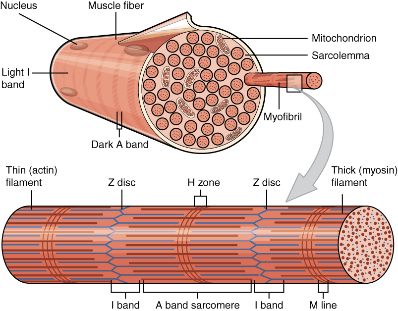
The sliding filament theory explains the mechanism of muscle contraction based on muscle proteins that slide past each other to generate movement. According to the sliding filament theory, the myosin (thick filaments) of muscle fibers slide past the actin (thin filaments) during muscle contraction, while the two groups of filaments remain at relatively constant length. The theory was independently introduced in 1954 by two research teams, one consisting of Andrew Huxley and Rolf Niedergerke from the University of Cambridge, and the other consisting of Hugh Huxley and Jean Hanson from the Massachusetts Institute of Technology. It was originally conceived by Hugh Huxley in 1953. Andrew Huxley and Niedergerke introduced it as a "very attractive" hypothesis. Before the 1950s there were several competing theories on muscle contraction, including electrical attraction, protein folding, and protein modification. The novel theory directly introduced a new concept called cross-bridge th ... [...More Info...] [...Related Items...] OR: [Wikipedia] [Google] [Baidu] |
Myofilament
Myofilaments are the three protein filaments of myofibrils in muscle cells. The main proteins involved are myosin, actin, and titin. Myosin and actin are the ''contractile proteins'' and titin is an elastic protein. The myofilaments act together in muscle contraction, and in order of size are a thick one of mostly myosin, a thin one of mostly actin, and a very thin one of mostly titin. Types of muscle tissue are striated skeletal muscle and cardiac muscle, obliquely striated muscle (found in some invertebrates), and non-striated smooth muscle. Various arrangements of myofilaments create different muscles. Striated muscle has transverse bands of filaments. In obliquely striated muscle, the filaments are staggered. Smooth muscle has irregular arrangements of filaments. Structure There are three different types of myofilaments: thick, thin, and elastic filaments. *Thick filaments consist primarily of a type of myosin, a motor protein – myosin II. Each thick filament is approx ... [...More Info...] [...Related Items...] OR: [Wikipedia] [Google] [Baidu] |
Cardiomyocytes
Cardiac muscle (also called heart muscle, myocardium, cardiomyocytes and cardiac myocytes) is one of three types of vertebrate muscle tissues, with the other two being skeletal muscle and smooth muscle. It is an involuntary, striated muscle that constitutes the main tissue of the wall of the heart. The cardiac muscle (myocardium) forms a thick middle layer between the outer layer of the heart wall (the pericardium) and the inner layer (the endocardium), with blood supplied via the coronary circulation. It is composed of individual cardiac muscle cells joined by intercalated discs, and encased by collagen fibers and other substances that form the extracellular matrix. Cardiac muscle contracts in a similar manner to skeletal muscle, although with some important differences. Electrical stimulation in the form of a cardiac action potential triggers the release of calcium from the cell's internal calcium store, the sarcoplasmic reticulum. The rise in calcium causes the cell's my ... [...More Info...] [...Related Items...] OR: [Wikipedia] [Google] [Baidu] |
MYOM2
Myomesin-2, also known as M-protein is a protein that in humans is encoded by the ''MYOM2'' gene. M-protein is expressed in adult cardiac muscle and fast skeletal muscle, and functions to stabilize the three-dimensional arrangement of proteins comprising M-band structures in a sarcomere. Structure Human M-protein is 165.0 kDa and 1465 amino acids in length. ''MYOM2'' is localized to the human chromosome 8p23.3. M-protein belong to the superfamily of cytoskeletal proteins having immunoglobulin/fibronectin repeats; M-protein contains two immunoglobulin C2-type repeats in the N-terminal region, five fibronectin type III repeats in the central region, and an additional four immunoglobulin C2-type repeats in the C-terminal region. M-protein is expressed only in striated muscle, including fast skeletal muscle and cardiac muscle. Function M-protein exhibits a different pattern of expression in cardiac and skeletal muscle, as well as fast- versus slow-skeletal muscle during development ... [...More Info...] [...Related Items...] OR: [Wikipedia] [Google] [Baidu] |
Myosin Binding Protein C, Cardiac
The myosin-binding protein C, cardiac-type is a protein that in humans is encoded by the ''MYBPC3'' gene. This isoform is expressed exclusively in heart muscle during human and mouse development, and is distinct from those expressed in slow skeletal muscle (MYBPC1) and fast skeletal muscle (MYBPC2). Structure cMyBP-C is a 140.5 kDa protein composed of 1273 amino acids. cMyBP-C is a myosin-associated protein that binds at 43 nm intervals along the myosin thick filament backbone, stretching for 200 nm on either side of the M-line within the crossbridge-bearing zone (C-region) of the A band in striated muscle. The approximate stoichiometry of cMyBP-C along the thick filament is 1 per 9-10 myosin molecules, or 37 cMyBP-C molecules per thick filament. In addition to myosin, cMyBP-C also binds titin and actin. The cMyBP-C isoform expressed in cardiac muscle differs from those expressed in slow and fast skeletal muscle (MYBPC1 and MYBPC2, respectively) by three features: (1) ... [...More Info...] [...Related Items...] OR: [Wikipedia] [Google] [Baidu] |
TNNI3
Troponin I, cardiac muscle is a protein that in humans is encoded by the ''TNNI3'' gene. It is a tissue-specific subtype of troponin I, which in turn is a part of the troponin complex image:Troponin Ribbon Diagram.png, 400px, Ribbon representation of the human cardiac troponin core complex (52 kDa core) in the calcium-saturated form. Blue = troponin C; green = troponin I; magenta = troponin T.; ; rendered with PyMOL Troponin, .... The ''TNNI3'' gene encoding cardiac troponin I (cTnI) is located at 19q13.4 in the human chromosomal genome. Human cTnI is a 24 kDa protein consisting of 210 amino acids with isoelectric point (pI) of 9.87. cTnI is exclusively expressed in adult cardiac muscle. Gene evolution left, Figure 1: A phylogenetic tree is derived from alignment of amino acid sequences. cTnI has diverged from the skeletal muscle isoforms of TnI (slow TnI and fast TnI) mainly with a unique N-terminal extension. The amino acid sequence of cTnI is strongly conserved among ma ... [...More Info...] [...Related Items...] OR: [Wikipedia] [Google] [Baidu] |
Ca2+/calmodulin-dependent Protein Kinase II
/calmodulin-dependent protein kinase II (CaM kinase II or CaMKII) is a serine/threonine-specific protein kinase that is regulated by the /calmodulin complex. CaMKII is involved in many signaling cascades and is thought to be an important mediator of learning and memory. CaMKII is also necessary for homeostasis and reuptake in cardiomyocytes, chloride transport in epithelia, positive T-cell selection, and CD8 T-cell activation. Misregulation of CaMKII is linked to Alzheimer's disease, Angelman syndrome, and heart arrhythmia. Types There are two types of CaM kinase: * Specialized CaM kinases, such as the myosin light chain kinase that phosphorylates myosin, causing smooth muscles to contract * Multifunctional CaM kinases, also collectively called ''CaM kinase II'', which play a role in neurotransmitter secretion, transcription factor regulation, and glycogen metabolism. Structure, function, and autoregulation CaMKII accounts for 1–2% of all proteins in the brain, and ha ... [...More Info...] [...Related Items...] OR: [Wikipedia] [Google] [Baidu] |
L-type Calcium Channel
The L-type calcium channel (also known as the dihydropyridine channel, or DHP channel) is part of the high-voltage activated family of voltage-dependent calcium channel. "L" stands for long-lasting referring to the length of activation. This channel has four isoforms: Cav1.1, Cav1.2, Cav1.3, and Cav1.4. L-type calcium channels are responsible for the excitation-contraction coupling of skeletal, smooth, cardiac muscle, and for aldosterone secretion in endocrine cells of the adrenal cortex. They are also found in neurons, and with the help of L-type calcium channels in endocrine cells, they regulate neurohormones and neurotransmitters. They have also been seen to play a role in gene expression, mRNA stability, neuronal survival, ischemic-induced axonal injury, synaptic efficacy, and both activation and deactivation of other ion channels. In cardiac myocytes, the L-type calcium channel passes inward Ca2+ current (ICaL) and triggers calcium release from the sarcoplasmic reticul ... [...More Info...] [...Related Items...] OR: [Wikipedia] [Google] [Baidu] |
Calcium
Calcium is a chemical element with the symbol Ca and atomic number 20. As an alkaline earth metal, calcium is a reactive metal that forms a dark oxide-nitride layer when exposed to air. Its physical and chemical properties are most similar to its heavier homologues strontium and barium. It is the fifth most abundant element in Earth's crust, and the third most abundant metal, after iron and aluminium. The most common calcium compound on Earth is calcium carbonate, found in limestone and the fossilised remnants of early sea life; gypsum, anhydrite, fluorite, and apatite are also sources of calcium. The name derives from Latin ''calx'' "lime", which was obtained from heating limestone. Some calcium compounds were known to the ancients, though their chemistry was unknown until the seventeenth century. Pure calcium was isolated in 1808 via electrolysis of its oxide by Humphry Davy, who named the element. Calcium compounds are widely used in many industries: in foods and pharma ... [...More Info...] [...Related Items...] OR: [Wikipedia] [Google] [Baidu] |


