|
Type Ia Sensory Fiber
A type Ia sensory fiber, or a primary afferent fiber is a type of afferent nerve fiber. It is the sensory fiber of a stretch receptor called the muscle spindle found in muscles, which constantly monitors the rate at which a muscle stretch changes. The information carried by type Ia fibers contributes to the sense of proprioception. Function of muscle spindles For the body to keep moving properly and with finesse, the nervous system has to have a constant input of sensory data coming from areas such as the muscles and joints. In order to receive a continuous stream of sensory data, the body has developed special sensory receptors called proprioceptors. Muscle spindles are a type of proprioceptor, and they are found inside the muscle itself. They lie parallel with the contractile fibers. This gives them the ability to monitor muscle length with precision. Types of sensory fibers This change in length of the spindle is transduced (transformed into electric membrane potentials ... [...More Info...] [...Related Items...] OR: [Wikipedia] [Google] [Baidu] |
Axons
An axon (from Greek ἄξων ''áxōn'', axis), or nerve fiber (or nerve fibre: see American and British English spelling differences#-re, -er, spelling differences), is a long, slender projection of a nerve cell, or neuron, in vertebrates, that typically conducts electrical impulses known as action potentials away from the Soma (biology), nerve cell body. The function of the axon is to transmit information to different neurons, muscles, and glands. In certain sensory neurons (pseudounipolar neurons), such as those for touch and warmth, the axons are called afferent nerve fibers and the electrical impulse travels along these from the peripheral nervous system, periphery to the cell body and from the cell body to the spinal cord along another branch of the same axon. Axon dysfunction can be the cause of many inherited and acquired neurological disorders that affect both the Peripheral nervous system, peripheral and Central nervous system, central neurons. Nerve fibers are Axon#Cl ... [...More Info...] [...Related Items...] OR: [Wikipedia] [Google] [Baidu] |
Gamma Motor Neuron
A gamma motor neuron (γ motor neuron), also called gamma motoneuron, or fusimotor neuron, is a type of lower motor neuron that takes part in the process of muscle contraction, and represents about 30% of ( Aγ) fibers going to the muscle. Like alpha motor neurons, their cell bodies are located in the anterior grey column of the spinal cord. They receive input from the reticular formation of the pons in the brainstem. Their axons are smaller than those of the alpha motor neurons, with a diameter of only 5 μm. Unlike the alpha motor neurons, gamma motor neurons do not directly adjust the lengthening or shortening of muscles. However, their role is important in keeping muscle spindles taut, thereby allowing the continued firing of alpha neurons, leading to muscle contraction. These neurons also play a role in adjusting the sensitivity of muscle spindles. The presence of myelination in gamma motor neurons allows a conduction velocity of 4 to 24 meters per second, significantl ... [...More Info...] [...Related Items...] OR: [Wikipedia] [Google] [Baidu] |
Type II Sensory Fiber
Type II sensory fiber (group Aβ) is a type of sensory fiber, the second of the two main groups of touch receptors. The responses of different type Aβ fibers to these stimuli can be subdivided based on their adaptation properties, traditionally into rapidly adapting (RA) or slowly adapting (SA) neurons. Type II sensory fibers are slowly-adapting (SA), meaning that even when there is no change in touch, they keep respond to stimuli and fire action potentials. In the body, Type II sensory fibers belong to pseudounipolar neurons. The most notable example are neurons with Merkel cell-neurite complexes on their dendrites (sense static touch) and Ruffini endings (sense stretch on the skin and over-extension inside joints). Under pathological conditions they may become hyper-excitable leading to stimuli that would usually elicit sensations of tactile touch causing pain. These changes are in part induced by PGE2 which is produced by COX1, and type II fibers with free nerve endings ... [...More Info...] [...Related Items...] OR: [Wikipedia] [Google] [Baidu] |
Intrafusal Muscle Fiber
Intrafusal muscle fibers are skeletal muscle fibers that serve as specialized sensory organs (proprioceptors). They detect the amount and rate of change in length of a muscle.Casagrand, Janet (2008) ''Action and Movement: Spinal Control of Motor Units and Spinal Reflexes.'' University of Colorado, Boulder. They constitute the muscle spindle, and are innervated by both sensory (afferent) and motor (efferent) fibers. Intrafusal muscle fibers are not to be confused with extrafusal muscle fibers, which contract, generating skeletal movement and are innervated by alpha motor neurons. Structure Types There are two types of intrafusal muscle fibers: nuclear bag fibers and nuclear chain fibers. They bear two types of sensory ending, known as annulospiral and flower-spray endings. Both ends of these fibers contract, but the central region only stretches and does not contract. Intrafusal muscle fibers are walled off from the rest of the muscle by an outer connective tissue sheath ... [...More Info...] [...Related Items...] OR: [Wikipedia] [Google] [Baidu] |
Muscle Contractions
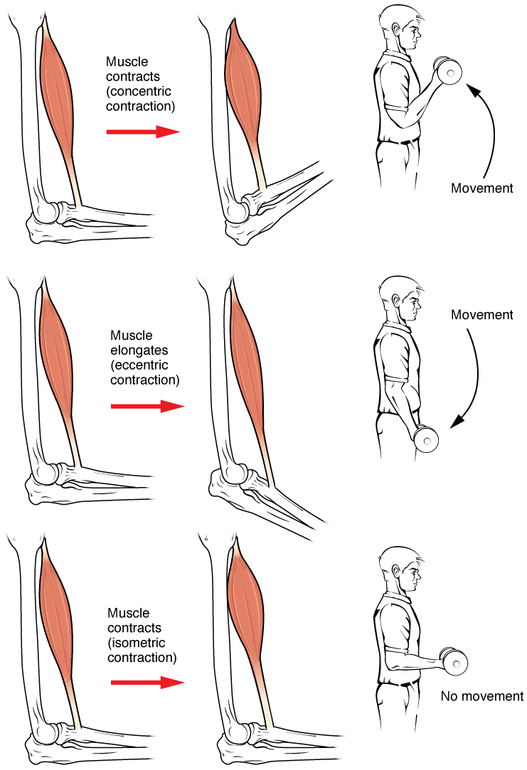
Muscle contraction is the activation of tension-generating sites within muscle cells. In physiology, muscle contraction does not necessarily mean muscle shortening because muscle tension can be produced without changes in muscle length, such as when holding something heavy in the same position. The termination of muscle contraction is followed by muscle relaxation, which is a return of the muscle fibers to their low tension-generating state. For the contractions to happen, the muscle cells must rely on the interaction of two types of filaments which are the thin and thick filaments. Thin filaments are two strands of actin coiled around each, and thick filaments consist of mostly elongated proteins called myosin. Together, these two filaments form myofibrils which are important organelles in the skeletal muscle system. Muscle contraction can also be described based on two variables: length and tension. A muscle contraction is described as isometric if the muscle tension changes b ... [...More Info...] [...Related Items...] OR: [Wikipedia] [Google] [Baidu] |
Cerebellum
The cerebellum (Latin for "little brain") is a major feature of the hindbrain of all vertebrates. Although usually smaller than the cerebrum, in some animals such as the mormyrid fishes it may be as large as or even larger. In humans, the cerebellum plays an important role in motor control. It may also be involved in some cognition, cognitive functions such as attention and language as well as emotion, emotional control such as regulating fear and pleasure responses, but its movement-related functions are the most solidly established. The human cerebellum does not initiate movement, but contributes to Motor coordination, coordination, precision, and accurate timing: it receives input from sensory systems of the spinal cord and from other parts of the brain, and integrates these inputs to fine-tune motor activity. Cerebellar damage produces disorders in Fine motor skill, fine movement, Equilibrioception, equilibrium, Human positions, posture, and motor learning in humans. Anatomica ... [...More Info...] [...Related Items...] OR: [Wikipedia] [Google] [Baidu] |
Cutaneous Nerve
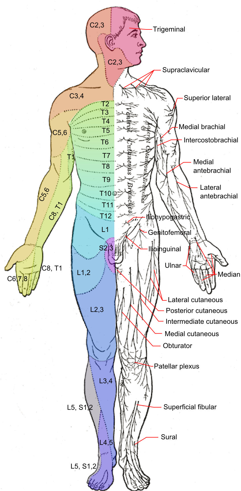
A cutaneous nerve is a nerve that provides nerve supply to the skin. Human anatomy In human anatomy, cutaneous nerves are primarily responsible for providing sensory innervation to the skin. In addition to sympathetic and autonomic afferent (sensory) fibers, most cutaneous nerves also contain sympathetic efferent (visceromotor) fibers, which innervate cutaneous blood vessels, sweat glands, and the arrector pilli muscles of hair follicles. These structures are important to the sympathetic nervous response. There are many cutaneous nerves in the human body, only some of which are named. Some of the larger cutaneous nerves are as follows: Upper body * In the arm (proper) ** Superior lateral cutaneous nerve of arm (Superior LCNOA) ** Inferior lateral cutaneous nerve of arm (Inferior LCNOA) ** Posterior cutaneous nerve of arm (PCNOA) ** Medial cutaneous nerve of arm (MCNOA) * In the forearm ** Lateral cutaneous nerve of forearm (LCNOF) ** Posterior cutaneous nerve of forearm (PCNOF ... [...More Info...] [...Related Items...] OR: [Wikipedia] [Google] [Baidu] |
Brain
A brain is an organ that serves as the center of the nervous system in all vertebrate and most invertebrate animals. It is located in the head, usually close to the sensory organs for senses such as vision. It is the most complex organ in a vertebrate's body. In a human, the cerebral cortex contains approximately 14–16 billion neurons, and the estimated number of neurons in the cerebellum is 55–70 billion. Each neuron is connected by synapses to several thousand other neurons. These neurons typically communicate with one another by means of long fibers called axons, which carry trains of signal pulses called action potentials to distant parts of the brain or body targeting specific recipient cells. Physiologically, brains exert centralized control over a body's other organs. They act on the rest of the body both by generating patterns of muscle activity and by driving the secretion of chemicals called hormones. This centralized control allows rapid and coordinated respon ... [...More Info...] [...Related Items...] OR: [Wikipedia] [Google] [Baidu] |
Stretch Reflex
The stretch reflex (myotatic reflex), or more accurately "muscle stretch reflex", is a muscle contraction in response to stretching within the muscle. The reflex functions to maintain the muscle at a constant length. The term deep tendon reflex is often used by many health workers and students to refer to this reflex. "Tendons have little to do with the response, other than being responsible for mechanically transmitting the sudden stretch from the reflex hammer to the muscle spindle. In addition, some muscles with stretch reflexes have no tendons (e.g., "jaw jerk" of the masseter muscle)". As an example of a spinal reflex, it results in a fast response that involves an afferent signal into the spinal cord and an efferent signal out to the muscle. The stretch reflex can be a monosynaptic reflex which provides automatic regulation of skeletal muscle length, whereby the signal entering the spinal cord arises from a change in muscle length or velocity. It can also include a polysyna ... [...More Info...] [...Related Items...] OR: [Wikipedia] [Google] [Baidu] |
Motor Neurons
A motor neuron (or motoneuron or efferent neuron) is a neuron whose cell body is located in the motor cortex, brainstem or the spinal cord, and whose axon (fiber) projects to the spinal cord or outside of the spinal cord to directly or indirectly control effector organs, mainly muscles and glands. There are two types of motor neuron – upper motor neurons and lower motor neurons. Axons from upper motor neurons synapse onto interneurons in the spinal cord and occasionally directly onto lower motor neurons. The axons from the lower motor neurons are efferent nerve fibers that carry signals from the spinal cord to the effectors. Types of lower motor neurons are alpha motor neurons, beta motor neurons, and gamma motor neurons. A single motor neuron may innervate many muscle fibres and a muscle fibre can undergo many action potentials in the time taken for a single muscle twitch. Innervation takes place at a neuromuscular junction and twitches can become superimposed as a result of ... [...More Info...] [...Related Items...] OR: [Wikipedia] [Google] [Baidu] |
Anterior Grey Column
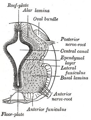
The anterior grey column (also called the anterior cornu, anterior horn of spinal cord, motor horn or ventral horn) is the front column of grey matter in the spinal cord. It is one of the three grey columns. The anterior grey column contains motor neurons that affect the skeletal muscles while the posterior grey column receives information regarding touch and sensation. The anterior grey column is the column where the cell bodies of alpha motor neurons are located. Structure The anterior grey column, directed forward, is broad and of a rounded or quadrangular shape. Its posterior part is termed the base, and its anterior part the head, but these are not differentiated from each other by any well-defined constriction. It is separated from the surface of the medulla spinalis by a layer of white substance which is traversed by the bundles of the anterior nerve roots. In the thoracic region, the postero-lateral part of the anterior column projects laterally as a triangular field, whi ... [...More Info...] [...Related Items...] OR: [Wikipedia] [Google] [Baidu] |


