|
Tuberculoma
A tuberculoma is a clinical manifestation of tuberculosis which conglomerates tubercles into a firm lump, and so can mimic cancer tumors of many types in medical imaging studies. They often arise within individuals in whom a primary tuberculosis infection is not well controlled. When tuberculomas arise intracranially, they represent a manifestation of CNS tuberculosis. Since these are evolutions of primary complex, the tuberculomas may contain caseum or calcifications. With the passage of time, '' Mycobacterium tuberculosis'' can transform into crystals of calcium. These can affect any organ such as the brain, intestine, ovaries, breast, lungs, esophagus, pancreas, bones, and many others. Even with guideline-directed treatment they often persist for months to years. Mechanism The exact mechanism of tuberculoma development has not been determined, although multiple theories have been proposed. It is possible that, following an initial tuberculosis infection resulting in bacte ... [...More Info...] [...Related Items...] OR: [Wikipedia] [Google] [Baidu] |
PET-CT Of A Tuberculoma
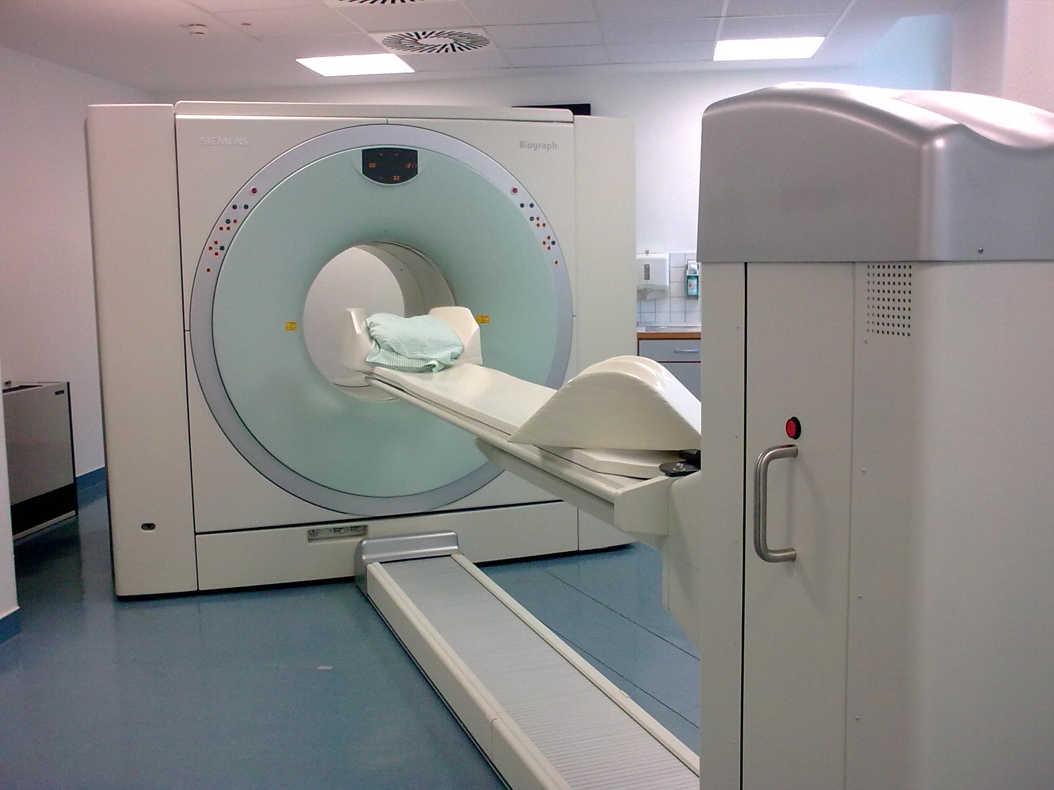
Positron emission tomography–computed tomography (better known as PET-CT or PET/CT) is a nuclear medicine technique which combines, in a single gantry, a positron emission tomography (PET) scanner and an x-ray computed tomography (CT) scanner, to acquire sequential images from both devices in the same session, which are combined into a single superposed ( co-registered) image. Thus, functional imaging obtained by PET, which depicts the spatial distribution of metabolic or biochemical activity in the body can be more precisely aligned or correlated with anatomic imaging obtained by CT scanning. Two- and three-dimensional image reconstruction may be rendered as a function of a common software and control system. PET-CT has revolutionized medical diagnosis in many fields, by adding precision of anatomic localization to functional imaging, which was previously lacking from pure PET imaging. For example, many diagnostic imaging procedures in oncology, surgical planning, radia ... [...More Info...] [...Related Items...] OR: [Wikipedia] [Google] [Baidu] |
Papilledema
Papilledema or papilloedema is optic disc swelling that is caused by increased intracranial pressure due to any cause. The swelling is usually bilateral and can occur over a period of hours to weeks. Unilateral presentation is extremely rare. In intracranial hypertension, the optic disc swelling most commonly occurs bilaterally. When papilledema is found on fundoscopy, further evaluation is warranted because vision loss can result if the underlying condition is not treated. Further evaluation with a CT or MRI of the brain and/or spine is usually performed. Recent research has shown that point-of-care ultrasound can be used to measure optic nerve sheath diameter for detection of increased intracranial pressure and shows good diagnostic test accuracy compared to CT. Thus, if there is a question of papilledema on fundoscopic examination or if the optic disc cannot be adequately visualized, ultrasound can be used to rapidly assess for increased intracranial pressure and help direct f ... [...More Info...] [...Related Items...] OR: [Wikipedia] [Google] [Baidu] |
Ring Enhancing Mass
A ring-enhancing lesion is an abnormal radiologic sign on MRI or CT scans obtained using radiocontrast. On the image, there is an area of decreased density (see radiodensity) surrounded by a bright rim from concentration of the enhancing contrast dye. This enhancement may represent breakdown of the blood-brain barrier and the development of an inflammatory capsule. This can be a finding in numerous disease states. In the brain, it can occur with an early brain abscess as well as in '' Nocardia'' infections associated with lung cavitary lesions. In patients with HIV, the major differential is between CNS lymphoma and CNS toxoplasmosis Toxoplasmosis is a parasitic disease caused by '' Toxoplasma gondii'', an apicomplexan. Infections with toxoplasmosis are associated with a variety of neuropsychiatric and behavioral conditions. Occasionally, people may have a few weeks or mont ..., with CT imaging being the appropriate next step to differentiate between the two conditions.Fauci ... [...More Info...] [...Related Items...] OR: [Wikipedia] [Google] [Baidu] |
Pathognomonic
Pathognomonic (rare synonym ''pathognomic'') is a term, often used in medicine, that means "characteristic for a particular disease". A pathognomonic sign is a particular sign whose presence means that a particular disease is present beyond any doubt. Labelling a sign or symptom "pathognomonic" represents a marked intensification of a "diagnostic" sign or symptom. The word is an adjective of Greek origin derived from πάθος ''pathos'' "disease" and γνώμων ''gnomon'' "indicator" (from γιγνώσκω ''gignosko'' "I know, I recognize"). Practical use While some findings may be classic, typical or highly suggestive in a certain condition, they may not occur ''uniquely'' in this condition and therefore may not directly imply a specific diagnosis. A pathognomonic sign or symptom has very high positive predictive value but does not need to have high sensitivity: for example it can sometimes be absent in a certain disease, since the term only implies that, when it is prese ... [...More Info...] [...Related Items...] OR: [Wikipedia] [Google] [Baidu] |
Hypodense
Radiodensity (or radiopacity) is opacity to the radio wave and X-ray portion of the electromagnetic spectrum: that is, the relative inability of those kinds of electromagnetic radiation to pass through a particular material. Radiolucency or hypodensity indicates greater passage (greater transradiancy) to X-ray photonsNovelline, Robert. ''Squire's Fundamentals of Radiology''. Harvard University Press. 5th edition. 1997. . and is the analogue of transparency and translucency with visible light. Materials that inhibit the passage of electromagnetic radiation are called radiodense or radiopaque, while those that allow radiation to pass more freely are referred to as radiolucent. Radiopaque volumes of material have white appearance on radiographs, compared with the relatively darker appearance of radiolucent volumes. For example, on typical radiographs, bones look white or light gray (radiopaque), whereas muscle and skin look black or dark gray, being mostly invisible (radiolucent). T ... [...More Info...] [...Related Items...] OR: [Wikipedia] [Google] [Baidu] |
Caseating
Caseous necrosis or caseous degeneration () is a unique form of cell death in which the tissue maintains a cheese-like appearance.Robbins and Cotran: Pathologic Basis of Disease, 8th Ed. 2010. Pg. 16 It is also a distinctive form of coagulative necrosis. The dead tissue appears as a soft and white proteinaceous dead cell mass. Etymology The word ''caseous'' means 'pertaining or related to cheese', and comes from the Latin word ''caseus'' 'cheese'. Necrosis refers to the fact that cells do not die in a programmed and orderly way as in apoptosis. Causes Frequently, caseous necrosis is encountered in the foci of tuberculosis infections. It can also be caused by syphilis and certain fungi. A similar appearance can be associated with histoplasmosis, cryptococcosis, and coccidioidomycosis. Pathophysiology This begins as infection is recognized by the body and macrophages begin walling off the microorganisms or pathogens. As macrophages release chemicals that digest cells, the cells beg ... [...More Info...] [...Related Items...] OR: [Wikipedia] [Google] [Baidu] |
Stereotactic Biopsy
Stereotactic biopsy, also known as stereotactic core biopsy, is a biopsy procedure that uses a computer and imaging performed in at least two planes to localize a target lesion (such as a tumor or microcalcifications in the breast) in three-dimensional space and guide the removal of tissue for examination by a pathologist under a microscope. Stereotactic core biopsy makes use of the underlying principle of parallax to determine the depth or "Z-dimension" of the target lesion. Stereotactic core biopsy is extensively used by radiologists specializing in breast imaging to obtain tissue samples containing microcalcifications, which can be an early sign of breast cancer Breast cancer is cancer that develops from breast tissue. Signs of breast cancer may include a lump in the breast, a change in breast shape, dimpling of the skin, milk rejection, fluid coming from the nipple, a newly inverted nipple, or a r .... Uses X-ray-guided stereotactic biopsy is used for impalpable lesio ... [...More Info...] [...Related Items...] OR: [Wikipedia] [Google] [Baidu] |
Cerebrospinal Fluid
Cerebrospinal fluid (CSF) is a clear, colorless body fluid found within the tissue that surrounds the brain and spinal cord of all vertebrates. CSF is produced by specialised ependymal cells in the choroid plexus of the ventricles of the brain, and absorbed in the arachnoid granulations. There is about 125 mL of CSF at any one time, and about 500 mL is generated every day. CSF acts as a shock absorber, cushion or buffer, providing basic mechanical and immunological protection to the brain inside the skull. CSF also serves a vital function in the cerebral autoregulation of cerebral blood flow. CSF occupies the subarachnoid space (between the arachnoid mater and the pia mater) and the ventricular system around and inside the brain and spinal cord. It fills the ventricles of the brain, cisterns, and sulci, as well as the central canal of the spinal cord. There is also a connection from the subarachnoid space to the bony labyrinth of the inner ear via the perilymphat ... [...More Info...] [...Related Items...] OR: [Wikipedia] [Google] [Baidu] |
Polymerase Chain Reaction
The polymerase chain reaction (PCR) is a method widely used to rapidly make millions to billions of copies (complete or partial) of a specific DNA sample, allowing scientists to take a very small sample of DNA and amplify it (or a part of it) to a large enough amount to study in detail. PCR was invented in 1983 by the American biochemist Kary Mullis at Cetus Corporation; Mullis and biochemist Michael Smith (chemist), Michael Smith, who had developed other essential ways of manipulating DNA, were jointly awarded the Nobel Prize in Chemistry in 1993. PCR is fundamental to many of the procedures used in genetic testing and research, including analysis of Ancient DNA, ancient samples of DNA and identification of infectious agents. Using PCR, copies of very small amounts of DNA sequences are exponentially amplified in a series of cycles of temperature changes. PCR is now a common and often indispensable technique used in medical laboratory research for a broad variety of applications ... [...More Info...] [...Related Items...] OR: [Wikipedia] [Google] [Baidu] |
Interferon Gamma Release Assay
Interferon-γ release assays (IGRA) are medical tests used in the diagnosis of some infectious diseases, especially tuberculosis. Interferon-γ (IFN-γ) release assays rely on the fact that T-lymphocytes will release IFN-γ when exposed to specific antigens. These tests are mostly developed for the field of tuberculosis diagnosis, but in theory, may be used in the diagnosis of other diseases which rely on cell-mediated immunity, e.g. cytomegalovirus and leishmaniasis. For example, in patients with cutaneous adverse drug reactions, challenge of peripheral blood lymphocytes with the drug causing the reaction produced a positive test result for half of the drugs tested. There are currently two IFN-γ release assays available for the diagnosis of tuberculosis: * QuantiFERON-TB Gold (licensed in US, Europe and Japan); and * T-SPOT.TB, a form of ELISpot, the variant of ELISA (licensed in Europe, US, Japan and China). The former test quantitates the amount of IFN-γ produced in response ... [...More Info...] [...Related Items...] OR: [Wikipedia] [Google] [Baidu] |
Tuberculosis - Tuberculoma With Cavitation (6596009867)
Tuberculosis (TB) is an infectious disease usually caused by '' Mycobacterium tuberculosis'' (MTB) bacteria. Tuberculosis generally affects the lungs, but it can also affect other parts of the body. Most infections show no symptoms, in which case it is known as latent tuberculosis. Around 10% of latent infections progress to active disease which, if left untreated, kill about half of those affected. Typical symptoms of active TB are chronic cough with blood-containing mucus, fever, night sweats, and weight loss. It was historically referred to as consumption due to the weight loss associated with the disease. Infection of other organs can cause a wide range of symptoms. Tuberculosis is spread from one person to the next through the air when people who have active TB in their lungs cough, spit, speak, or sneeze. People with Latent TB do not spread the disease. Active infection occurs more often in people with HIV/AIDS and in those who smoke. Diagnosis of active TB is ... [...More Info...] [...Related Items...] OR: [Wikipedia] [Google] [Baidu] |
Arachnoiditis
Arachnoiditis is an inflammatory condition of the arachnoid mater or 'arachnoid', one of the membranes known as meninges that surround and protect the nerves of the central nervous system, including the brain and spinal cord. The arachnoid can become inflamed because of adverse reactions to chemicals, infection from bacteria or viruses, as the result of direct injury to the spine, chronic compression of spinal nerves, complications from spinal surgery or other invasive spinal procedures, or the accidental intrathecal injection of steroids intended for the epidural space. Inflammation can sometimes lead to the formation of scar tissue and adhesion that can make the spinal nerves "stick" together,NINDS Arachnoiditis Information Page |


