|
Trochlea Of Superior Oblique
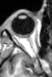
The trochlea of superior oblique is a pulley-like structure in the eye. The tendon of the superior oblique muscle passes through it. Situated on the superior nasal aspect of the frontal bone, it is the only cartilage found in the normal orbit. The word ''trochlea'' comes from the Greek word for pulley. Actions of the superior oblique muscle In order to understand the actions of the superior oblique muscle, it is useful to imagine the eyeball as a sphere that is constrained – like the trackball of a computer mouse – in such a way that only certain rotational movements are possible. Allowable movements for the superior oblique are (1) rotation in a vertical plane – looking down and up (''depression'' and ''elevation'' of the eyeball) and (2) rotation in the plane of the face (''intorsion'' and ''extorsion'' of the eyeball). The body of the superior oblique muscle is located ''behind'' the eyeball, but the tendon (which is redirected by the trochlea) approaches the eyeball fr ... [...More Info...] [...Related Items...] OR: [Wikipedia] [Google] [Baidu] |
Superior Rectus Muscle
The superior rectus muscle is a muscle in the orbit. It is one of the extraocular muscles. It is innervated by the superior division of the oculomotor nerve (III). In the primary position (looking straight ahead), its primary function is elevation, although it also contributes to intorsion and adduction. It is associated with a number of medical conditions, and may be weak, paralysed, overreactive, or even congenitally absent in some people. Structure The superior rectus muscle originates from the annulus of Zinn. It inserts into the anterosuperior surface of the eye. This insertion has a width of around 11 mm. It is around 8 mm from the corneal limbus. Nerve supply The superior rectus muscle is supplied by the superior division of the oculomotor nerve (III). Relations The superior rectus muscle is related to the other extraocular muscles, particularly to the medial rectus muscle and the lateral rectus muscle. The insertion of the superior rectus muscle is around 7.5 mm ... [...More Info...] [...Related Items...] OR: [Wikipedia] [Google] [Baidu] |
Sclera
The sclera, also known as the white of the eye or, in older literature, as the tunica albuginea oculi, is the opaque, fibrous, protective, outer layer of the human eye containing mainly collagen and some crucial elastic fiber. In humans, and some other vertebrates, the whole sclera is white, contrasting with the coloured iris, but in most mammals, the visible part of the sclera matches the colour of the iris, so the white part does not normally show while other vertebrates have distinct colors for both of them. In the development of the embryo, the sclera is derived from the neural crest. In children, it is thinner and shows some of the underlying pigment, appearing slightly blue. In the elderly, fatty deposits on the sclera can make it appear slightly yellow. People with dark skin can have naturally darkened sclerae, the result of melanin pigmentation. The human eye is relatively rare for having a pale sclera (relative to the iris). This makes it easier for one individual to ide ... [...More Info...] [...Related Items...] OR: [Wikipedia] [Google] [Baidu] |
Human Eye
The human eye is a sensory organ, part of the sensory nervous system, that reacts to visible light and allows humans to use visual information for various purposes including seeing things, keeping balance, and maintaining circadian rhythm. The eye can be considered as a living optical device. It is approximately spherical in shape, with its outer layers, such as the outermost, white part of the eye (the sclera) and one of its inner layers (the pigmented choroid) keeping the eye essentially light tight except on the eye's optic axis. In order, along the optic axis, the optical components consist of a first lens (the cornea—the clear part of the eye) that accomplishes most of the focussing of light from the outside world; then an aperture (the pupil) in a diaphragm (the iris—the coloured part of the eye) that controls the amount of light entering the interior of the eye; then another lens (the crystalline lens) that accomplishes the remaining focussing of light into ... [...More Info...] [...Related Items...] OR: [Wikipedia] [Google] [Baidu] |
Cartilage
Cartilage is a resilient and smooth type of connective tissue. In tetrapods, it covers and protects the ends of long bones at the joints as articular cartilage, and is a structural component of many body parts including the rib cage, the neck and the bronchial tubes, and the intervertebral discs. In other taxa, such as chondrichthyans, but also in cyclostomes, it may constitute a much greater proportion of the skeleton. It is not as hard and rigid as bone, but it is much stiffer and much less flexible than muscle. The matrix of cartilage is made up of glycosaminoglycans, proteoglycans, collagen fibers and, sometimes, elastin. Because of its rigidity, cartilage often serves the purpose of holding tubes open in the body. Examples include the rings of the trachea, such as the cricoid cartilage and carina. Cartilage is composed of specialized cells called chondrocytes that produce a large amount of collagenous extracellular matrix, abundant ground substance that is rich in pro ... [...More Info...] [...Related Items...] OR: [Wikipedia] [Google] [Baidu] |
Superior Oblique Muscle
The superior oblique muscle, or obliquus oculi superior, is a fusiform muscle originating in the upper, medial side of the orbit (i.e. from beside the nose) which abducts, depresses and internally rotates the eye. It is the only extraocular muscle innervated by the trochlear nerve (the fourth cranial nerve). Structure The superior oblique muscle loops through a pulley-like structure (the trochlea of superior oblique) and inserts into the sclera on the posterotemporal surface of the eyeball. It is the pulley system that gives superior oblique its actions, causing depression of the eyeball despite being inserted on the superior surface. The superior oblique arises immediately above the margin of the optic foramen, superior and medial to the origin of the superior rectus, and, passing forward, ends in a rounded tendon, which plays in a fibrocartilaginous ring or pulley attached to the trochlear fossa of the frontal bone. The contiguous surfaces of the tendon and ring are lined by ... [...More Info...] [...Related Items...] OR: [Wikipedia] [Google] [Baidu] |
Human Eye
The human eye is a sensory organ, part of the sensory nervous system, that reacts to visible light and allows humans to use visual information for various purposes including seeing things, keeping balance, and maintaining circadian rhythm. The eye can be considered as a living optical device. It is approximately spherical in shape, with its outer layers, such as the outermost, white part of the eye (the sclera) and one of its inner layers (the pigmented choroid) keeping the eye essentially light tight except on the eye's optic axis. In order, along the optic axis, the optical components consist of a first lens (the cornea—the clear part of the eye) that accomplishes most of the focussing of light from the outside world; then an aperture (the pupil) in a diaphragm (the iris—the coloured part of the eye) that controls the amount of light entering the interior of the eye; then another lens (the crystalline lens) that accomplishes the remaining focussing of light into ... [...More Info...] [...Related Items...] OR: [Wikipedia] [Google] [Baidu] |
Pulley
A pulley is a wheel on an axle or shaft that is designed to support movement and change of direction of a taut cable or belt, or transfer of power between the shaft and cable or belt. In the case of a pulley supported by a frame or shell that does not transfer power to a shaft, but is used to guide the cable or exert a force, the supporting shell is called a block, and the pulley may be called a sheave. A pulley may have a groove or grooves between flanges around its circumference to locate the cable or belt. The drive element of a pulley system can be a rope, cable, belt, or chain. The earliest evidence of pulleys dates back to Ancient Egypt in the Twelfth Dynasty (1991-1802 BCE) and Mesopotamia in the early 2nd millennium BCE. In Roman Egypt, Hero of Alexandria (c. 10-70 CE) identified the pulley as one of six simple machines used to lift weights. Pulleys are assembled to form a block and tackle in order to provide mechanical advantage to apply large forces. Pulleys are ... [...More Info...] [...Related Items...] OR: [Wikipedia] [Google] [Baidu] |
Optic Nerve
In neuroanatomy, the optic nerve, also known as the second cranial nerve, cranial nerve II, or simply CN II, is a paired cranial nerve that transmits visual system, visual information from the retina to the brain. In humans, the optic nerve is derived from optic stalks during the seventh week of development and is composed of retinal ganglion cell axons and glial cells; it extends from the optic disc to the optic chiasma and continues as the optic tract to the lateral geniculate nucleus, Pretectal area, pretectal nuclei, and superior colliculus. Structure The optic nerve has been classified as the second of twelve paired cranial nerves, but it is technically part of the central nervous system, rather than the peripheral nervous system because it is derived from an out-pouching of the diencephalon (optic stalks) during embryonic development. As a consequence, the fibers of the optic nerve are covered with myelin produced by oligodendrocytes, rather than Schwann cells of the per ... [...More Info...] [...Related Items...] OR: [Wikipedia] [Google] [Baidu] |
Tarsus (eyelids)
The tarsi (tarsal plates) are two comparatively thick, elongated plates of dense connective tissue, about in length for the upper eyelid and 5 mm for the lower eyelid; one is found in each eyelid, and contributes to its form and support. They are located directly above the lid margins. The tarsus has a lower and upper part making up the palpebrae. Superior The ''superior tarsus'' (''tarsus superior''; superior tarsal plate), the larger, is of a semilunar form, about in breadth at the center, and gradually narrowing toward its extremities. It is adjoined by the superior tarsal muscle. To the anterior surface of this plate the aponeurosis of the levator palpebræ superioris is attached. Inferior The ''inferior tarsus'' (''tarsus inferior''; inferior tarsal plate) is smaller, is thin, is elliptical in form, and has a vertical diameter of about . The free or ciliary margins of these plates are thick and straight. Relations The attached or orbital margins are connected to the ... [...More Info...] [...Related Items...] OR: [Wikipedia] [Google] [Baidu] |
Inferior Rectus Muscle
The inferior rectus muscle is a muscle in the orbit near the eye. It is one of the four recti muscles in the group of extraocular muscles. It originates from the common tendinous ring, and inserts into the anteroinferior surface of the eye. It depresses the eye (downwards). Structure The inferior rectus muscle originates from the common tendinous ring (annulus of Zinn). It inserts into the anteroinferior surface of the eye. This insertion has a width of around 10.5 mm. It is around 7 mm from the corneal limbus. Blood supply The inferior rectus muscle is supplied by an inferior muscular branch of the ophthalmic artery. It may also be supplied by a branch of the infraorbital artery. It is drained by the corresponding veins: the inferior muscular branch of the ophthalmic vein, and sometimes a branch of the infraorbital vein. Nerve supply The inferior rectus muscle is supplied by the inferior division of the oculomotor nerve (III). Development The inferior rectus muscle devel ... [...More Info...] [...Related Items...] OR: [Wikipedia] [Google] [Baidu] |
Annulus Of Zinn
The common tendinous ring, also known as the annulus of Zinn, or annular tendon, is a ring of fibrous tissue surrounding the optic nerve at its entrance at the apex of the orbit. It is the common origin of the four recti muscles of the group of extraocular muscles. It can be used to divide the regions of the superior orbital fissure. The arteries surrounding the optic nerve form a vascular structure known as the circle of Zinn-Haller, or sometimes as the ''circle of Zinn''. The following structures pass through the tendinous ring (superior to inferior): * Superior division of the oculomotor nerve (CNIII) * Nasociliary nerve (branch of ophthalmic nerve) * Inferior division of the oculomotor nerve (CNIII) * Abducens nerve (CNVI) * Optic nerve Parts The common tendinous ring spans the superior orbital fissure and can be described as having two parts – an inferior tendon which gives origin to the inferior rectus muscle, and to part of the lateral rectus muscle; and a superi ... [...More Info...] [...Related Items...] OR: [Wikipedia] [Google] [Baidu] |
Levator Palpebrae Superioris Muscle
The levator palpebrae superioris ( la, elevating muscle of upper eyelid) is the muscle in the orbit that elevates the upper eyelid. Structure The levator palpebrae superioris originates from inferior surface of the lesser wing of the sphenoid bone, just above the optic foramen. It broadens and decreases in thickness (becomes thinner) and becomes the levator aponeurosis. This portion inserts on the skin of the upper eyelid, as well as the superior tarsal plate. It is a skeletal muscle. The superior tarsal muscle, a smooth muscle, is attached to the levator palpebrae superioris, and inserts on the superior tarsal plate as well. Blood supply The levator palebrae superioris receives its blood supply from branches of the ophthalmic artery, specifically, muscular branches and the supraorbital artery. Blood is drained into the superior ophthalmic vein. Nerve supply The levator palpebrae superioris receives motor innervation from the superior division of the oculomotor nerve. The smoo ... [...More Info...] [...Related Items...] OR: [Wikipedia] [Google] [Baidu] |


