|
Trichosporon Asahii
''Trichosporon asahii ''is a non-Candida yeast that has been reported to cause infections in immunocompromised patients. ''T. asahii'' is the most prominent human pathogen in its genus, causing more than half of all Trichosporon infections. First discovered and named in 1929, The currently accepted nomenclature of ''T. asahii'' was validated in 1994. Disease The clinical manifestations of ''T. asahii'' infection are non-specific and vary depending on the site of infection. The most common types of infection were urinary tract infections, fungemia, and disseminated infection. Cutaneous infections have also been reported. Identification and culture ''T. asahii'' grows readily on routine laboratory media, producing white, yellow, or cream, yeast-like colonies on Sabouraud dextrose agar. This fungus has a rapid growth rate and colonies mature in 5 days. When grown on cornmeal-Tween 80 agar, true hypha A hypha (; ) is a long, branching, filamentous structure of a fungus, ... [...More Info...] [...Related Items...] OR: [Wikipedia] [Google] [Baidu] |
Yeast
Yeasts are eukaryotic, single-celled microorganisms classified as members of the fungus kingdom. The first yeast originated hundreds of millions of years ago, and at least 1,500 species are currently recognized. They are estimated to constitute 1% of all described fungal species. Yeasts are unicellular organisms that evolved from multicellular ancestors, with some species having the ability to develop multicellular characteristics by forming strings of connected budding cells known as pseudohyphae or false hyphae. Yeast sizes vary greatly, depending on species and environment, typically measuring 3–4 µm in diameter, although some yeasts can grow to 40 µm in size. Most yeasts reproduce asexually by mitosis, and many do so by the asymmetric division process known as budding. With their single-celled growth habit, yeasts can be contrasted with molds, which grow hyphae. Fungal species that can take both forms (depending on temperature or other conditions) are ca ... [...More Info...] [...Related Items...] OR: [Wikipedia] [Google] [Baidu] |
Trichosporon
''Trichosporon'' is a genus of anamorphic fungi in the family Trichosporonaceae. All species of ''Trichosporon'' are yeasts with no known teleomorphs (sexual states). Most are typically isolated from soil, but several species occur as a natural part of the skin microbiota of humans and other animals. Proliferation of ''Trichosporon'' yeasts in the hair can lead to an unpleasant but non-serious condition known as white piedra. ''Trichosporon'' species can also cause severe opportunistic infections ( trichosporonosis) in immunocompromised individuals. Taxonomy The genus was first described by the German dermatologist Gustav Behrend in 1890, based on yeasts isolated from the hairs of a moustache where they were causing the condition known as "white piedra". Behrend called his new species '' Trichosporon ovoides''. Friedrich Küchenmeister and Rabenhorst had, however, previously described a species in 1867 from the hairs of a wig. They thought that the organism was an alga and plac ... [...More Info...] [...Related Items...] OR: [Wikipedia] [Google] [Baidu] |
Sabouraud Agar
Sabouraud agar or Sabouraud dextrose agar (SDA) is a type of agar growth medium containing peptones. It is used to cultivate dermatophytes and other types of fungi, and can also grow filamentous bacteria such as '' Nocardia''. It has utility for research and clinical care. It was created by, and is named after, Raymond Sabouraud in 1892. In 1977 the formulation was adjusted by Chester W. Emmons when the pH level was brought closer to the neutral range and the dextrose concentration lowered to support the growth of other microorganisms. The acidic pH (5.6) of traditional Sabouraud agar inhibits bacterial growth. Typical composition Sabouraud agar is commercially available and typically contains:University of Sydney''Recipes'' * 40 g/L dextrose * 10 g/L peptone * 20 g/L agar * pH 5.6 Medical Use Clinical laboratories can use this growth medium to diagnose and further speciate fungal infections, allowing medical professionals to provide appropriate treatment with antifunga ... [...More Info...] [...Related Items...] OR: [Wikipedia] [Google] [Baidu] |
Hypha
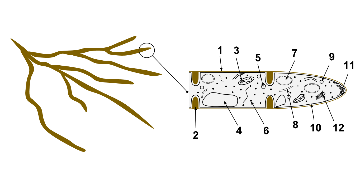
A hypha (; ) is a long, branching, filamentous structure of a fungus, oomycete, or actinobacterium. In most fungi, hyphae are the main mode of vegetative growth, and are collectively called a mycelium. Structure A hypha consists of one or more cells surrounded by a tubular cell wall. In most fungi, hyphae are divided into cells by internal cross-walls called "septa" (singular septum). Septa are usually perforated by pores large enough for ribosomes, mitochondria, and sometimes nuclei to flow between cells. The major structural polymer in fungal cell walls is typically chitin, in contrast to plants and oomycetes that have cellulosic cell walls. Some fungi have aseptate hyphae, meaning their hyphae are not partitioned by septa. Hyphae have an average diameter of 4–6 µm. Growth Hyphae grow at their tips. During tip growth, cell walls are extended by the external assembly and polymerization of cell wall components, and the internal production of new cell membrane. The S ... [...More Info...] [...Related Items...] OR: [Wikipedia] [Google] [Baidu] |
Blastoconidium
A blastoconidium (plural blastoconidia) is an asexual holoblastic conidia formed through the blowing out or budding process of a yeast cell, which is a type of asexual reproduction that results in a bud arising from a parent cell. The production of a blastoconidium can occur along a true hyphae, pseudohyphae, or a singular yeast cell. The word "conidia" comes from the Greek word ''konis'' and ''eidos, konis'' meaning dust and ''eidos'' meaning like. The term "bud" comes from the Greek word ''blastos,'' which means bud. Yeasts such as ''Candida albicans'' and ''Cryptococcus neoformans'' produce these budded cells known as blastoconidia. Formation of a blastoconidium The mitotic budding process through which blastoconidia are formed consists of three steps. The first step is bud emergence, in which the outer cell wall of the parental yeast thins. At the same time, there is growth of new cell wall and plasma membrane components. The next step is bud growth, a process that is regulat ... [...More Info...] [...Related Items...] OR: [Wikipedia] [Google] [Baidu] |

