|
Trabecula
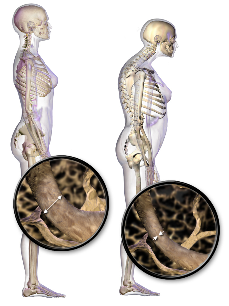
A trabecula (: trabeculae, from Latin for 'small beam') is a small, often microscopic, biological tissue, tissue element in the form of a small Beam (structure), beam, strut or rod that supports or anchors a framework of parts within a body or organ. A trabecula generally has a mechanical function, and is usually composed of dense collagenous tissue (such as the trabeculae of spleen, trabecula of the spleen). It can be composed of other material such as muscle and bone. In the heart, muscles form trabeculae carneae and septomarginal trabecula, septomarginal trabeculae, and the left atrial appendage has a tubular trabeculated structure. Bone#Trabeculae, Cancellous bone is formed from groupings of trabeculated bone tissue. In cross section, trabeculae of a Bone#Trabeculae, cancellous bone can look like septum, septa, but in three dimensions they are topologically distinct, with trabeculae being roughly rod or pillar-shaped and septa being sheet-like. When crossing fluid-filled ... [...More Info...] [...Related Items...] OR: [Wikipedia] [Google] [Baidu] |
Bone
A bone is a rigid organ that constitutes part of the skeleton in most vertebrate animals. Bones protect the various other organs of the body, produce red and white blood cells, store minerals, provide structure and support for the body, and enable mobility. Bones come in a variety of shapes and sizes and have complex internal and external structures. They are lightweight yet strong and hard and serve multiple functions. Bone tissue (osseous tissue), which is also called bone in the uncountable sense of that word, is hard tissue, a type of specialised connective tissue. It has a honeycomb-like matrix internally, which helps to give the bone rigidity. Bone tissue is made up of different types of bone cells. Osteoblasts and osteocytes are involved in the formation and mineralisation of bone; osteoclasts are involved in the resorption of bone tissue. Modified (flattened) osteoblasts become the lining cells that form a protective layer on the bone surface. The mine ... [...More Info...] [...Related Items...] OR: [Wikipedia] [Google] [Baidu] |
Osteoporosis
Osteoporosis is a systemic skeletal disorder characterized by low bone mass, micro-architectural deterioration of bone tissue leading to more porous bone, and consequent increase in Bone fracture, fracture risk. It is the most common reason for a broken bone among the Old age, elderly. Bones that commonly break include the vertebrae in the Vertebral column, spine, the bones of the forearm, the wrist, and the hip. Until a broken bone occurs there are typically no symptoms. Bones may weaken to such a degree that a break may occur with minor stress or spontaneously. After the broken bone heals, some people may have chronic pain and a decreased ability to carry out normal activities. Osteoporosis may be due to lower-than-normal peak bone mass, maximum bone mass and greater-than-normal bone loss. Bone loss increases after menopause in women due to lower levels of estrogen, and after andropause in older men due to lower levels of testosterone. Osteoporosis may also occur due to a ... [...More Info...] [...Related Items...] OR: [Wikipedia] [Google] [Baidu] |
Trabecular Meshwork Of The Eye
The trabecular meshwork is an area of tissue in the eye located around the base of the cornea, near the ciliary body, and is responsible for draining the aqueous humor from the eye via the anterior chamber (the chamber on the front of the eye covered by the cornea). The tissue is spongy and lined by trabeculocytes; it allows fluid to drain into a set of tubes called Schlemm's canal which is lined by endothelium with blood and lymphatic properties that allow aqueous humor to flow into the blood system. Structure The meshwork is divided up into three parts, with characteristically different ultrastructures: #''Inner uveal meshwork'' - Closest to the anterior chamber angle, contains thin cord-like trabeculae, orientated predominantly in a radial fashion, enclosing trabeculae spaces larger than the corneoscleral meshwork. #''Corneoscleral meshwork'' - Contains a large amount of elastin, arranged as a series of thin, flat, perforated sheets arranged in a laminar pattern; consi ... [...More Info...] [...Related Items...] OR: [Wikipedia] [Google] [Baidu] |
Trabeculae Carneae
The trabeculae carneae (columnae carneae or meaty ridges) are rounded or irregular muscular columns which project from the inner surface of the right and left ventricle of the heart.Moore, K.L., & Agur, A.M. (2007). ''Essential Clinical Anatomy: Third Edition.'' Baltimore: Lippincott Williams & Wilkins. 90-94. These are different from the pectinate muscles, which are present in the atria of the heart. In development, trabeculae carneae are among the first of the cardiac structures to develop in the embryonic cardiac tube. Further, throughout development some trabeculae carneae condense to form the myocardium, papillary muscles, chordae tendineae, and septum. Types There are two kinds: * Some are attached along their entire length on one side and merely form prominent ridges, * Others are fixed at their extremities but free in the middle, as in the moderator band in the right ventricle, or the papillary muscles that holds chordae tendinae, which are connected to cusps of valve ... [...More Info...] [...Related Items...] OR: [Wikipedia] [Google] [Baidu] |
Trabeculae Of Spleen
The fibroelastic coat of the spleen invests the organ, and at the hilum is reflected inward upon the vessels in the form of sheaths. From these sheaths, as well as from the inner surface of the fibroelastic coat, numerous small fibrous bands, the trabeculae of the spleen (or splenic trabeculae), emerge from all directions; these uniting, constitute the frame-work of the spleen. The spleen therefore consists of a number of small spaces or areolae, formed by the trabeculae; in these areolae is contained the splenic pulp. See also * Spleen * Trabecula A trabecula (: trabeculae, from Latin for 'small beam') is a small, often microscopic, biological tissue, tissue element in the form of a small Beam (structure), beam, strut or rod that supports or anchors a framework of parts within a body or ... References Spleen (anatomy) {{Portal bar, Anatomy ... [...More Info...] [...Related Items...] OR: [Wikipedia] [Google] [Baidu] |
Septomarginal Trabecula
The moderator band (also known as septomarginal trabecula) is a band of cardiac muscle found in the right ventricle of the heart. It is well-marked in sheep and some other animals, including humans. It extends from the base of the anterior papillary muscle of the tricuspid valve to the ventricular septum. Structure The moderator band is located in the right ventricle. The moderator band connects the base of the anterior papillary muscles of the tricuspid valve to the ventricular septum. Function The moderator band is important because it carries part of the right bundle branch of the atrioventricular bundle of the conduction system of the heart to the anterior papillary muscle. This shortcut across the chamber of the ventricle ensures equal conduction time in the left and right ventricles, allowing for coordinated contraction of the anterior papillary muscle. Clinical significance The moderator band is often used by radiologists and obstetricians to more easily identif ... [...More Info...] [...Related Items...] OR: [Wikipedia] [Google] [Baidu] |
Wolff's Law
Wolff's law, developed by the German anatomist and surgeon Julius Wolff (surgeon), Julius Wolff (1836–1902) in the 19th century, states that bone in a healthy animal will adapt to the loads under which it is placed. If loading on a particular bone increases, the bone will remodel itself over time to become stronger to resist that sort of loading. The internal architecture of the trabeculae undergoes adaptive changes, followed by secondary changes to the external cortical portion of the bone, perhaps becoming thicker as a result. The inverse is true as well: if the loading on a bone decreases, the bone will become less dense and weaker due to the lack of the stimulus required for continued bone remodeling, remodeling.Wolff J. "The Law of Bone Remodeling". Berlin Heidelberg New York: Springer, 1986 (translation of the German 1892 edition) This reduction in bone density (osteopenia) is known as stress shielding and can occur as a result of a hip replacement (or other prosthesis). ... [...More Info...] [...Related Items...] OR: [Wikipedia] [Google] [Baidu] |
Trabeculae Of Corpus Spongiosum Of Penis
The fibrous envelope of the corpus cavernosum urethrae is thinner, whiter in color, and more elastic than that of the corpora cavernosa penis. It is called the trabeculae of corpus spongiosum of penis. The trabeculae are more delicate, nearly uniform in size, and the meshes between them smaller than in the corpora cavernosa penis: their long diameters, for the most part, corresponding with that of the penis. The external envelope or outer coat of the corpus cavernosum urethrae is formed partly of unstriped muscular fibers, and a layer of the same tissue immediately surrounds the canal of the urethra. See also * Trabeculae of corpora cavernosa of penis From the internal surface of the fibrous envelope of the corpora cavernosa penis A corpus cavernosum penis (singular) (from Latin, characterised by "cavities/ hollows" of the penis, : corpora cavernosa) is one of a pair of sponge-like regions of ... References Mammal male reproductive system Human penis anatomy { ... [...More Info...] [...Related Items...] OR: [Wikipedia] [Google] [Baidu] |
Trabeculae Of Corpora Cavernosa Of Penis
From the internal surface of the fibrous envelope of the corpora cavernosa penis A corpus cavernosum penis (singular) (from Latin, characterised by "cavities/ hollows" of the penis, : corpora cavernosa) is one of a pair of sponge-like regions of erectile tissue, which contain most of the blood in the penis of several animals ..., as well as from the sides of the septum, numerous bands or cords are given off, which cross the interior of these corpora cavernosa in all directions, subdividing them into a number of separate compartments, and giving the entire structure a spongy appearance. These bands and cords are called the trabeculae of corpora cavernosa of penis, and consist of white fibrous tissue, elastic fibers, and plain muscular fibers. In them are contained numerous arteries and nerves. The component fibers which form the trabeculae are larger and stronger around the circumference than at the centers of the corpora cavernosa; they are also thicker behind than in front. ... [...More Info...] [...Related Items...] OR: [Wikipedia] [Google] [Baidu] |
Left Atrial Appendage
The atrium (; : atria) is one of the two upper chambers in the heart that receives blood from the circulatory system. The blood in the atria is pumped into the heart ventricles through the atrioventricular mitral and tricuspid heart valves. There are two atria in the human heart – the left atrium receives blood from the pulmonary circulation, and the right atrium receives blood from the venae cavae of the systemic circulation. During the cardiac cycle, the atria receive blood while relaxed in diastole, then contract in systole to move blood to the ventricles. Each atrium is roughly cube-shaped except for an ear-shaped projection called an atrial appendage, previously known as an auricle. All animals with a closed circulatory system have at least one atrium. The atrium was formerly called the 'auricle'. That term is still used to describe this chamber in some other animals, such as the ''Mollusca''. Auricles in this modern terminology are distinguished by having thicker muscu ... [...More Info...] [...Related Items...] OR: [Wikipedia] [Google] [Baidu] |
Heart
The heart is a muscular Organ (biology), organ found in humans and other animals. This organ pumps blood through the blood vessels. The heart and blood vessels together make the circulatory system. The pumped blood carries oxygen and nutrients to the tissue, while carrying metabolic waste such as carbon dioxide to the lungs. In humans, the heart is approximately the size of a closed fist and is located between the lungs, in the middle compartment of the thorax, chest, called the mediastinum. In humans, the heart is divided into four chambers: upper left and right Atrium (heart), atria and lower left and right Ventricle (heart), ventricles. Commonly, the right atrium and ventricle are referred together as the right heart and their left counterparts as the left heart. In a healthy heart, blood flows one way through the heart due to heart valves, which prevent cardiac regurgitation, backflow. The heart is enclosed in a protective sac, the pericardium, which also contains a sma ... [...More Info...] [...Related Items...] OR: [Wikipedia] [Google] [Baidu] |
Vertebra
Each vertebra (: vertebrae) is an irregular bone with a complex structure composed of bone and some hyaline cartilage, that make up the vertebral column or spine, of vertebrates. The proportions of the vertebrae differ according to their spinal segment and the particular species. The basic configuration of a vertebra varies; the vertebral body (also ''centrum'') is of bone and bears the load of the vertebral column. The upper and lower surfaces of the vertebra body give attachment to the intervertebral discs. The posterior part of a vertebra forms a vertebral arch, in eleven parts, consisting of two pedicles (pedicle of vertebral arch), two laminae, and seven processes. The laminae give attachment to the ligamenta flava (ligaments of the spine). There are vertebral notches formed from the shape of the pedicles, which form the intervertebral foramina when the vertebrae articulate. These foramina are the entry and exit conduits for the spinal nerves. The body of the vertebr ... [...More Info...] [...Related Items...] OR: [Wikipedia] [Google] [Baidu] |


