|
Tonometry
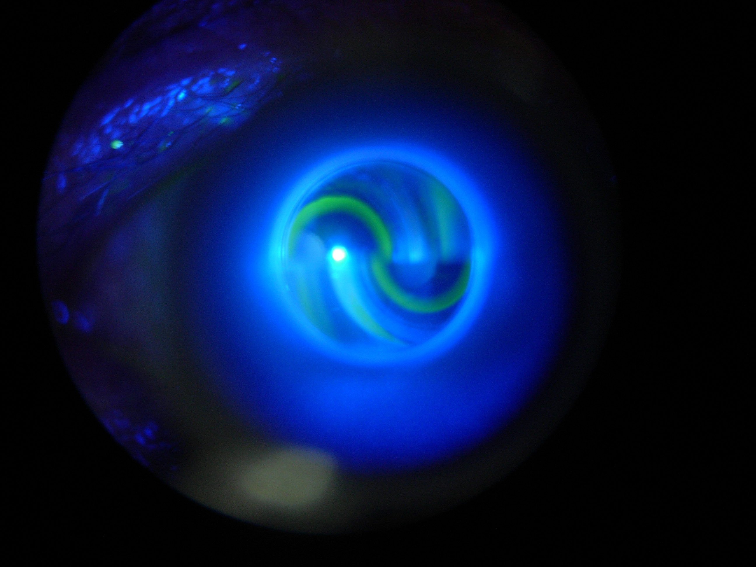
Tonometry is the procedure eye care professionals perform to determine the intraocular pressure (IOP), the fluid pressure inside the eye. It is an important test in the evaluation of patients at risk from glaucoma. Most tonometers are calibrated to measure pressure in millimeters of mercury ( mmHg), with the normal eye pressure range between . Methods Applanation tonometry In applanation tonometry the intraocular pressure (IOP) is inferred from the force required to flatten (applanate) a constant area of the cornea, for the Imbert-Fick law. The Maklakoff tonometer was an early example of this method, while the Goldmann tonometer is the most widely used version in current practice. Because the probe makes contact with the cornea, a topical anesthetic, such as proxymetacaine, is introduced on to the surface of the eye in the form of an eye drop. Goldmann tonometry Goldmann tonometry is considered to be the gold standard IOP test and is the most widely accepted method. A spec ... [...More Info...] [...Related Items...] OR: [Wikipedia] [Google] [Baidu] |
TONOMETER DIATON 2011
Tonometry is the procedure eye care professionals perform to determine the intraocular pressure (IOP), the fluid pressure inside the eye. It is an important test in the evaluation of patients at risk from glaucoma. Most tonometers are calibrated to measure pressure in millimeters of mercury (mmHg), with the normal eye pressure range between . Methods Applanation tonometry In applanation tonometry the intraocular pressure (IOP) is inferred from the force required to flatten (applanate) a constant area of the cornea, for the Imbert-Fick law. The Maklakoff tonometer was an early example of this method, while the Goldmann tonometer is the most widely used version in current practice. Because the probe makes contact with the cornea, a topical anesthetic, such as proxymetacaine, is introduced on to the surface of the eye in the form of an eye drop. Goldmann tonometry Goldmann tonometry is considered to be the gold standard IOP test and is the most widely accepted method. A speci ... [...More Info...] [...Related Items...] OR: [Wikipedia] [Google] [Baidu] |
Intraocular Pressure
Intraocular pressure (IOP) is the fluid pressure inside the eye. Tonometry is the method eye care professionals use to determine this. IOP is an important aspect in the evaluation of patients at risk of glaucoma. Most tonometers are calibrated to measure pressure in millimeters of mercury ( mmHg). Physiology Intraocular pressure is determined by the production and drainage of aqueous humour by the ciliary body and its drainage via the trabecular meshwork and uveoscleral outflow. The reason for this is because the vitreous humour in the posterior segment has a relatively fixed volume and thus does not affect intraocular pressure regulation. An important quantitative relationship (Goldmann's equation) is as follows: :P_o = \frac + P_v Where: * P_o is the IOP in millimeters of mercury (mmHg) * F the rate of aqueous humour formation in microliters per minute (μL/min) * U the resorption of aqueous humour through the uveoscleral route (μL/min) * C is the facility of outflow in micr ... [...More Info...] [...Related Items...] OR: [Wikipedia] [Google] [Baidu] |
Glaucoma
Glaucoma is a group of eye diseases that result in damage to the optic nerve (or retina) and cause vision loss. The most common type is open-angle (wide angle, chronic simple) glaucoma, in which the drainage angle for fluid within the eye remains open, with less common types including closed-angle (narrow angle, acute congestive) glaucoma and normal-tension glaucoma. Open-angle glaucoma develops slowly over time and there is no pain. Peripheral vision may begin to decrease, followed by central vision, resulting in blindness if not treated. Closed-angle glaucoma can present gradually or suddenly. The sudden presentation may involve severe eye pain, blurred vision, mid-dilated pupil, redness of the eye, and nausea. Vision loss from glaucoma, once it has occurred, is permanent. Eyes affected by glaucoma are referred to as being glaucomatous. Risk factors for glaucoma include increasing age, high pressure in the eye, a family history of glaucoma, and use of steroid medication. F ... [...More Info...] [...Related Items...] OR: [Wikipedia] [Google] [Baidu] |
Schiøtz Tonometer
Schiøtz tonometer is an indentation tonometer, used to measure the intraocular pressure (IOP) by measuring the depth produced on the surface of the cornea The cornea is the transparent front part of the eye that covers the iris, pupil, and anterior chamber. Along with the anterior chamber and lens, the cornea refracts light, accounting for approximately two-thirds of the eye's total optical power ... by a load of a known weight. The indentation of corneal surface is related to the IOP. Parts The Schiotz tonometer consists of a ''curved footplate'' which is placed on the cornea of a supine patient. A weighted ''plunger'' attached to the footplate sinks into the cornea. A ''scale'' then gives a reading depending on how much the plunger sinks into the cornea, and a ''conversion table'' converts the scale reading into IOP measured in mmHg. Footplates have to be cool, dry and sterilized before use. Eponym It was invented by the Norwegian ophthalmologist Hjalmar August Schiøtz, ... [...More Info...] [...Related Items...] OR: [Wikipedia] [Google] [Baidu] |
Imbert-Fick Law
Armand Imbert (1850-1922) and Adolf Fick (1829-1901) both demonstrated, independently of each other, that in ocular tonometry the tension of the wall can be neutralized when the application of the tonometer produces a flat surface instead of a convex one, and the reading of the tonometer (P) then equals (T) the IOP," whence all forces cancel each other. This principle was used by Hans Goldmann (1899–1991) who referred to it as the Imbert-Fick "law", thus giving his newly marketed tonometer (with the help of the Haag-Streit Company) a quasi-scientific basis; it is mentioned in the ophthalmic and optometric literature, but not in any books of physics. According to Goldmann, "The law states that the pressure in a sphere filled with liquid and surrounded by an infinitely thin membrane is measured by the counterpressure which just flattens the membrane." "The law presupposes that the membrane is without thickness and without rigidity...practically without any extensibility." A sph ... [...More Info...] [...Related Items...] OR: [Wikipedia] [Google] [Baidu] |
Eye Care Professional
An eye care professional (ECP) is an individual who provides a service related to the eyes or vision. It is any healthcare worker involved in eye care, from one with a small amount of post-secondary training to practitioners with a doctoral level of education. Types Ophthalmologist Ophthalmologists are Doctors of Medicine (M.D./D.O.)(physicians) who specialize in eye care - this includes optical, medical and surgical eye care. They have a general medical degree, not a degree in eye care specifically.” In the US, this usually includes four years of college, four years of medical school, one year surgical internship and three years of eye specific training (ophthalmology residency). Some surgeons complete additional training (fellowship) in specific areas of the eye. Ophthalmologists are qualified to manage any eye disease, perform invasive eye surgery (including injections) and provide general medical care (non eye related) also. While Ophthalmologists can provide comprehensiv ... [...More Info...] [...Related Items...] OR: [Wikipedia] [Google] [Baidu] |
Tear Film
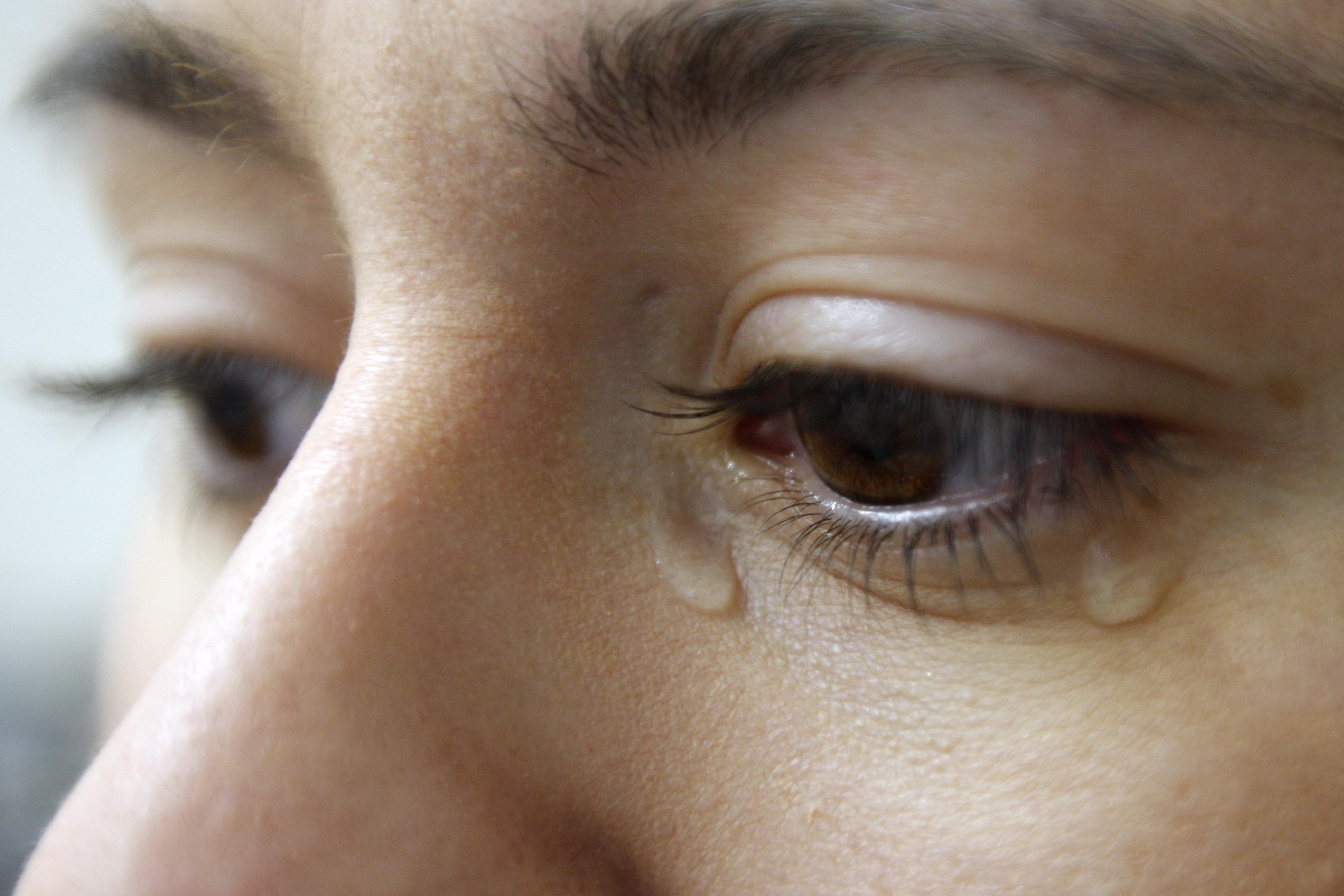
Tears are a clear liquid secreted by the lacrimal glands (tear gland) found in the eyes of all Mammal, land mammals. Tears are made up of water, electrolytes, proteins, lipids, and mucins that form layers on the surface of eyes. The different types of tears—basal, reflex, and emotional—vary significantly in composition. The functions of tears include lubricating the eyes (basal tears), removing irritants (reflex tears), and also aiding the immune system. Tears also occur as a part of the body's natural pain response. Emotional secretion of tears may serve a biological function by excreting stress-inducing hormones built up through times of emotional distress. Tears have Crying, symbolic significance among humans. Physiology Chemical composition Tears are made up of three layers: lipid, aqueous, and mucous. Tears are composed of water, salt (chemistry), salts, antibody, antibodies, and lysozymes (antibacterial enzymes); though composition varies among different tear type ... [...More Info...] [...Related Items...] OR: [Wikipedia] [Google] [Baidu] |
Sclera
The sclera, also known as the white of the eye or, in older literature, as the tunica albuginea oculi, is the opaque, fibrous, protective, outer layer of the human eye containing mainly collagen and some crucial elastic fiber. In humans, and some other vertebrates, the whole sclera is white, contrasting with the coloured iris, but in most mammals, the visible part of the sclera matches the colour of the iris, so the white part does not normally show while other vertebrates have distinct colors for both of them. In the development of the embryo, the sclera is derived from the neural crest. In children, it is thinner and shows some of the underlying pigment, appearing slightly blue. In the elderly, fatty deposits on the sclera can make it appear slightly yellow. People with dark skin can have naturally darkened sclerae, the result of melanin pigmentation. The human eye is relatively rare for having a pale sclera (relative to the iris). This makes it easier for one individual to ide ... [...More Info...] [...Related Items...] OR: [Wikipedia] [Google] [Baidu] |
Tarsal Plate
The tarsi (tarsal plates) are two comparatively thick, elongated plates of dense connective tissue, about in length for the upper eyelid and 5 mm for the lower eyelid; one is found in each eyelid, and contributes to its form and support. They are located directly above the lid margins. The tarsus has a lower and upper part making up the palpebrae. Superior The ''superior tarsus'' (''tarsus superior''; superior tarsal plate), the larger, is of a semilunar form, about in breadth at the center, and gradually narrowing toward its extremities. It is adjoined by the superior tarsal muscle. To the anterior surface of this plate the aponeurosis of the levator palpebræ superioris is attached. Inferior The ''inferior tarsus'' (''tarsus inferior''; inferior tarsal plate) is smaller, is thin, is elliptical in form, and has a vertical diameter of about . The free or ciliary margins of these plates are thick and straight. Relations The attached or orbital margins are connected to the ... [...More Info...] [...Related Items...] OR: [Wikipedia] [Google] [Baidu] |
Eyelid
An eyelid is a thin fold of skin that covers and protects an eye. The levator palpebrae superioris muscle retracts the eyelid, exposing the cornea to the outside, giving vision. This can be either voluntarily or involuntarily. The human eyelid features a row of eyelashes along the eyelid margin, which serve to heighten the protection of the eye from dust and foreign debris, as well as from perspiration. "Palpebral" (and "blepharal") means relating to the eyelids. Its key function is to regularly spread the tears and other secretions on the eye surface to keep it moist, since the cornea must be continuously moist. They keep the eyes from drying out when asleep. Moreover, the blink reflex protects the eye from foreign bodies. The appearance of the human upper eyelid often varies between different populations. The prevalence of an epicanthic fold covering the inner corner of the eye account for the majority of East Asian and Southeast Asian populations, and is also found i ... [...More Info...] [...Related Items...] OR: [Wikipedia] [Google] [Baidu] |


