|
Tobey–Ayer Test
The Tobey–Ayer test is used for lateral sinus thrombosis by monitoring cerebrospinal fluid pressure during a lumbar puncture. No increase of cerebrospinal fluid pressure during compression of the internal jugular vein on the affected side, and an exaggerated response on the patent side, is suggestive of lateral sinus thrombosis. History Tobey–Ayer test was the first specific test for lateral sinus thrombosis. It was created by Tobey, G. L. and Ayer, J. B. in 1925 when they first introduced modifications to the Queckenstedt's maneuver Queckenstedt's maneuver is a clinical test, formerly used for diagnosing spinal stenosis. The test is performed by placing the patient in the lateral decubitus position, thereafter the clinician performs a lumbar puncture. The opening pressure ... test used at the time to diagnose obstruction to spinal cerebrospinal fluid flow. References Medical tests {{med-sign-stub ... [...More Info...] [...Related Items...] OR: [Wikipedia] [Google] [Baidu] |
Thrombosis
Thrombosis (from Ancient Greek "clotting") is the formation of a blood clot inside a blood vessel, obstructing the flow of blood through the circulatory system. When a blood vessel (a vein or an artery) is injured, the body uses platelets (thrombocytes) and fibrin to form a blood clot to prevent blood loss. Even when a blood vessel is not injured, blood clots may form in the body under certain conditions. A clot, or a piece of the clot, that breaks free and begins to travel around the body is known as an embolus. Thrombosis may occur in veins (venous thrombosis) or in arteries (arterial thrombosis). Venous thrombosis (sometimes called DVT, deep vein thrombosis) leads to a blood clot in the affected part of the body, while arterial thrombosis (and, rarely, severe venous thrombosis) affects the blood supply and leads to damage of the tissue supplied by that artery (ischemia and necrosis). A piece of either an arterial or a venous thrombus can break off as an embolus, which could ... [...More Info...] [...Related Items...] OR: [Wikipedia] [Google] [Baidu] |
Cerebrospinal Fluid
Cerebrospinal fluid (CSF) is a clear, colorless body fluid found within the tissue that surrounds the brain and spinal cord of all vertebrates. CSF is produced by specialised ependymal cells in the choroid plexus of the ventricles of the brain, and absorbed in the arachnoid granulations. There is about 125 mL of CSF at any one time, and about 500 mL is generated every day. CSF acts as a shock absorber, cushion or buffer, providing basic mechanical and immunological protection to the brain inside the skull. CSF also serves a vital function in the cerebral autoregulation of cerebral blood flow. CSF occupies the subarachnoid space (between the arachnoid mater and the pia mater) and the ventricular system around and inside the brain and spinal cord. It fills the ventricles of the brain, cisterns, and sulci, as well as the central canal of the spinal cord. There is also a connection from the subarachnoid space to the bony labyrinth of the inner ear via the perilymphat ... [...More Info...] [...Related Items...] OR: [Wikipedia] [Google] [Baidu] |
Lumbar Puncture
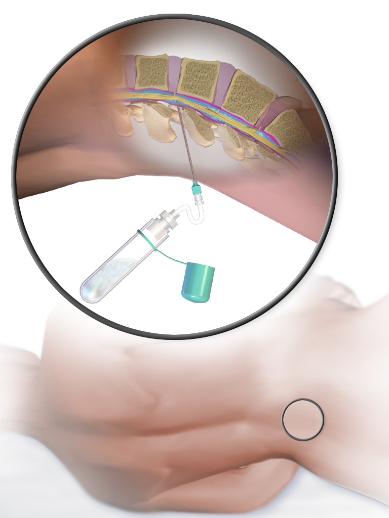
Lumbar puncture (LP), also known as a spinal tap, is a medical procedure in which a needle is inserted into the spinal canal, most commonly to collect cerebrospinal fluid (CSF) for diagnostic testing. The main reason for a lumbar puncture is to help diagnose diseases of the central nervous system, including the brain and spine. Examples of these conditions include meningitis and subarachnoid hemorrhage. It may also be used therapeutically in some conditions. Increased intracranial pressure (pressure in the skull) is a contraindication, due to risk of brain matter being compressed and pushed toward the spine. Sometimes, lumbar puncture cannot be performed safely (for example due to a bleeding diathesis, severe bleeding tendency). It is regarded as a safe procedure, but post-dural-puncture headache is a common side effect if a small atraumatic needle is not used. The procedure is typically performed under local anesthesia using a aseptic technique, sterile technique. A hypodermic ... [...More Info...] [...Related Items...] OR: [Wikipedia] [Google] [Baidu] |
Internal Jugular Vein
The internal jugular vein is a paired jugular vein that collects blood from the brain and the superficial parts of the face and neck. This vein runs in the carotid sheath with the common carotid artery and vagus nerve. It begins in the posterior compartment of the jugular foramen, at the base of the skull. It is somewhat dilated at its origin, which is called the ''superior bulb''. This vein also has a common trunk into which drains the anterior branch of the retromandibular vein, the facial vein, and the lingual vein. It runs down the side of the neck in a vertical direction, being at one end lateral to the internal carotid artery, and then lateral to the common carotid artery, and at the root of the neck, it unites with the subclavian vein to form the brachiocephalic vein (innominate vein); a little above its termination is a second dilation, the ''inferior bulb''. Above, it lies upon the rectus capitis lateralis, behind the internal carotid artery and the nerves passing ... [...More Info...] [...Related Items...] OR: [Wikipedia] [Google] [Baidu] |
Queckenstedt's Maneuver
Queckenstedt's maneuver is a clinical test, formerly used for diagnosing spinal stenosis. The test is performed by placing the patient in the lateral decubitus position, thereafter the clinician performs a lumbar puncture. The opening pressure is measured. Then, the clinician's assistant compresses both jugular veins (if increased intracranial pressure is not suspected then one may exert pressure on both external jugular veins but usually pressure is first exerted on the abdomen, this pressure causes an engorgement of spinal veins and in turn rapidly increases cerebrospinal fluid pressure), which leads to a rise in the intracranial pressure. Given normal anatomy, the intracranial pressure will be reflected as a rapidly rising pressure measured from the lumbar needle, within 10–12 seconds. If there is a stenosis in the spine, there will be a damped, delayed response in the lumbar pressure, thus a positive Queckenstedt's maneuver. Nowadays this test has been made mostly superflu ... [...More Info...] [...Related Items...] OR: [Wikipedia] [Google] [Baidu] |


