|
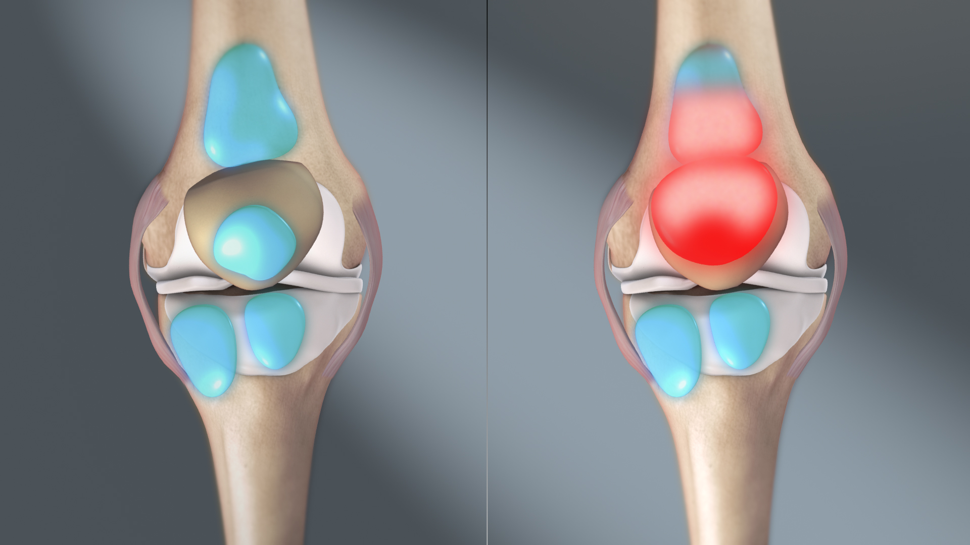
Tibial Tubercle
The tuberosity of the tibia or tibial tuberosity or tibial tubercle is an elevation on the proximal, anterior aspect of the tibia, just below where the anterior surfaces of the lateral and medial tibial condyles end. Structure The tuberosity of the tibia gives attachment to the patellar ligament, which attaches to the patella from where the suprapatellar ligament forms the distal tendon of the quadriceps femoris muscles. The quadriceps muscles consist of the rectus femoris, vastus lateralis, vastus medialis, and vastus intermedius. These quadriceps muscles are innervated by the femoral nerve. KneeHipPain (1998) The tibial tuberosity thus forms the terminal part of the large structure that acts as a lever to extend the knee-joint and prevents the knee from collapsing when the foot strikes the ground. The t ... [...More Info...] [...Related Items...] OR: [Wikipedia] [Google] [Baidu] |
Tibia
The tibia (; ), also known as the shinbone or shankbone, is the larger, stronger, and anterior (frontal) of the two bones in the leg below the knee in vertebrates (the other being the fibula, behind and to the outside of the tibia); it connects the knee with the ankle. The tibia is found on the medial side of the leg next to the fibula and closer to the median plane. The tibia is connected to the fibula by the interosseous membrane of leg, forming a type of fibrous joint called a syndesmosis with very little movement. The tibia is named for the flute '' tibia''. It is the second largest bone in the human body, after the femur. The leg bones are the strongest long bones as they support the rest of the body. Structure In human anatomy, the tibia is the second largest bone next to the femur. As in other vertebrates the tibia is one of two bones in the lower leg, the other being the fibula, and is a component of the knee and ankle joints. The ossification or formation of the ... [...More Info...] [...Related Items...] OR: [Wikipedia] [Google] [Baidu] |
Vastus Medialis
The vastus medialis (vastus internus or teardrop muscle) is an extensor muscle located medially in the thigh that extends the knee. The vastus medialis is part of the quadriceps muscle group. Structure The vastus medialis is a muscle present in the anterior compartment of thigh, and is one of the four muscles that make up the quadriceps muscle. The others are the vastus lateralis, vastus intermedius and rectus femoris. It is the most medial of the "vastus" group of muscles. The vastus medialis arises medially along the entire length of the femur, and attaches with the other muscles of the quadriceps in the quadriceps tendon. The vastus medialis muscle originates from a continuous line of attachment on the femur, which begins on the front and middle side (anteromedially) on the intertrochanteric line of the femur. It continues down and back (posteroinferiorly) along the pectineal line and then descends along the inner (medial) lip of the linea aspera and onto the media ... [...More Info...] [...Related Items...] OR: [Wikipedia] [Google] [Baidu] |
Bursitis
Bursitis is the inflammation of one or more bursae (fluid filled sacs) of synovial fluid in the body. They are lined with a synovial membrane that secretes a lubricating synovial fluid. There are more than 150 bursae in the human body. The bursae rest at the points where internal functionaries, such as muscles and tendons, slide across bone. Healthy bursae create a smooth, almost frictionless functional gliding surface making normal movement painless. When bursitis occurs, however, movement relying on the inflamed bursa becomes difficult and painful. Moreover, movement of tendons and muscles over the inflamed bursa aggravates its inflammation, perpetuating the problem. Muscle can also be stiffened. Signs and symptoms Bursitis commonly affects superficial bursae. These include the subacromial, prepatellar, retrocalcaneal, and ''pes anserinus'' bursae of the shoulder, knee, heel and shin, etc. (see below). Symptoms vary from localized warmth and erythema to joint pain and stif ... [...More Info...] [...Related Items...] OR: [Wikipedia] [Google] [Baidu] |
Orthopedic Cast
An orthopedic cast, or simply cast, is a shell, frequently made from plaster or fiberglass, that encases a limb (or, in some cases, large portions of the body) to stabilize and hold anatomical structures—most often a broken bone (or bones), in place until healing is confirmed. It is similar in function to a splint. Plaster bandages consist of a cotton bandage that has been combined with plaster of paris, which hardens after it has been made wet. Plaster of Paris is calcined gypsum (roasted gypsum), ground to a fine powder by milling. When water is added, the more soluble form of calcium sulfate returns to the relatively insoluble form, and heat is produced. :2 (CaSO4·½ H2O) + 3 H2O → 2 (CaSO4.2H2O) + Heat The setting of unmodified plaster starts about 10 minutes after mixing and is complete in about 45 minutes; however, the cast is not fully dry for 72 hours. Current bandages of synthetic materials are often used, often knitted fiberglass bandages impregnated with poly ... [...More Info...] [...Related Items...] OR: [Wikipedia] [Google] [Baidu] |
Avulsion Fracture
An avulsion fracture is a bone fracture which occurs when a fragment of bone tears away from the main mass of bone as a result of physical trauma. This can occur at the ligament by the application of forces external to the body (such as a fall or pull) or at the tendon by a muscular contraction that is stronger than the forces holding the bone together. Generally muscular avulsion is prevented by the neurological limitations placed on muscle contractions. Highly trained athletes can overcome this neurological inhibition of strength and produce a much greater force output capable of breaking or avulsing a bone. Types Dental avulsion Traumatic complete displacement of a tooth from its socket in alveolar bone. It is a serious dental emergency in which prompt management (within 20–40 minutes of injury) affects the prognosis of the tooth. Tuberosity avulsion of the 5th metatarsal left, Proximal fractures of 5th metatarsa The tuberosity avulsion fracture (also known as ... [...More Info...] [...Related Items...] OR: [Wikipedia] [Google] [Baidu] |
Adolescence
Adolescence () is a transitional stage of physical and psychological development that generally occurs during the period from puberty to adulthood (typically corresponding to the age of majority). Adolescence is usually associated with the teenage years, but its physical, psychological or cultural expressions may begin earlier and end later. Puberty now typically begins during preadolescence, particularly in females. Physical growth (particularly in males) and cognitive development can extend past the teens. Age provides only a rough marker of adolescence, and scholars have not agreed upon a precise definition. Some definitions start as early as 10 and end as late as 25 or 26. The World Health Organization definition officially designates an adolescent as someone between the ages of 10 and 19. Biological development Puberty in general Puberty is a period of several years in which rapid physical growth and psychological changes occur, culminating in sexual maturity. The a ... [...More Info...] [...Related Items...] OR: [Wikipedia] [Google] [Baidu] |
Vastus Intermedius
The vastus intermedius () (Cruraeus) arises from the front and lateral surfaces of the body of the femur in its upper two-thirds, sitting under the rectus femoris muscle and from the lower part of the lateral intermuscular septum. Its fibers end in a superficial aponeurosis, which forms the deep part of the quadriceps femoris tendon. The vastus medialis and vastus intermedius appear to be inseparably united, but when the rectus femoris has been reflected during dissection a narrow interval will be observed extending upward from the medial border of the patella between the two muscles, and the separation may be continued as far as the lower part of the intertrochanteric line, where, however, the two muscles are frequently continuous. Due to being the deeper middle-most of the quadriceps muscle group, the intermedius is the most difficult to stretch once maximum knee flexion is attained. It cannot be further stretched by hip extension as the rectus femoris can, nor is it access ... [...More Info...] [...Related Items...] OR: [Wikipedia] [Google] [Baidu] |
Vastus Lateralis
The vastus lateralis (), also called the vastus externus, is the largest and most powerful part of the quadriceps femoris, a muscle in the thigh. Together with other muscles of the quadriceps group, it serves to extend the knee joint, moving the lower leg forward. It arises from a series of flat, broad tendons attached to the femur, and attaches to the outer border of the patella. It ultimately joins with the other muscles that make up the quadriceps in the quadriceps tendon, which travels over the knee to connect to the tibia. The vastus lateralis is the recommended site for intramuscular injection in infants less than 7 months old and those unable to walk, with loss of muscular tone.Mann, E. (2016). ''Injection (Intramuscular): Clinician Information.'' The Johanna Briggs Institute. Structure The vastus lateralis muscle arises from several areas of the femur, including the upper part of the intertrochanteric line; the lower, anterior borders of the greater trochanter, to the ... [...More Info...] [...Related Items...] OR: [Wikipedia] [Google] [Baidu] |
Lateral Condyle Of Tibia
The lateral condyle is the lateral portion of the upper extremity of tibia. It serves as the insertion for the biceps femoris muscle (small slip). Most of the tendon of the biceps femoris inserts on the fibula The fibula or calf bone is a leg bone on the lateral side of the tibia, to which it is connected above and below. It is the smaller of the two bones and, in proportion to its length, the most slender of all the long bones. Its upper extremity .... See also * Gerdy's tubercle * Medial condyle of tibia Additional images File:Gray258.png, Bones of the right leg. Anterior surface. File:Slide2bib.JPG, Right knee in extension. Deep dissection. Posterior view. File:Slide2cocc.JPG, Right knee in extension. Deep dissection. Posterior view. References External links * * * () Bones of the lower limb Tibia {{musculoskeletal-stub ... [...More Info...] [...Related Items...] OR: [Wikipedia] [Google] [Baidu] |
Rectus Femoris
The rectus femoris muscle is one of the four quadriceps muscles of the human body. The others are the vastus medialis, the vastus intermedius (deep to the rectus femoris), and the vastus lateralis. All four parts of the quadriceps muscle attach to the patella (knee cap) by the quadriceps tendon. The rectus femoris is situated in the middle of the front of the thigh; it is fusiform in shape, and its superficial fibers are arranged in a bipenniform manner, the deep fibers running straight ( la, rectus) down to the deep aponeurosis. Its functions are to flex the thigh at the hip joint and to extend the leg at the knee joint. Structure It arises by two tendons: one, the anterior or straight, from the anterior inferior iliac spine; the other, the posterior or reflected, from a groove above the rim of the acetabulum. The two unite at an acute angle and spread into an aponeurosis that is prolonged downward on the anterior surface of the muscle, and from this the muscular fibers ari ... [...More Info...] [...Related Items...] OR: [Wikipedia] [Google] [Baidu] |
Quadriceps Femoris Muscle
The quadriceps femoris muscle (, also called the quadriceps extensor, quadriceps or quads) is a large muscle group that includes the four prevailing muscles on the front of the thigh. It is the sole extensor muscle of the knee, forming a large fleshy mass which covers the front and sides of the femur. The name derives . Structure Parts The quadriceps femoris muscle is subdivided into four separate muscles (the 'heads'), with the first superficial to the other three over the femur (from the trochanters to the condyles): *The rectus femoris muscle occupies the middle of the thigh, covering most of the other three quadriceps muscles. It originates on the ilium. It is named for its straight course. *The vastus lateralis muscle is on the ''lateral side'' of the femur (i.e. on the outer side of the thigh). *The vastus medialis muscle is on the ''medial side'' of the femur (i.e. on the inner part thigh). *The vastus intermedius muscle lies between vastus lateralis and vastus medi ... [...More Info...] [...Related Items...] OR: [Wikipedia] [Google] [Baidu] |


