|
Throat Diagram
In vertebrate anatomy, the throat is the front part of the neck, internally positioned in front of the vertebrae. It contains the pharynx and larynx. An important section of it is the epiglottis, separating the esophagus from the trachea (windpipe), preventing food and drinks being inhaled into the lungs. The throat contains various blood vessels, pharyngeal muscles, the nasopharyngeal tonsil, the tonsils, the palatine uvula, the trachea, the esophagus, and the vocal cords. Mammal throats consist of two bones, the hyoid bone and the clavicle. The "throat" is sometimes thought to be synonymous for the fauces. It works with the mouth, ears and nose, as well as a number of other parts of the body. Its pharynx is connected to the mouth, allowing speech to occur, and food and liquid to pass down the throat. It is joined to the nose by the nasopharynx at the top of the throat, and to the ear by its Eustachian tube. The throat's trachea carries inhaled air to the bronchi of the lungs. T ... [...More Info...] [...Related Items...] OR: [Wikipedia] [Google] [Baidu] |
Bone
A bone is a Stiffness, rigid Organ (biology), organ that constitutes part of the skeleton in most vertebrate animals. Bones protect the various other organs of the body, produce red blood cell, red and white blood cells, store minerals, provide structure and support for the body, and enable animal locomotion, mobility. Bones come in a variety of shapes and sizes and have complex internal and external structures. They are lightweight yet strong and hard and serve multiple Function (biology), functions. Bone tissue (osseous tissue), which is also called bone in the mass noun, uncountable sense of that word, is hard tissue, a type of specialized connective tissue. It has a honeycomb-like matrix (biology), matrix internally, which helps to give the bone rigidity. Bone tissue is made up of different types of bone cells. Osteoblasts and osteocytes are involved in the formation and mineralization (biology), mineralization of bone; osteoclasts are involved in the bone resorption, resor ... [...More Info...] [...Related Items...] OR: [Wikipedia] [Google] [Baidu] |
Tonsilloliths
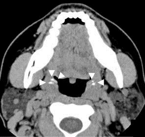
Tonsil stones, also known as tonsilloliths, are mineralizations of debris within the crevices of the tonsils. When not mineralized, the presence of debris is known as chronic caseous tonsillitis (CCT). Symptoms may include bad breath. Generally there is no pain, though there may be the feeling of something present. Risk factors may include recurrent throat infections. Tonsil stones contain a biofilm composed of a number of different bacteria. While they most commonly occur in the palatine tonsils, they may also occur in the lingual tonsils. Tonsil stones have been recorded weighing from 0.3g to 42g. They are often discovered during medical imaging for other reasons. If tonsil stones do not bother the patient, no treatment is needed. Otherwise gargling salt water and manual removal may be tried. Chlorhexidine or cetylpyridinium chloride may also be tried. Surgical treatment may include partial or complete tonsil removal. Up to 10% of people have tonsil stones. Biological se ... [...More Info...] [...Related Items...] OR: [Wikipedia] [Google] [Baidu] |
Tracheotomy
Tracheotomy (, ), or tracheostomy, is a surgical airway management procedure which consists of making an incision (cut) on the anterior aspect (front) of the neck and opening a direct airway through an incision in the trachea (windpipe). The resulting stoma (hole) can serve independently as an airway or as a site for a tracheal tube or tracheostomy tube to be inserted; this tube allows a person to breathe without the use of the nose or mouth. Etymology and terminology The etymology of the word ''tracheotomy'' comes from two Greek words: the root ''tom-'' (from Greek τομή ''tomḗ'') meaning "to cut", and the word ''trachea'' (from Greek τραχεία ''tracheía''). The word ''tracheostomy'', including the root ''stom-'' (from Greek στόμα ''stóma'') meaning "mouth," refers to the making of a semi-permanent or permanent opening, and to the opening itself. Some sources offer different definitions of the above terms. Part of the ambiguity is due to the uncertainty of ... [...More Info...] [...Related Items...] OR: [Wikipedia] [Google] [Baidu] |
Strep Throat
Streptococcal pharyngitis, also known as streptococcal sore throat (strep throat), is pharyngitis (an infection of the pharynx, the back of the throat) caused by ''Streptococcus pyogenes'' a gram-positive, group A streptococcus. Common symptoms include fever, sore throat, red tonsils, and enlarged lymph nodes in the front of the neck. A headache and nausea or vomiting may also occur. Some develop a sandpaper-like rash which is known as scarlet fever. Symptoms typically begin one to three days after exposure and last seven to ten days. Strep throat is spread by respiratory droplets from an infected person, spread by talking, coughing or sneezing, or by touching something that has droplets on it and then touching the mouth, nose, or eyes. It may be spread directly through touching infected sores. It may also be spread by contact with skin infected with group A strep. The diagnosis is made based on the results of a rapid antigen detection test or throat culture. Some people may ... [...More Info...] [...Related Items...] OR: [Wikipedia] [Google] [Baidu] |
Jugular Vein
The jugular veins are veins that take deoxygenated blood from the head back to the heart via the superior vena cava. The internal jugular vein descends next to the internal carotid artery and continues posteriorly to the sternocleidomastoid muscle. Structure and Function There are two sets of jugular veins: external and internal. The left and right external jugular veins drain into the subclavian veins. The internal jugular veins join with the subclavian veins more medially to form the brachiocephalic veins. Finally, the left and right brachiocephalic veins join to form the superior vena cava, which delivers deoxygenated blood to the right atrium of the heart. The Jugular veins help carry blood from the heart to and from the brain. An average human brain weighs about 3 pounds, and gets about 15%-20% of the blood that the heart pumps out. It is important for the brain to get enough blood for many reasons. The jugu ... [...More Info...] [...Related Items...] OR: [Wikipedia] [Google] [Baidu] |
Adenoid
In anatomy, the adenoid, also known as the pharyngeal tonsil or nasopharyngeal tonsil, is the superior-most of the tonsils. It is a mass of lymphatic tissue located behind the nasal cavity, in the roof of the nasopharynx, where the nose blends into the throat. In children, it normally forms a soft mound in the roof and back wall of the nasopharynx, just above and behind the uvula. The term ''adenoid'' is also used to represent adenoid hypertrophy, the abnormal growth of the pharyngeal tonsils. Structure The adenoid is a mass of lymphatic tissue located behind the nasal cavity, in the roof of the nasopharynx, where the nose blends into the throat. The adenoid, unlike the palatine tonsils, has pseudostratified epithelium. The adenoids are part of the so-called Waldeyer ring of lymphoid tissue which also includes the palatine tonsils, the lingual tonsils and the tubal tonsils. Development Adenoids develop from a subepithelial infiltration of lymphocytes after the 16th week o ... [...More Info...] [...Related Items...] OR: [Wikipedia] [Google] [Baidu] |
Stomach
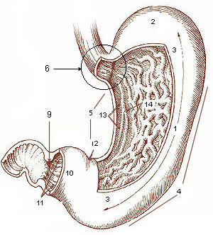
The stomach is a muscular, hollow organ in the gastrointestinal tract of humans and many other animals, including several invertebrates. The stomach has a dilated structure and functions as a vital organ in the digestive system. The stomach is involved in the gastric phase of digestion, following chewing. It performs a chemical breakdown by means of enzymes and hydrochloric acid. In humans and many other animals, the stomach is located between the oesophagus and the small intestine. The stomach secretes digestive enzymes and gastric acid to aid in food digestion. The pyloric sphincter controls the passage of partially digested food ( chyme) from the stomach into the duodenum, where peristalsis takes over to move this through the rest of intestines. Structure In the human digestive system, the stomach lies between the oesophagus and the duodenum (the first part of the small intestine). It is in the left upper quadrant of the abdominal cavity. The top of the stomach lies ag ... [...More Info...] [...Related Items...] OR: [Wikipedia] [Google] [Baidu] |
Bronchi
A bronchus is a passage or airway in the lower respiratory tract that conducts air into the lungs. The first or primary bronchi pronounced (BRAN-KAI) to branch from the trachea at the carina are the right main bronchus and the left main bronchus. These are the widest bronchi, and enter the right lung, and the left lung at each hilum. The main bronchi branch into narrower secondary bronchi or lobar bronchi, and these branch into narrower tertiary bronchi or segmental bronchi. Further divisions of the segmental bronchi are known as 4th order, 5th order, and 6th order segmental bronchi, or grouped together as subsegmental bronchi. The bronchi, when too narrow to be supported by cartilage, are known as bronchioles. No gas exchange takes place in the bronchi. Structure The trachea (windpipe) divides at the carina into two main or primary bronchi, the left bronchus and the right bronchus. The carina of the trachea is located at the level of the sternal angle and the fifth thoracic verte ... [...More Info...] [...Related Items...] OR: [Wikipedia] [Google] [Baidu] |
Eustachian Tube
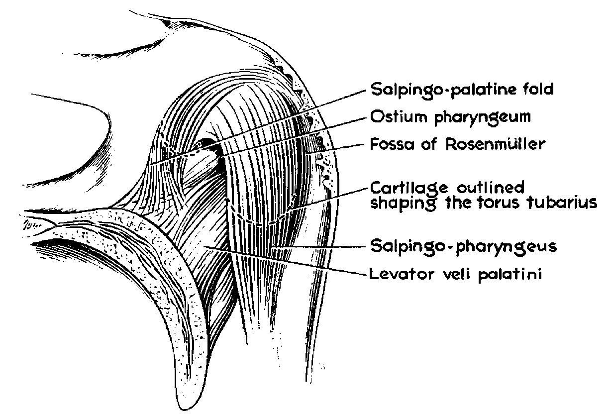
In anatomy, the Eustachian tube, also known as the auditory tube or pharyngotympanic tube, is a tube that links the nasopharynx to the middle ear, of which it is also a part. In adult humans, the Eustachian tube is approximately long and in diameter. It is named after the sixteenth-century Italian anatomist Bartolomeo Eustachi. In humans and other tetrapods, both the middle ear and the ear canal are normally filled with air. Unlike the air of the ear canal, however, the air of the middle ear is not in direct contact with the atmosphere outside the body; thus, a pressure difference can develop between the atmospheric pressure of the ear canal and the middle ear. Normally, the Eustachian tube is collapsed, but it gapes open with swallowing and with positive pressure, allowing the middle ear's pressure to adjust to the atmospheric pressure. When taking off in an aircraft, the ambient air pressure goes from higher (on the ground) to lower (in the sky). The air in the middle ear ... [...More Info...] [...Related Items...] OR: [Wikipedia] [Google] [Baidu] |
Nasopharynx
The pharynx (plural: pharynges) is the part of the throat behind the mouth and nasal cavity, and above the oesophagus and trachea (the tubes going down to the stomach and the lungs). It is found in vertebrates and invertebrates, though its structure varies across species. The pharynx carries food and air to the esophagus and larynx respectively. The flap of cartilage called the epiglottis stops food from entering the larynx. In humans, the pharynx is part of the digestive system and the conducting zone of the respiratory system. (The conducting zone—which also includes the nostrils of the nose, the larynx, trachea, bronchi, and bronchioles—filters, warms and moistens air and conducts it into the lungs). The human pharynx is conventionally divided into three sections: the nasopharynx, oropharynx, and laryngopharynx. It is also important in vocalization. In humans, two sets of pharyngeal muscles form the pharynx and determine the shape of its lumen. They are arranged as an in ... [...More Info...] [...Related Items...] OR: [Wikipedia] [Google] [Baidu] |
Nose
A nose is a protuberance in vertebrates that houses the nostrils, or nares, which receive and expel air for respiration alongside the mouth. Behind the nose are the olfactory mucosa and the sinuses. Behind the nasal cavity, air next passes through the pharynx, shared with the digestive system, and then into the rest of the respiratory system. In humans, the nose is located centrally on the face and serves as an alternative respiratory passage especially during suckling for infants. The protruding nose that completely separate from the mouth part is a characteristic found only in therian mammals. It has been theorized that this unique mammalian nose evolved from the anterior part of the upper jaw of the reptilian-like ancestors ( synapsids). Air treatment Acting as the first interface between the external environment and an animal's delicate internal lungs, a nose conditions incoming air, both as a function of thermal regulation and filtration during respiration, as well as ... [...More Info...] [...Related Items...] OR: [Wikipedia] [Google] [Baidu] |


