|
The Journal Of Cell Biology
The ''Journal of Cell Biology'' is a peer-reviewed scientific journal published by Rockefeller University Press. History In the early 1950s, a small group of biologists began to explore intracellular anatomy using the emerging technology of electron microscopy. Many of these researchers were at The Rockefeller Institute of Medicine, the predecessor of The Rockefeller University. As their work progressed to publication, they were disappointed with the limited quality of halftone image reproduction in the printed journals of the time, and frustrated by the narrow editorial policies of existing journals regarding their image-based results. In 1954, the Director of the Rockefeller Institute, Detlev Bronk, convened a luncheon to discuss the creation of a new journal as a venue for publication of this type of work. The first issue of ''The Journal of Biophysical and Biochemical Cytology'' was published less than a year later on January 25, 1955. A subscription cost $15 per year. The ... [...More Info...] [...Related Items...] OR: [Wikipedia] [Google] [Baidu] |
Jodi Nunnari
Jodi Nunnari, Ph.D. is an American cell biologist and pioneer in the field of mitochondrial biology. She is currently an investigator at the biotechnology company Altos, and Distinguished Professor in the Department of Molecular and Cellular Biology at the University of California, Davis and editor-in-chief of the ''Journal of Cell Biology''. Nunnari served as president of the American Society for Cell Biology in 2018. Nunnari was born in Cleveland, Ohio, and studied chemistry at the College of Wooster before obtaining a Ph.D. in pharmacology from Vanderbilt University, working with Lee Limbird. As a postdoctoral fellow with Peter Walter at the University of California, San Francisco, Nunnari pioneered the use of green fluorescent protein to visualize mitochondria in budding yeast, helping to establish the field of mitochondrial dynamics. Since obtaining an independent position at the University of California, Davis, Nunnari and colleagues have investigated the molecular mechanism ... [...More Info...] [...Related Items...] OR: [Wikipedia] [Google] [Baidu] |
Biochemical
Biochemistry or biological chemistry is the study of chemical processes within and relating to living organisms. A sub-discipline of both chemistry and biology, biochemistry may be divided into three fields: structural biology, enzymology and metabolism. Over the last decades of the 20th century, biochemistry has become successful at explaining living processes through these three disciplines. Almost all areas of the life sciences are being uncovered and developed through biochemical methodology and research.Voet (2005), p. 3. Biochemistry focuses on understanding the chemical basis which allows biological molecules to give rise to the processes that occur within living cells and between cells, Karp (2009), p. 2. in turn relating greatly to the understanding of tissues and organs, as well as organism structure and function. Miller (2012). p. 62. Biochemistry is closely related to molecular biology, which is the study of the molecular mechanisms of biological phenome ... [...More Info...] [...Related Items...] OR: [Wikipedia] [Google] [Baidu] |
Bernie Gilula
{{disambiguation ...
Bernie may refer to: Places in the United States * Bernie, Missouri, a city * Griffithsville, West Virginia, also called Bernie People * Bernie (given name) ** Bernie Sanders, United States senator and 2016 and 2020 Democratic presidential candidate * Bernie (surname) Films * ''Bernie'' (1996 film), a French film * ''Bernie'' (2011 film), directed by Richard Linklater * ''Weekend At Bernie's'' (1989 film), directed by Ted Kotcheff Television * ''Bernie'', a British comedy series running from 1978 to 1980 featuring Bernie Winters See also * Bern (other) * Berne (other) * Berny (other) Berny is a given name, usually a short form (hypocorism) of Bernard. It may refer to: * Bernardina Berny Boxem-Lenferink (born 1948), Dutch retired middle-distance runner * Berny Burke (born 1996), Costa Rican footballer * Bernabé Berny Peña ... [...More Info...] [...Related Items...] OR: [Wikipedia] [Google] [Baidu] |
Cadherins
Cadherins (named for "calcium-dependent adhesion") are a type of cell adhesion molecule (CAM) that is important in the formation of adherens junctions to allow cells to adhere to each other . Cadherins are a class of type-1 transmembrane proteins, and they are dependent on calcium (Ca2+) ions to function, hence their name. Cell-cell adhesion is mediated by extracellular cadherin domains, whereas the intracellular cytoplasmic tail associates with numerous adaptors and signaling proteins, collectively referred to as the cadherin adhesome. The cadherin family is essential in maintaining the cell-cell contact and regulating cytoskeletal complexes. The cadherin superfamily includes cadherins, protocadherins, desmogleins, desmocollins, and more. In structure, they share ''cadherin repeats'', which are the extracellular Ca2+-binding domains. There are multiple classes of cadherin molecules, each designated with a prefix (in general, noting the types of tissue with which it is associated). ... [...More Info...] [...Related Items...] OR: [Wikipedia] [Google] [Baidu] |
Adherens Junctions
Adherens junctions (or zonula adherens, intermediate junction, or "belt desmosome") are protein complexes that occur at cell–cell junctions, cell–matrix junctions in epithelial and endothelial tissues, usually more basal than tight junctions. An adherens junction is defined as a cell junction whose cytoplasmic face is linked to the actin cytoskeleton. They can appear as bands encircling the cell (zonula adherens) or as spots of attachment to the extracellular matrix (focal adhesion). Adherens junctions uniquely disassemble in uterine epithelial cells to allow the blastocyst to penetrate between epithelial cells. A similar cell junction in non-epithelial, non-endothelial cells is the fascia adherens. It is structurally the same, but appears in ribbonlike patterns that do not completely encircle the cells. One example is in cardiomyocytes. Proteins Adherens junctions are composed of the following proteins: * cadherins. The cadherins are a family of transmembrane proteins ... [...More Info...] [...Related Items...] OR: [Wikipedia] [Google] [Baidu] |
Claudins
Claudins are a family of proteins which, along with occludin, are the most important components of the tight junctions ( zonulae occludentes). Tight junctions establish the paracellular barrier that controls the flow of molecules in the intercellular space between the cells of an epithelium. They have four transmembrane domains, with the N-terminus and the C-terminus in the cytoplasm. Structure Claudins are small (20–24/27 kilodalton (kDa)) transmembrane proteins which are found in many organisms, ranging from nematodes to human beings. They all have a very similar structure. Claudins span the cellular membrane 4 times, with the N-terminal end and the C-terminal end both located in the cytoplasm, and two extracellular loops which show the highest degree of conservation. Claudins have both cis and trans interactions between cell membranes. Cis-interactions is when claudins on the same membrane interact, one way they interact is by transmembrane domain having molecular inte ... [...More Info...] [...Related Items...] OR: [Wikipedia] [Google] [Baidu] |
Occludin
Occludin is an enzyme ( EC 1.6) that oxidizes NADH. It was first identified in epithelial cells as a 65 kDa integral plasma-membrane protein localized at the tight junctions. Together with Claudins, and zonula occludens-1 (ZO-1), occludin has been considered a staple of tight junctions, and although it was shown to regulate the formation, maintenance, and function of tight junctions, its precise mechanism of action remained elusive and most of its actions were initially attributed to conformational changes following selective phosphorylation, and its redox-sensitive dimerization. However, mounting evidence demonstrated that occludin is not only present in epithelial/endothelial cells, but is also expressed in large quantities in cells that do not have tight junctions but have very active metabolism: pericytes, neurons and astrocytes, oligodendrocytes, dendritic cells, monocytes/macrophages lymphocytes, and myocardium. Recent work, using molecular modeling, supported by biochemical ... [...More Info...] [...Related Items...] OR: [Wikipedia] [Google] [Baidu] |
Tight Junctions
Tight junctions, also known as occluding junctions or ''zonulae occludentes'' (singular, ''zonula occludens''), are multiprotein junctional complexes whose canonical function is to prevent leakage of solutes and water and seals between the epithelial cells. They also play a critical role maintaining the structure and permeability of endothelial cells. Tight junctions may also serve as leaky pathways by forming selective channels for small cations, anions, or water. The corresponding junctions that occur in invertebrates are septate junctions. Structure Tight junctions are composed of a branching network of sealing strands, each strand acting independently from the others. Therefore, the efficiency of the junction in preventing ion passage increases exponentially with the number of strands. Each strand is formed from a row of transmembrane proteins embedded in both plasma membranes, with extracellular domains joining one another directly. There are at least 40 different protei ... [...More Info...] [...Related Items...] OR: [Wikipedia] [Google] [Baidu] |
Intermediate Filaments
Intermediate filaments (IFs) are cytoskeletal structural components found in the cells of vertebrates, and many invertebrates. Homologues of the IF protein have been noted in an invertebrate, the cephalochordate ''Branchiostoma''. Intermediate filaments are composed of a family of related proteins sharing common structural and sequence features. Initially designated 'intermediate' because their average diameter (10 nm) is between those of narrower microfilaments (actin) and wider myosin filaments found in muscle cells, the diameter of intermediate filaments is now commonly compared to actin microfilaments (7 nm) and microtubules (25 nm). Animal intermediate filaments are subcategorized into six types based on similarities in amino acid sequence and protein structure. Most types are cytoplasmic, but one type, Type V is a nuclear lamin. Unlike microtubules, IF distribution in cells show no good correlation with the distribution of either mitochondria or ... [...More Info...] [...Related Items...] OR: [Wikipedia] [Google] [Baidu] |
Microtubules
Microtubules are polymers of tubulin that form part of the cytoskeleton and provide structure and shape to eukaryotic cells. Microtubules can be as long as 50 micrometres, as wide as 23 to 27 nm and have an inner diameter between 11 and 15 nm. They are formed by the polymerization of a dimer of two globular proteins, alpha and beta tubulin into protofilaments that can then associate laterally to form a hollow tube, the microtubule. The most common form of a microtubule consists of 13 protofilaments in the tubular arrangement. Microtubules play an important role in a number of cellular processes. They are involved in maintaining the structure of the cell and, together with microfilaments and intermediate filaments, they form the cytoskeleton. They also make up the internal structure of cilia and flagella. They provide platforms for intracellular transport and are involved in a variety of cellular processes, including the movement of secretory vesicles, or ... [...More Info...] [...Related Items...] OR: [Wikipedia] [Google] [Baidu] |
Chloroplast
A chloroplast () is a type of membrane-bound organelle known as a plastid that conducts photosynthesis mostly in plant and algal cells. The photosynthetic pigment chlorophyll captures the energy from sunlight, converts it, and stores it in the energy-storage molecules ATP and NADPH while freeing oxygen from water in the cells. The ATP and NADPH is then used to make organic molecules from carbon dioxide in a process known as the Calvin cycle. Chloroplasts carry out a number of other functions, including fatty acid synthesis, amino acid synthesis, and the immune response in plants. The number of chloroplasts per cell varies from one, in unicellular algae, up to 100 in plants like '' Arabidopsis'' and wheat. A chloroplast is characterized by its two membranes and a high concentration of chlorophyll. Other plastid types, such as the leucoplast and the chromoplast, contain little chlorophyll and do not carry out photosynthesis. Chloroplasts are highly dynamic—they ... [...More Info...] [...Related Items...] OR: [Wikipedia] [Google] [Baidu] |
Mitochondrial
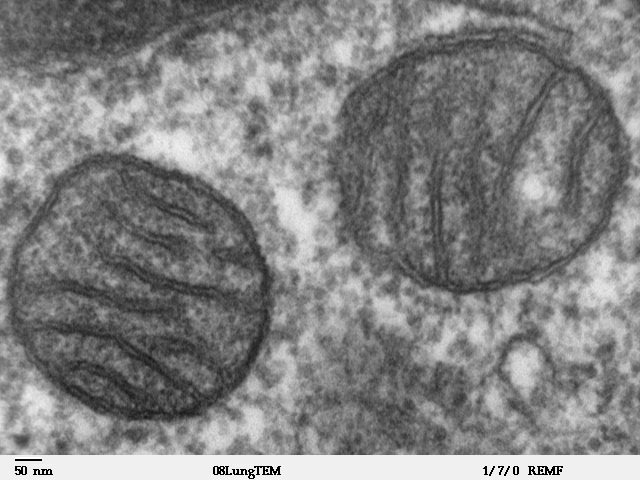
A mitochondrion (; ) is an organelle found in the cells of most Eukaryotes, such as animals, plants and fungi. Mitochondria have a double membrane structure and use aerobic respiration to generate adenosine triphosphate (ATP), which is used throughout the cell as a source of chemical energy. They were discovered by Albert von Kölliker in 1857 in the voluntary muscles of insects. The term ''mitochondrion'' was coined by Carl Benda in 1898. The mitochondrion is popularly nicknamed the "powerhouse of the cell", a phrase coined by Philip Siekevitz in a 1957 article of the same name. Some cells in some multicellular organisms lack mitochondria (for example, mature mammalian red blood cells). A large number of unicellular organisms, such as microsporidia, parabasalids and diplomonads, have reduced or transformed their mitochondria into other structures. One eukaryote, '' Monocercomonoides'', is known to have completely lost its mitochondria, and one multicellular organis ... [...More Info...] [...Related Items...] OR: [Wikipedia] [Google] [Baidu] |


